. 2004 Dec;15(12):5318-28.
doi: 10.1091/mbc.e04-05-0385. Epub 2004 Sep 22.

Characterization of the TPX2 domains involved in microtubule nucleation and spindle assembly in Xenopus egg extracts

Affiliations

Characterization of the TPX2 domains involved in microtubule nucleation and spindle assembly in Xenopus egg extracts

Stéphane Brunet et al. Mol Biol Cell. 2004 Dec.

Abstract

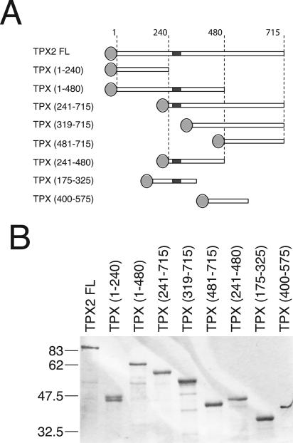
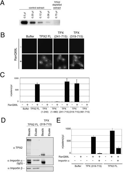
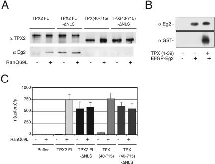
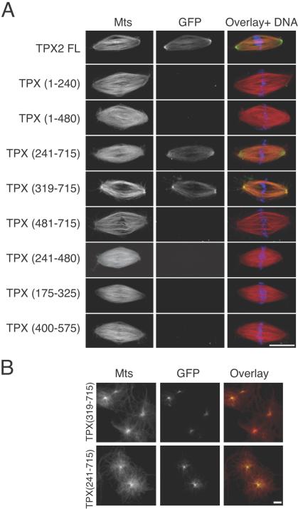
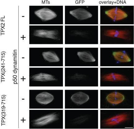
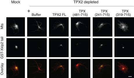
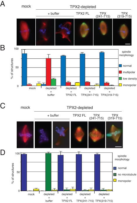
TPX2 has multiple functions during mitosis, including microtubule nucleation around the chromosomes and the targeting of Xklp2 and Aurora A to the spindle. We have performed a detailed domain functional analysis of TPX2 and found that a large N-terminal domain containing the Aurora A binding peptide interacts directly with and nucleates microtubules in pure tubulin solutions. However, it cannot substitute the endogenous TPX2 to support microtubule nucleation in response to Ran guanosine triphosphate (GTP) and spindle assembly in egg extracts. By contrast, a large C-terminal domain of TPX2 that does not bind directly to pure microtubules and does not bind Aurora A kinase rescues microtubule nucleation in response to RanGTP and spindle assembly in TPX2-depleted extract. These and previous results suggest that under physiological conditions, TPX2 is essential for microtubule nucleation around chromatin and functions in a network of other molecules, some of which also are regulated by RanGTP.


Figures

Figure 1.
Figure 1.
TPX2 recombinant proteins used in this study. (A) Schematic representation of the GFP-fusion TPX2 fragments generated for this study. Light gray discs represent EGFP tag. TPX2 functional NLS (NLS284) is a dark gray square. (B) Coomassie-stained gel of the GFP-fusion TPX2 fragments purified from E. coli (see Materials and Methods). Molecular masses of marker proteins are indicated on the left.
Figure 2.
Figure 2.
Microtubule binding of GFP-TPX2 fragments in vitro. (A) Behavior of TPX2 FL, TPX (1-480), and TPX (319-715) in a microtubule-pelleting assay. Equivalent volumes of the reaction before sedimentation (Tot), the microtubule pellet (Pel), the microtubule supernatant (SN), and the control pellet (without microtubules) (Cont) were analyzed by SDS-PAGE. Proteins were visualized with Coomassie Brilliant Blue (TPX fragments are indicated with a bar and tubulin dimers with a spot). (B) Microtubule binding properties of the GFP-TPX2 fragments studied by FLAP. Glass bottomed wells were coated with taxol-stabilized microtubules labeled with Cy5 (left). GFP-TPX2 fragments were added to the wells in an oxygen-depleted medium. Before photoconversion, no signal is detected in the photoconversion channel. After illumination of a restricted area (100-μm2 square), GFP-fragments that bind to the microtubules (here GFP-TPX2 full length) emit a signal in the photoconversion channel. (C) Typical decay curve of the photoconversion signal (here measured for GFP-TPX2 full length). (D) Half-life of the interaction of GFP-TPX2 fragments with microtubules. Ten curves were analyzed for GFP-TPX2 full length, 21 for TPX (1-240), 30 for TPX (1-480), 13 for TPX (241-480), and 15 for TPX (241-715). The average half-life value for each fragment is indicated on the corresponding bar. Error bars represent SD.
Figure 3.
Figure 3.
Microtubule nucleation activity of GFP-TPX2 fragments in vitro. (A) Representative images of microtubules and aggregates formed after incubation of GFP-TPX2 fragments (as indicated) with tubulin. Bar, 10 μm. (B) Quantification of the structures observed as in A. For each TPX2 fragment, the number of structures associated with microtubules (white bars) or not associated with microtubules (black bars) was counted in at least 10 fields in two independent experiments. Error bars correspond to the variance between the two experiments.
Figure 4.
Figure 4.
Microtubule nucleation activity of GFP-TPX2 fragments in Xenopus egg extract. (A) Western blot probed with the anti-TPX2 antibody showing the efficiency of TPX2 depletion from the egg extract. Indicated volumes of TPX2 depleted and control extracts were loaded on SDS-PAGE gel. (B) Representative images of microtubule asters formed upon addition of GFP-TPX2 FL or GFP-TPX2 fragments to TPX2 depleted extract, in the presence or absence of RanQ69L. Bar, 10 μm. (C) Quantification of the experiment shown in B. The values represent the averages from three independent experiments. Error bars represent SD. (D) GFP-TPX2 FL and GFP-TPX (319-715) were incubated in mitotic egg extract and immunoprecipitated with an anti GFP antibody. Immunoprecipitated proteins were eluted with a high salt buffer (eluate) and sample buffer (beads) successively. The blot was probed with anti-importin α and β antibodies. (E) Quantification of microtubule asters formed in TPX2-depleted extract containing RanQ69L after addition of either TPX2 full-length or TPX (319-715) in the presence or absence of exogenous importin α. Importin α inhibits microtubule aster formation induced by TPX2 full length and TPX (319-715). The averages from two independent experiments are shown. Error bars represent the variance between the two experiments.
Figure 5.
Figure 5.
The interaction of TPX2 with Aurora A/Eg2 is not required for TPX2 directed microtubule nucleation in the egg extract. (A) GFP-TPX2 FL, GFP-TPX (40-715), and the corresponding proteins mutated in the NLS [GFP-TPX2 FL-ΔNLS and GFP-TPX (40-715)-ΔNLS] were incubated in mitotic egg extract in the presence or absence of RanQ69L and immunoprecipitated with an anti-GFP antibody. Immunoprecipitated proteins were analyzed by Western blot by using anti-GFP and anti-Eg2 antibodies. TPX (40-715) does not interact with Aurora A/Eg2 even in the presence of RanQ69L. (B) GST-TPX (1-39) and GFP-Eg2 were added to a mitotic egg extract as indicated. Immunoprecipitation with an anti-GFP antibody revealed that the first N-terminal amino acids of TPX2 are sufficient for interaction with GFP-Eg2. Western blots probed with anti-Eg2 and anti-GST are shown. (C) Quantification of asters formed in TPX2-depleted extract upon addition of buffer, GFP-TPX2 FL, GFP-TPX (40-715), GFP-TPX2 FL-ΔNLS, and GFP-TPX (40-715)-ΔNLS. The graph shows the data obtained in one representative experiment counting the number of asters in ten randomly selected microscope fields (the experiment was repeated twice). The bars represent the internal SD of the experiment.
Figure 6.
Figure 6.
Localization of GFP-TPX2 fragments on spindles and asters assembled in Xenopus egg extracts. (A) GFP-TPX2 fragments (0.1 μM) were added to egg extracts as they were cycled back into mitosis. Their localization was examined on spindles assembled around sperm nuclei. In the overlays, microtubules are in red, GFP-fragments in green, and chromatin in blue. Bar, 10 μm. (B) TPX (241-715) and TPX (319-715) (0.1 μM) were added to a TPX2-depleted extract. Microtubule asters formation was induced by addition of purified centrosomes. In the overlays, microtubules are in red, and GFP-fragments in green.
Figure 7.
Figure 7.
P50 dynamitin disrupts GFP-TPX2 full length, GFP-TPX (241-715), and GFP-TPX (319-715) association with the spindle. Recombinant p50 dynamitin (0.7 mg/ml) and TPX (241-715) or TPX (319-715) or TPX2 full length (0.1 μM) were added to cycled spindle reactions. In overlays, microtubules are in red, GFP-fragments in green, and chromatin in blue. Bar, 10 μm.
Figure 8.
Figure 8.
GFP-TPX (319-715) is sufficient to localize GST-Xklp2-tail to the center of centrosomal asters. Purified centrosomes were incubated for 30 min in TPX2-depleted or mock-depleted mitotic egg extracts containing GST-Xklp2 tail (2 μM) and GFP-TPX2 fragments (0.1 μM). GST-Xklp2-tail was detected with an anti-GST antibody. Microtubules are in red, and the GST-Xklp2 tail in green. Bar, 10 μm.
Figure 9.
Figure 9.
GFP-TPX (319-715) rescues spindle assembly around sperm nuclei and DNA-coated beads in TPX2-depleted extract. (A) Morphology of spindles assembled around sperm nuclei in mock-depleted extract and in TPX2-depleted extract to which buffer, GFP-TPX2 full length, GFP-TPX (241-715), or GFP-TPX (319-715) was added. Microtubules are in red, chromatin in blue, and the GFP-proteins in green. Bar, 10 μm. (B) Quantification of the different types of structures formed under various depletion and add-back conditions as in A. Blue bars, normal spindles; red bars, multipolar spindles; green bars, bipolar spindles with low microtubule density; and yellow bars, monopolar spindles. The experiment was performed in three different egg extracts. The bars represent the SD. (C) Morphology of spindles assembled around DNA-coated beads in mock-depleted extract and in TPX2-depleted extract to which buffer, GFP-TPX2 full length, GFP-TPX (241-715), or GFP-TPX (319-715) was added. Microtubules are in red, chromatin in blue, and the GFP-proteins in green. Bar, 10 μm. (D) Quantification of different types of structures formed under the various depletion and addback conditions as in B. Magenta bars, normal spindles; green bars, beads without microtubules; and yellow bars, monopolar spindles. The experiment was performed in two different egg extracts. The bars correspond to the variance between the two experiments.

Similar articles

Cited by

References

    1. Bayliss, R., Sardon, T., Vernos, I., and Conti, E. (2003). Structural basis of Aurora-A activation by TPX2 at the mitotic spindle. Mol. Cell 12, 851–862. - PubMed
    1. Bornens, M., and Moudjou, M. (1999). Studying the composition and function of centrosomes in vertebrates. Methods Cell Biol. 61, 13–34. - PubMed
    1. Dasso, M. (2001). Running on Ran: nuclear transport and the mitotic spindle. Cell 104, 321–324. - PubMed
    1. Dasso, M. (2002). The Ran GTPase: theme and variations. Curr. Biol. 12, R502–R508. - PubMed
    1. Desai, A., Murray, A., Mitchison, T.J., and Walczak, C.E. (1999). The use of Xenopus egg extracts to study mitotic spindle assembly and function in vitro. Methods Cell Biol. 61, 385–412. - PubMed

Publication types

MeSH terms

LinkOut - more resources