. 2004 Sep 20;200(6):783-95.
doi: 10.1084/jem.20040254.

Lymph node fibroblastic reticular cells construct the stromal reticulum via contact with lymphocytes

Affiliations

Lymph node fibroblastic reticular cells construct the stromal reticulum via contact with lymphocytes

Tomoya Katakai et al. J Exp Med. .

Abstract

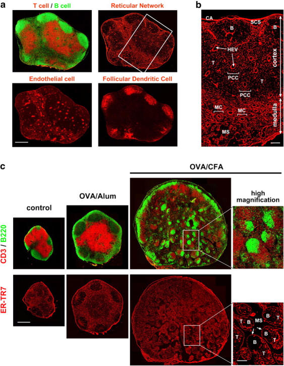
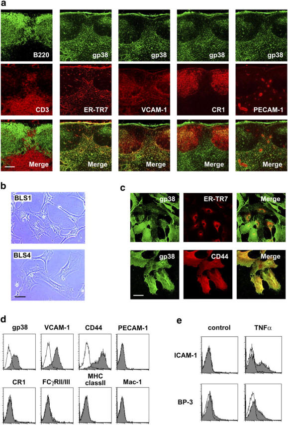
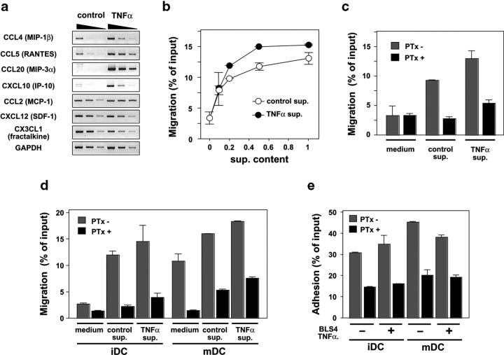
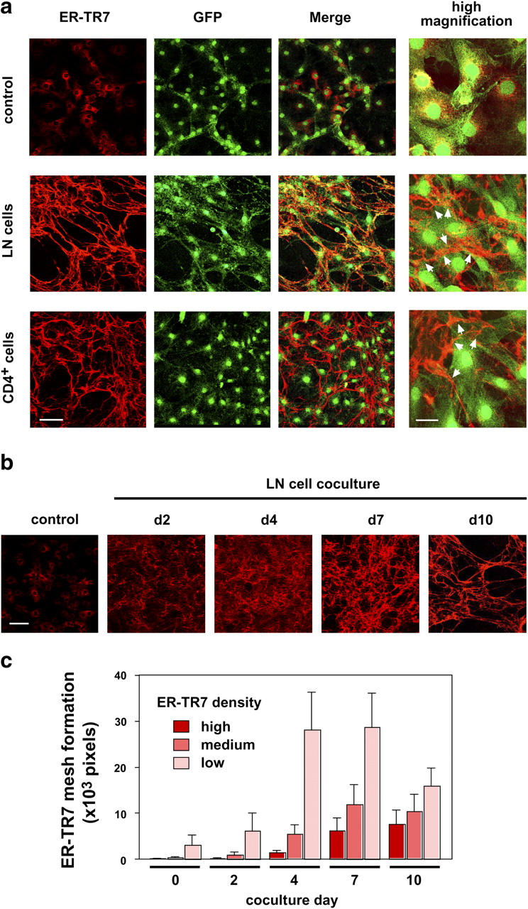
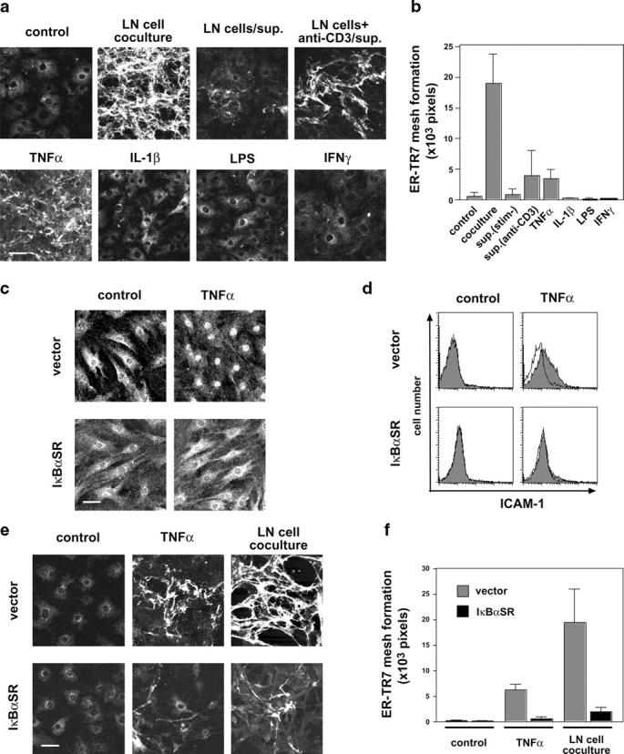
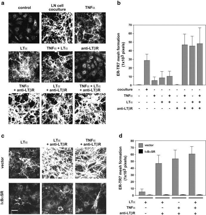
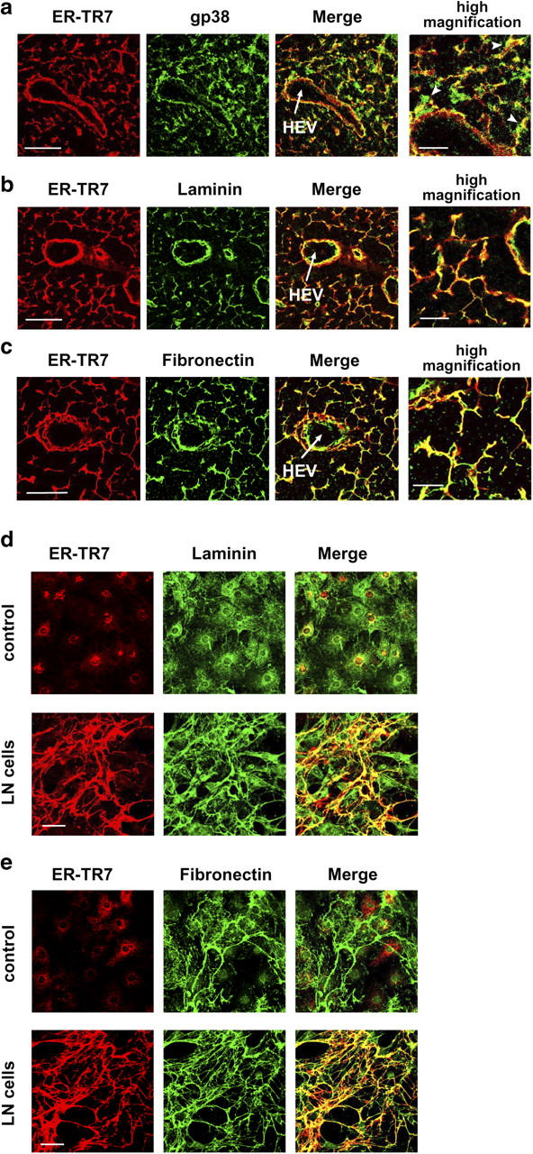
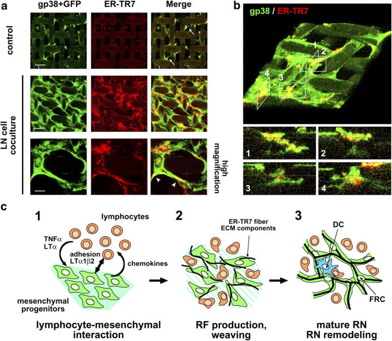
The sophisticated microarchitecture of the lymph node, which is largely supported by a reticular network of fibroblastic reticular cells (FRCs) and extracellular matrix, is essential for immune function. How FRCs form the elaborate network and remodel it in response to lymphocyte activation is not understood. In this work, we established ERTR7(+)gp38(+)VCAM-1(+) FRC lines and examined the production of the ER-TR7 antigen. Multiple chemokines produced by FRCs induced T cell and dendritic cell chemotaxis and adhesion to the FRC surface. FRCs can secrete the ER-TR7 antigen as an extracellular matrix component to make a reticular meshwork in response to contact with lymphocytes. The formation of the meshwork is induced by stimulation with tumor necrosis factor-alpha or lymphotoxin-alpha in combination with agonistic antibody to lymphotoxin-beta receptor in a nuclear factor-kappaB (RelA)-dependent manner. These findings suggest that signals from lymphocytes induce FRCs to form the network that supports the movement and interactions of immune effectors within the lymph node.


Figures

Figure 1.
Figure 1.
Architecture of the ER-TR7+-RN in mouse LN, and the dynamic remodeling during immune responses. (a) Whole views of LN stromal populations. Serial frozen sections were stained with several antibodies to detect CD3 (T cells), B220 (B cells), ER-TR7 antigen (reticular fibroblasts), PECAM-1 (endothelial cells), or CR1 (FDCs). The images shown are composites of multiple high-magnification images at distinct positions of an LN assembled on silica. Bar, 400 μm. (b) Higher magnification view of RN (inset in panel a) shows subreticular structures or compartments. B, B zone [follicle]; CA, capsule; HEV, high endothelial venule; MC, medullary cord; MS, medullary sinus; PCC, paracortical cord; SCS, subcapsular sinus; T, T zone. Bar, 100 μm. (c) Immunizations with OVA plus adjuvant induce dynamic remodeling of the RN within popliteal LNs. Serial frozen sections of the popliteal LNs from untreated, OVA + alum-, or OVA + CFA-immunized mice were stained with antibodies against CD3, B220, or ER-TR7. Three whole LN images are shown at the same magnification. Higher magnification view of the inset region in the medulla of CFA-immunized LN shows some nodular islands containing discrete B and T zones, enclosed by a single FRC layer (arrows). Bars: 500 μm for whole views and 100 μm for high-magnification views.
Figure 2.
Figure 2.
Phenotypic features of FRCs and the establishment of BLS lines. (a) FRCs in the LN are ER-TR7+gp38+VCAM-1+ cells. Serial frozen sections were stained for various markers. Bar, 100 μm. (b) Typical fibroblastic morphology of BLS1 and BLS4 cells. Phase-contrast views of growing cells on plastic dishes are shown. Bar, 50 μm. (c) Intracellular localization of ER-TR7 compared to surface expression of gp38 and CD44 in BLS4 cells. Cells grown on chamber slides were stained with antibodies after fixation and permeabilization. Bar, 50 μm. (d) Cell surface expression of gp38, VCAM-1, and CD44 in BLS4 cells. EDTA-harvested cells were stained for the indicated surface markers and analyzed by flow cytometry. (e) Expression of ICAM-1 and BP-3 is induced by TNFα. Cells were stimulated with TNFα and subsequently stained with anti–ICAM-1 or anti–BP-3 antibody for flow cytometry.
Figure 3.
Figure 3.
BLS cells support the migration and adhesion of T cells and DCs. (a) BLS4 cells express various chemokines with or without TNFα stimulus. Transcripts for the indicated chemokines were detected by semi-quantitative RT-PCR analysis. (b) BLS4 supernatant (sup.) induces the chemotaxis of CD4+ T cells. The chemotactic activity in diluted culture supernatants from BLS4 cells (prestimulated or not prestimulated with TNFα) was analyzed by a chemotaxis assay. (c) Chemotaxis of CD4+ T cells to the BLS supernatants is Gαi dependent. T cells were pretreated or not pretreated with PTx and subjected to the chemotaxis assay. Two-fold diluted supernatants were used as attractant. (d) BLS4 supernatant induces DC chemotaxis. Immature (iDC) or mature (mDC) DCs were pretreated or not pretreated with PTx and subjected to the chemotaxis assay. (e) BLS4 cells support DC adhesion to their surface. DCs were pretreated or not pretreated with PTx and subjected to the adhesion assay.
Figure 4.
Figure 4.
BLS cells construct the ER-TR7 meshwork via contact with lymphocytes. (a) ER-TR7 meshwork formation by BLS4 cells cocultured with LN cells or CD4+ T cells. BLS4-GFP monolayers on chamber slides were cocultured with LN cells or CD4+ T cells for 10 d. Cells were stained with anti–ER-TR7 antibody after fixation and permeabilization, and examined by confocal microscopy. Higher-magnification views show that the ER-TR7 meshwork was frequently positioned in the cell periphery outside the cells (arrowheads). Bars: 100 μm for low- and 30 μm for high-magnification views. (b) BLS4 cells gradually constructed ER-TR7 meshwork during ∼1 wk in the coculture system. Cells were stained for ER-TR7 after the indicated number of days of coculturing. Bar, 100 μm. (c) Quantification of ER-TR7 meshwork formation. Total pixels at a certain density window (high: 0–50; medium: 51–100; low: 101–150) of more than five images were calculated and are shown as mean ± SD. We defined high and medium signals as the mature ER-TR7 fibers.
Figure 5.
Figure 5.
TNFα–NF-κB pathway participates in the ER-TR7 meshwork formation. (a and b) Soluble factors from activated T cells or TNFα partially induce ER-TR7 meshwork in BLS4 cultures. BLS4 cells on chamber slides were cocultured with LN cells or stimulated with LN culture supernatants (in the presence or absence of anti-CD3 antibody) or various cytokines for 10 d and stained with anti–ER-TR7 antibody. Quantification of mature ER-TR7 meshwork (high and medium density) is shown as mean ± SD (b). (c and d) IκBαSR prevents the nuclear translocation of RelA (p65) and expression of ICAM-1 in BLS4 cells. Stable transfectants for control vector or IκBαSR were stimulated with TNFα for 1 h and stained with anti-RelA antibody for confocal microscopic analysis (c) or anti–ICAM-1 antibody for flow cytometry (d). (e and f) IκBαSR inhibits ER-TR7 meshwork formation in BLS4 cultures. Meshwork formation of vector- or IκBαSR-transfectant was examined after 10 d of TNFα stimulation or coculturing with LN cells. Bar, 50 μm. Quantification of mature ER-TR7 meshwork is shown as mean ± SD (f).
Figure 6.
Figure 6.
Dual signaling via TNFR and LTβR triggers ER-TR7 meshwork formation. (a) Combined stimulation using agonistic anti-LTβR antibody and TNFα or LTα induces full construction of the ER-TR7 meshwork. BLS4 cells on chamber slides were stimulated with TNFα, LTα, or anti-LTβR antibody, or their combinations, and stained for ER-TR7. Quantification of mature ER-TR7 meshwork is shown as mean ± SD (b). (c and d) IκBaSR inhibits ER-TR7 meshwork formation induced by dual stimulation with TNFR and LTβR. Meshwork formation of transfectants in response to the combinatorial stimulation was examined and quantified. Bar, 50 μm.
Figure 7.
Figure 7.
ER-TR7 antigen is secreted as an ECM component. (a) FRC cell body marked by gp38 ensheathes ER-TR7+-RF (arrowheads). (b and c) ER-TR7 is colocalized with laminin (b) and fibronectin (c) in the RN in vivo. LN sections were stained for ER-TR7 and gp38, laminin, or fibronectin. Bars: 50 μm for lower- and 20 μm for higher-magnification views. (d and e) Colocalization of ER-TR7 and laminin (d), or fibronectin (e) in cocultures of BLS4 monolayers with LN cells. Bars, 50 μm.
Figure 8.
Figure 8.
Three-dimensional FRC/RF network construction. (a) Reconstitution of RN-like structure in semi–three-dimensional nylon-mesh culture. BLS4-GFP cells grown on nylon-mesh supports were cocultured with LN cells for 10 d and stained for ER-TR7 and gp38. Note that weak nonspecific background signals were observed on the nylon supports. Bars: top and middle, 50 μm and bottom, 20 μm. (b) BLS4 cells enwrap ER-TR7 fibers. A three-dimensional reconstitution image of the semi–three-dimensional mesh culture stained for gp38 and ER-TR7 was produced from the z-stacks of image data (top, large angled view). Optical sections in the z-plane of appropriate positions (–4) are shown (bottom). (c) Model of RN formation. (1) Accumulation of lymphocytes induces alteration of mesenchymal progenitors such that they produce chemokines for further lymphocyte attraction and RF components via the secretion of TNFα or LTα (or related cytokines) and likely also via direct contacts mediated by adhesion molecules and LTα1β2. (2) Lymphocytes and differentiated FRCs gradually degrade preexisting matrix, and FRCs weave RF meshwork from the newly produced components. (3) Mature RN forms and antigen presentation occur on the stromal reticulum.

Similar articles

Cited by

References

    1. Zinkernagel, R.M., S. Ehl, P. Aichele, S. Oehen, T. Kündig, and H. Hengartner. 1997. Antigen localization regulates immune responses in a dose- and time-dependent fashion: a geographical view of immune reactivity. Immunol. Revs. 156:199–209. - PubMed
    1. Fu, Y.-X., and D.D. Chaplin. 1999. Development and maturation of secondary lymphoid tissues. Annu. Rev. Immunol. 17:399–433. - PubMed
    1. Cyster, J.G. 1999. Chemokine and cell migration in secondary lymphoid organs. Science. 286:2098–2102. - PubMed
    1. Young, B., and J.H. Heath. 2000. Wheater's Functional Histology. 4th ed. Harcourt Publishers Limited, London.
    1. Cyster, J.G., K.M. Ansel, K. Reif, E.H. Ekland, P.L. Hyman, H.L. Tang, S.A. Luther, and V.N. Ngo. 2000. Follicular stromal cells and lymphocyte homing to follicles. Immunol. Revs. 176:181–193. - PubMed

Publication types

Substances