. 2004 Oct;16(10):2573-85.
doi: 10.1105/tpc.104.024810. Epub 2004 Sep 14.

Stimulus-dependent, promoter-specific binding of transcription factor WRKY1 to Its native promoter and the defense-related gene PcPR1-1 in Parsley

Affiliations

Stimulus-dependent, promoter-specific binding of transcription factor WRKY1 to Its native promoter and the defense-related gene PcPR1-1 in Parsley

Franziska Turck et al. Plant Cell. 2004 Oct.

Abstract

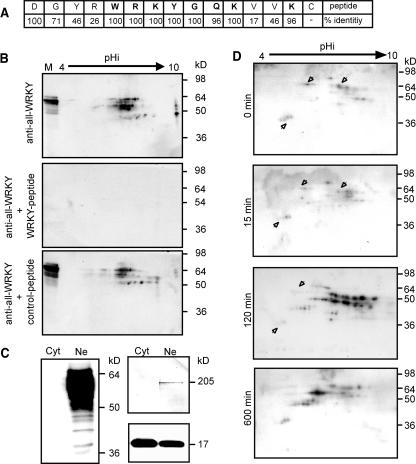
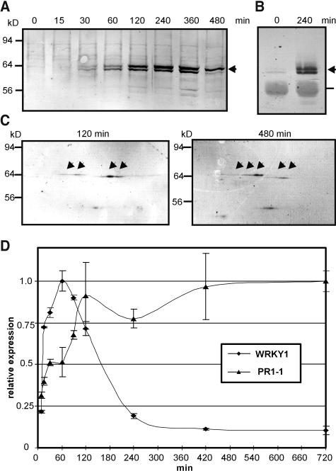
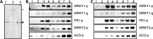
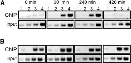
WRKY transcription factors form a large family that plays a role in plant responses to biotic stress and during senescence. Defining in vivo relevant WRKY/promoter relationships has been hampered by the factors' indiscriminate binding to known W box DNA elements and their possible genetic redundance. Employing chromatin immunoprecipitations (ChIP) of cultured cells, we show that parsley (Petroselinum crispum) WRKY1 protein binds to the W boxes of its native promoter as well as to that of PcWRKY3 and the defense-related PR10-class marker gene Pathogenesis-Related1-1 (PcPR1-1). Although present at low concentrations in resting cells, WRKY1 does not appear to play a role in the immediate early gene response upon elicitation because it does not bind to the promoter at this time. Paradoxically, in vivo binding at the PcWRKY1 promoter correlates more with downregulation of gene expression, whereas previous overexpression studies suggested an activating function of WRKY1 on PcWRKY1 expression. By contrast, PcPR1-1 expression remains strong when its promoter is occupied in vivo by WRKY1. Unexpectedly, ChIP revealed that W boxes at promoter sites are constitutively occupied by other WRKY transcription factors, indicating that site recruitment does not seem to play a major role in their regulation. Rather, WRKY proteins very likely act in a network of mutually competing participants with temporal displacement occurring at defined preoccupied sites by other family members in a stimulus-dependent manner.


Figures

Figure 1.
Figure 1.
Characterization of WRKY Protein Pools. (A) Immunogenic peptide for the generation of anti-all-WRKY serum. Rabbits were immunized with the depicted 15–amino acid long peptide that spans the most conserved part of the WRKY domain. The overall identities of sequential amino acids to all 74 known Arabidopsis WRKY proteins are indicated. (B) Specificity of the generated serum. Arabidopsis nuclear extracts were separated by two-dimensional PAGE and probed with the anti-all-WRKY serum alone (anti-all-WRKY, top panel), in the presence of immunogenic peptide (+ WRKY-peptide, middle panel), or in the presence of the same amount of unrelated peptide (+ control-peptide, bottom panel). At the left border of the gel, a mixture of prestained protein gel markers and Arabidopsis nuclear extract was loaded for one-dimensional separation (M). (C) Cytoslic/nuclear distribution of WRKY proteins. The anti-all-WRKY serum was used to probe parsley cytosolic (Cyt) and nuclear (Ne) extracts from 1 h pep25 stimulated parsley cells for the presence of WRKY proteins (left panel). The quality of cell fractionation was assessed by probing parallel blots with monoclonal antibodies against the Ser-5 phosphorylated heptapeptide repeat YSPTSPS of RNA polymerase II (right, top panel) or a serum raised against PcPR1-1 (right, bottom panel). (D) Preformed and induced WRKY protein pools in parsley. Nuclear extracts from resting parsley cells (0 min, first panel) or cells stimulated for 15 min (15 min, second panel), 120 min (120 min, third panel), or 600 min (600 min, fourth panel) with pep25 elicitor were separated by two-dimensional PAGE and analyzed by protein gel blots using the anti-all-WRKY serum. Arrowheads denote proteins showing reduced levels in elicitor treated cells. Molecular mass range is indicated at the right. The entire time course was analyzed twice, and time points from 0 to 120 min were analyzed five times in independent experiments.
Figure 2.
Figure 2.
Alterations of WRKY1 in Stimulated Parsley Cells. (A) Direct protein gel blot analysis of parsley nuclear extracts during an 8 h time course of stimulation with pep25 elicitor using a WRKY1 peptide specific antiserum. The position of the WRKY1 protein doublet is marked by an arrow. Other visible signals on the blot correspond with background detected by the serum because they are not competed out by the immunogenic peptide (see Methods). Molecular mass range is indicated at the left. The experiment was performed five times with similar results. (B) Protein gel blot analysis of immuno-enriched nuclear extracts. After immunoprecipition of WRKY1 from nuclear extracts of resting or 2 h elicitor treated cells with the help of cyanogen bromide-agarose coupled anti-WRKY1–specific serum, precipitates were separated on one-dimensional gels and reprobed with the same antibodies. WRKY1 protein doublet bands are marked by an arrow and IgG background by a line. The experiment was performed twice with similar results. (C) Two-dimensional protein gel blot of WRKY1. Nuclear extracts of parsley cells that were pep25 elicitor treated for the indicated time were separated on two-dimensional gels. Pictures show portions of the original gels covering the pH range of 7 to 9. The position of WRKY1 signals are indicated by arrows. This experiment was performed twice with similar results. (D) Analysis of WRKY1 and PR1-1 expression by RT-PCR. Changes in mRNA levels were assayed over a time course of elicitor stimulation of parsley cells with pep25 as indicated. Expression levels were analyzed by SYBR Green-based real-time PCR and expression values normalized to those of PcUbiquitin4. Values on the graph are normalized to the point of highest expression for each gene. Expression analyses of the full time course were performed twice with similar results.
Figure 3.
Figure 3.
Detection of WRKY1 in Vivo Target Sites. (A) Analysis of WRKY1 protein. Chromatin was prepared from cells treated with pep25 elicitor for 2 h (lane 1) or from cells subsequently fixed with formaldehyde (0.5%) for 7 min (lane 2) and analyzed by protein gel blots for the detection of WRKY1 protein. The cross-links from a parallel sample as in lane 2 were reversed by heat treatment (lane 3) and analyzed. The position of free WRKY1 is indicated by an arrowhead and the position of cross-linked complexes at the height of the gel pocket by a straight line. This experiment was performed three times with similar results. (B) Analysis of DNA after anti-WRKY1 immunoprecipitation. Chromatin preparations from elicitor-treated and formaldehyde fixed cells as in (A) were immunoprecipitated with WRKY1-specific antibody. The DNA was recovered after reversal of the cross-links and analyzed for the enrichment of WRKY1 promoter (WRKY1-p), WRKY1 intragenic region (WRKY1-g), PR1-1 promoter (PR1-p), WRKY3 promoter (WRKY3-p), and parsley ACO promoter (ACO-p) by PCR. Immunoprecipitations were performed with anti-WRKY1 preimmune serum (lane 1), with affinity-purified anti-WRKY1 antibodies in the presence of immunogenic peptide (lane 2), with anti-WRKY1 antibodies in the presence of the same amount of unrelated peptide (lane 3), or with anti-WRKY1 antibodies alone (lane 4). As a control for quantities amplified during the PCR, sequential twofold dilutions of input material from chromatin preparations were processed in parallel. Lane 5, 1.25 × 10⁻⁴; lane 6, 2.5 × 10⁻⁴; lane 7, 0.5 × 10⁻³; lane 8, 1.0 × 10⁻³. (C) Analysis of DNA after anti-all-WRKY immunoprecipitation. Parallel incubations were performed as in (B) except that anti-all-WRKY serum and immunogenic peptide were used in the incubations. The input dilutions were as follows: lane 5, 0.5 × 10⁻³; lane 6, 1.0 × 10⁻³; lane 7, 2.0 × 10⁻³; lane 8, 4.0 × 10⁻³. The experiment was performed twice with similar results.
Figure 4.
Figure 4.
Resolution of ChIP. (A) Schematic representation of the parsley WRKY1 locus. PcWRKY1 promoter and terminator regions are indicated by horizontal lines and transcribed sequences by closed boxes (untranslated regions), open boxes (exons), and bent lines (introns). The positions of W box consensus motifs are indicated by gray transversal lines, and the position of the well-characterized functional WBC cluster is labeled. The positions and relative sizes of the different PCR fragments covering the locus are indicated by labeled boxes (a to j). (B) WRKY1 ChIP resolution scan. Formaldehyde cross-linked chromatin preparations (derived from 1 h elicitor treated cells) were immunoprecipitated with anti-WRKY1 preimmune serum (top panels, lane 1), anti-WRKY1–specific affinity-purified antibodies in the presence of immunogenic peptide (top panels, lane 2), anti-WRKY1 antibodies in the presence of unrelated peptide (top panels, lane 3), or with anti-WRKY1 antibodies alone (top panels, lane 4). DNA was recovered and analyzed on agarose gels after performing PCR with the different primer combinations yielding the appropriate fragments as indicated above the panels. As a control for quantities amplified during PCR, sequential twofold dilutions of input material from chromatin preparations were processed in parallel and are shown in the bottom panels. Lane 1, 1.25 × 10⁻⁴; lane 2, 2.5 × 10⁻⁴; lane 3, 0.5 × 10⁻³; lane 3, 1.0 × 10⁻³. (C) All-WRKY ChIP scan. ChIP and analysis were performed as in (B) except that the anti-all-WRKY serum and specific peptide were used in the incubations. Parallel PCR of input dilutions are shown in the bottom panels. Lane 1, 0.5 × 10⁻³; lane 2, 1.0 × 10⁻³; lane 3, 2.0 × 10⁻³; lane 4, 4.0 × 10⁻³. The experiment was performed three times with similar results.
Figure 5.
Figure 5.
Elicitor-Dependent Time-Course Analysis of WRKY Factor Binding to the WRKY1 Promoter. (A) In vivo WRKY1 binding to parsley WRKY1. Pep25-treated parsley cells were formaldehyde fixed over a time course of 12 h and analyzed by ChIP using anti-WRKY1 preimmune serum (lane 1), anti-WRKY1–specific antibodies in the presence of immunogenic peptide (lane 2), anti-WRKY1–specific antibodies in the presence of unrelated peptide (lane 3), or with anti-WRKY1–specific antibodies alone (lane 4). PCR was performed using primer combinations e (top panel set) and j (bottom panel set). Diluted samples from ChIP inputs were analyzed by PCR in parallel (bottom row of each set, lane 1, 1.25 × 10⁻⁴; lane 2, 2.5 × 10⁻⁴; lane 3, 0.5 × 10⁻³; lane 3, 1.0 × 10⁻³). (B) Overall WRKY factor binding to parsley WRKY1. ChIP was performed from the same chromatin samples as in (A) with anti-all-WRKY preimmune serum (lane 1), anti-all-WRKY immune serum in the presence of immunogenic peptide (lane 2), anti-all-WRKY immune serum in the presence of unrelated peptide (lane 3), or with anti-all-WRKY immune serum alone (lane 4). PCR was performed using primer combinations e (top panel set), j (middle panel set), and h (bottom panel set). Diluted samples from ChIP inputs were analyzed by PCR in parallel (bottom row of each panel set, lane 1, 0.5 × 10⁻³; lane 2, 1.0 × 10⁻³; lane 3, 2.0 × 10⁻³; lane 4, 4.0 × 10⁻³). The experiment covering the entire time course was performed twice with similar results, whereas the time points from 0 to 240 min were analyzed in five independent experiments with similar results.
Figure 6.
Figure 6.
Elicitor-Dependent Time-Course Analysis of WRKY Factor Binding in Vivo to the Parsley PR1-1 Promoter. (A) WRKY1-specific binding. Pep25 treated parsley cells were formaldehyde treated over a time course of 7 h and analyzed by ChIP using anti-WRKY1 preimmune serum (lane 1), anti-WRKY1–specific antibodies in the presence of immunogenic peptide (lane 2), anti-WRKY1–specific antibodies in the presence of unrelated peptide (lane 3), or with anti-WRKY1–specific antibodies alone (lane 4). PCR was performed using a primer combination that covered the W box–containing region of PR1-1. Diluted input samples from ChIP were analyzed by PCR in parallel (bottom row of panel set, lane 1, 1.25 × 10⁻⁴; lane 2, 2.5 × 10⁻⁴; lane 3, 0.5 × 10⁻³; lane 3, 1.0 × 10⁻³). (B) Overall WRKY factor binding. Samples were processed and analyzed as in (A) except that anti-all-WRKY serum and immunogenic peptide were used in the incubations. Diluted samples from ChIP inputs were analyzed by PCR in parallel (bottom row of each panel set, lane 1, 0.5 × 10⁻³; lane 2, 1.0 × 10⁻³; lane 3, 2.0 × 10⁻³; lane 4, 4.0 × 10⁻³). The entire time-course experiment was performed twice with similar results, whereas time points from 0 to 240 min were analyzed in five independent experiments with similar results.

Similar articles

Cited by

References

    1. Asai, T., Tena, G., Plotnikova, J., Willmann, M.R., Chiu, W.L., Gomez-Gomez, L., Boller, T., Ausubel, F.M., and Sheen, J. (2002). MAP kinase signalling cascade in Arabidopsis innate immunity. Nature 415, 977–983. - PubMed
    1. Barolo, S., and Posakony, J.W. (2002). Three habits of highly effective signaling pathways: Principles of transcriptional control by developmental cell signaling. Genes Dev. 16, 1167–1181. - PubMed
    1. Cahill, M.A., Ernst, W.H., Janknecht, R., and Nordheim, A. (1994). Regulatory squelching. FEBS Lett. 344, 105–108. - PubMed
    1. Chen, C., and Chen, Z. (2000). Isolation and characterization of two pathogen- and salicylic acid-induced genes encoding WRKY DNA-binding proteins from tobacco. Plant Mol. Biol. 42, 387–396. - PubMed
    1. Chen, C., and Chen, Z. (2002). Potentiation of developmentally regulated plant defense response by AtWRKY18, a pathogen-induced Arabidopsis transcription factor. Plant Physiol. 129, 706–716. - PMC - PubMed

Publication types

LinkOut - more resources