Comparative Study
. 2004 Sep;78(18):9872-89.
doi: 10.1128/JVI.78.18.9872-9889.2004.

Molecular, biological, and in vivo characterization of the guinea pig cytomegalovirus (CMV) homologs of the human CMV matrix proteins pp71 (UL82) and pp65 (UL83)

Affiliations
Comparative Study

Molecular, biological, and in vivo characterization of the guinea pig cytomegalovirus (CMV) homologs of the human CMV matrix proteins pp71 (UL82) and pp65 (UL83)

Alistair McGregor et al. J Virol. 2004 Sep.

Abstract

We recently identified the genes encoding the guinea pig cytomegalovirus (GPCMV) homologs of the upper and lower matrix proteins of human CMV, pp71 (UL82) and pp65 (UL83), which we designated GP82 and GP83, respectively. Transient-expression studies with a GP82 plasmid demonstrated that the encoded protein targets the nucleus and that the infectivity and plaquing efficiency of cotransfected GPCMV viral DNA was enhanced by GP82. The transactivation function of GP82 was not limited to GPCMV, but was also observed for a heterologous virus, herpes simplex virus type 1 (HSV-1). This was confirmed by its ability to complement the growth of an HSV-1 VP16 transactivation-defective mutant virus in an HSV viral DNA cotransfection assay. Study of a GP82 "knockout" virus (and its attendant rescuant), generated on a GPCMV bacterial artificial chromosome construct, confirmed the essential nature of the gene. Conventional homologous recombination was used to generate a GP83 mutant to examine the role of GP83 in the viral life cycle. Comparison of the one-step growth kinetics of the GP83 mutant (vAM409) and wild-type GPCMV indicated that GP83 protein is not required for viral replication in tissue culture. The role of GP83 in vivo was examined by comparing the pathogenesis of wild-type GPCMV, vAM409, and a control virus, vAM403, in guinea pigs. The vAM409 mutant was significantly attenuated for dissemination in immunocompromised strain 2 guinea pigs, suggesting that the GP83 protein is essential for full pathogenicity in vivo.


Figures

FIG. 1.
FIG. 1.
Molecular comparison of CMV UL82 homologs. (A) Phylogenetic comparison of GP82 and other CMV UL82 (pp71) homologs. GP82 diverges down the same pathway as rodent UL82 homologs (MCMV and RCMV). Neighbor-joining tree methodology used the following Clustal multiple alignment parameters: open gap penalty, 10; extend gap penalty, 0.1; delay divergent, 40%; gap distance, 8; similarity matrix, blosum. (B) Alignment of GPCMV GP82 protein coding sequence with primate UL82 homologs. Amino acid residues conserved across species are indicated: double dot, conserved residue; asterisk, identical residue. Conserved DIDs (shaded regions marked I and II) (23) and LXCXD motifs (shaded regions marked III) (29) important in primate UL82 function are shaded.
FIG. 2.
FIG. 2.
Immunofluorescence assay of the transient expression of FLAG-tagged GP82. Expression plasmid FLAG82GP (5 μg) was transfected onto subconfluent monolayers on glass coverslips in individual wells of a six-well plate, as described in Materials and Methods. At 36 h posttransfection, cells were fixed and GP82 expression was assayed by immunofluorescence assay using anti-FLAG mouse monoclonal and secondary anti-mouse-fluorescein isothiocyanate conjugate as described in Materials and Methods. (A) FLAG82GP plasmid; (B) mock-transfected cells.
FIG. 3.
FIG. 3.
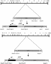
Strategy for the generation of GPCMV GP83 and GP82 mutant viruses. (A) The BamHI subfragment of the HindIII A fragment of the GPCMV genome, encoding the entire GP83 ORF and part of the coding sequences of GP82 and GP84 homolog genes, was cloned into pBluescript to generate pKTS409. To generate the GP83 knockout virus, a shuttle plasmid was generated by collapsing the internal XmnI sites towards the start of the GP83 coding sequence to create an out-of-frame, approximately 250-bp deletion. A BamHI cassette encoding a gpt/EGFP cassette was then introduced into a unique BglII site towards the 3′ end of the ORF. The modified vector was designated pKTS409XmGG and was cotransfected with GPCMV viral DNA onto GPL cells to generate GP83 null virus (vAM409) under gpt+ selection conditions. (B) To generate a GP82 knockout virus, a shuttle vector was generated by introducing a blunt-ended gpt/EGFP cassette into a unique EcoRV site at the start of the GP82 coding sequence to disrupt the GP82 coding sequence. The modified plasmid (pGP82GG) was cotransfected with viral DNA onto GPL cells to generate progeny virus, and mutant virus was selected under gpt+ selection.
FIG. 4.
FIG. 4.
GP82 enhancement of generation of GPCMV from viral DNA transfections. A fixed amount of GPCMV viral DNA (2 μg) was cotransfected onto one well of a six-well dish of GPL cells (106 cells) in the presence of expression plasmids (5 μg) encoding GP82 (A), GP83 (B), VP16 (C), or mock transfected (D). Virus was allowed to develop over a 2-week time period and then stained for viral CPE as described in Materials and Methods. During this time period, viral CPE was only detected in GP82-cotransfected cells, indicating the ability of GP82 to accelerate the infectivity of GPCMV DNA. These results demonstrate the transactivation function of the GP82 ORF, the first demonstration of a transactivation function for a nonhuman CMV.
FIG. 5.
FIG. 5.
GP82 enhancement of growth of the HSV-1 VP16 transactivation-deficient mutant. HSV viral DNA (1780 VP16 mutant), 2 μg, was cotransfected onto one well of a six-well dish of GPL cells (106 cells) in the presence of expression plasmids (5 μg) encoding GP82 (A), GP83 (B), or VP16 (C). Virus was allowed to develop over a 2-week time period and then stained for viral CPE as described in Materials and Methods. Viral CPE was only detected in VP16 and GP82-transfected cells. Additional control wells were MI (D) and HSV 1780 DNA, cotransfected in the presence of 3 mM HMBA (E). Demonstration of CPE in the presence of GP82 expression for the VP16 transactivation-deficient mutant indicates the transactivation function of the GP82 ORF, but not GP83.
FIG. 6.
FIG. 6.
Generation of GP82-GPCMV BAC mutant. (A) Overall GPCMV BAC GP82 knockout strategy (steps 1 to 7). GP82 coding plasmid was modified to introduce a Km drug resistance cassette to disrupt the GP82 coding sequence (pNEBGP82Km). Modified GP82 plasmid was linearized by restriction digestion and band purified by gel electrophoresis. Linearized GP82Km plasmid (pNEBGP82Km) was introduced into ET recombination-induced GPCMV BAC-containing DH10B cells. GP82 GPCMV BAC mutant colonies were selected on chloramphenicol-kanamycin LB agar plates grown overnight at 37°C. Mutant BACs were verified by restriction profile analysis (EcoRI and HindIII) and by Southern blotting. (B) Restriction profile and Southern analyses of GPCMV GP82 BAC mutant versus wild-type GPCMV BAC. Left panel, HindIII profile. Lane 1, wild-type BAC; lane 2, GP82 mutant. Mutagenesis as predicted introduced a novel HindIII restriction site into a HindIII A region of the genome, resulting in a shift in the band by gel electrophoresis and Southern analysis (arrows). Right panel, Southern blot analyses of the GP82 BAC mutant. Blot of HindIII restriction digest of wild-type and GP82 GPCMV BAC DNA. Lane 1, wild-type BAC DNA; lane 2, GP82 mutant. Probing of blot with GP82 probe confirmed polymorphism introduced by pGETrec-mediated mutagenesis. Position of molecular weight markers is indicated. (C) Confirmation of essential nature of the GP82 gene. GP82 BAC mutant DNA and wild-type GPCMV BAC DNA were transfected onto GPL cells in separate experiments, and virus production was monitored by EGFP expression. Absence of EGFP-positive CPE and absence of virus from culture supernatants in GP82-transfected cells confirmed the essential nature of the GP82 gene. (D) Rescue of the GP82 BAC mutant. Cotransfection of GP82 BAC DNA with GP82 expression plasmid restored replication competence (right panel), compared to GP82 BAC DNA alone (left panel). Monolayers were photographed 10 days posttransfection and visualized for EGFP fluorescence.
FIG. 6.
FIG. 6.
Generation of GP82-GPCMV BAC mutant. (A) Overall GPCMV BAC GP82 knockout strategy (steps 1 to 7). GP82 coding plasmid was modified to introduce a Km drug resistance cassette to disrupt the GP82 coding sequence (pNEBGP82Km). Modified GP82 plasmid was linearized by restriction digestion and band purified by gel electrophoresis. Linearized GP82Km plasmid (pNEBGP82Km) was introduced into ET recombination-induced GPCMV BAC-containing DH10B cells. GP82 GPCMV BAC mutant colonies were selected on chloramphenicol-kanamycin LB agar plates grown overnight at 37°C. Mutant BACs were verified by restriction profile analysis (EcoRI and HindIII) and by Southern blotting. (B) Restriction profile and Southern analyses of GPCMV GP82 BAC mutant versus wild-type GPCMV BAC. Left panel, HindIII profile. Lane 1, wild-type BAC; lane 2, GP82 mutant. Mutagenesis as predicted introduced a novel HindIII restriction site into a HindIII A region of the genome, resulting in a shift in the band by gel electrophoresis and Southern analysis (arrows). Right panel, Southern blot analyses of the GP82 BAC mutant. Blot of HindIII restriction digest of wild-type and GP82 GPCMV BAC DNA. Lane 1, wild-type BAC DNA; lane 2, GP82 mutant. Probing of blot with GP82 probe confirmed polymorphism introduced by pGETrec-mediated mutagenesis. Position of molecular weight markers is indicated. (C) Confirmation of essential nature of the GP82 gene. GP82 BAC mutant DNA and wild-type GPCMV BAC DNA were transfected onto GPL cells in separate experiments, and virus production was monitored by EGFP expression. Absence of EGFP-positive CPE and absence of virus from culture supernatants in GP82-transfected cells confirmed the essential nature of the GP82 gene. (D) Rescue of the GP82 BAC mutant. Cotransfection of GP82 BAC DNA with GP82 expression plasmid restored replication competence (right panel), compared to GP82 BAC DNA alone (left panel). Monolayers were photographed 10 days posttransfection and visualized for EGFP fluorescence.
FIG. 7.
FIG. 7.
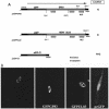
Nuclear localization of GP83 and HCMV pp65-EGFP fusion proteins. (A) Map of expression constructs. The peGFP-C1 and modified EGFP plasmids encoding C-terminal fusions of either GP83 ORF (peGFPGP83) or HCMV pp65 (peGFPUL83) are described in Materials and Methods. Restriction sites utilized for cloning purposes are indicated, as well as putative NLS predicted by coding sequence analysis. (B) Evaluation of fluorescence following transfection. Expression plasmids (5 μg) were transfected onto separate confluent monolayers of a six-well dish, and expression was allowed to proceed for 24 h prior to cell fixation with 4% paraformaldehyde and detection of autofluorescence as described in Materials and Methods. Transfection data are shown for peGFPGP83 (a and b, duplicate samples), peGFPUL83 (c), and the EGFP control plasmid, peGFP-C1 (d).
FIG. 8.
FIG. 8.
Analysis of the genomic configuration of the GP83 deletion virus. (A) Southern blot analysis of the vAM409 GPCMV recombinant. Viral DNA digested with BamHI was run on an agarose gel (0.7%), transferred to nylon membrane, and hybridized with a GP83 probe (pKTS409 [49]). Lane 1, wild type; lane 2, vAM409 DNA. The probe hybridized with a 3.4-kb BamHI subfragment of EcoRI C in wild-type DNA, but it hybridized with a larger 5.7-kb fragment in vAM409, indicating generation of recombinant virus containing a 2.3-kb gpt/EGFP cassette. (B) PCR analysis of recombinant genome and wild-type virus to verify incorporation of out-of-frame deletion (XmnI internal GP83 collapse). PCR of the region flanking the internal XmnI sites on GPCMV GP83 in both vAM409 and wild-type GPCMV demonstrated a deletion of ∼250 bp introduced into the GP83 mutant (vAM409). Lane 1, kilobase ladder; lane 2, wild-type CMV DNA; lanes 3 and 4, duplicate PCR analyses of vAM409 DNA.
FIG. 9.
FIG. 9.
One-step growth curve of the vAM409 mutant (▪) versus wild-type virus (▴). Wild-type GPCMV and the GP83 mutant were used to infect separate confluent cell culture monolayers at an MOI of 0.5 PFU/cell. After adsorption for 1 h at 37°C, the cells were washed and overlaid with medium and a zero time point sample was harvested. The remainder of the sample was then incubated at 37°C, with additional samples taken at the indicated times (12, 24, 36, 60, and 72 h postinfection). The viral titer of each time point was determined by plaque titration assay on GPL cells. Data shown are reproducible, representative results of several experiments.
FIG. 10.
FIG. 10.
Expression analyses of vAM409 and wild-type GPCMV. (A) Western blot analysis of wild-type and vAM409 (GP83 deletion) virus. Virus particles were purified from infected cell supernatants (49) and subjected to SDS-12% polyacrylamide gel electrophoresis, transferred onto nylon membrane, and probed with either anti-GPCMV antiserum or monospecific anti-GP83 polyclonal antisera (49). Anti-GPCMV antiserum was immunoreactive with multiple protein species in both wild-type and vAM409 particles (lanes 1 and 2). The band corresponding to GP83 protein (∼70 kDa) was detected only in the wild-type-infected virus particles (lane 3) by using the monospecific antiserum and was missing from vAM409 particles (lane 4). Lysates prepared from mock-infected cells were not immunoreactive with this antibody (49). Positions of molecular mass markers (kilodalton ladder) are indicated. (B) RT-PCR analysis of GP82 transcription. GP82-specific primers as described in Materials and Methods were used to amplify RNA (2 μg) from uninfected, wild-type GPCMV-infected, or vAM409-infected cells at 72 h postinfection. Guinea pig rRNA primers (2) were used as a positive control. Lane 1, kilobase ladder; lane 2, GP82 RT-PCR product, wild-type-infected cells; lane 3, rRNA amplification product, wild-type-infected cells; lane 4, GP82 RT-PCR product, vAM409-infected cells; lane 5, rRNA amplification product, vAM-409-infected cells; lane 6, GP82 RT-PCR product, uninfected cell RNA; lane 7, rRNA amplification product, uninfected cell RNA; lane 8, GP82 RT-PCR amplification product, no-reverse transcriptase control, wild-type-infected cells. RNA from both wild-type- and vAM-infected cells was also negative to rRNA RT-PCR amplification in the absence of reverse transcriptase (data not shown).
FIG. 11.
FIG. 11.
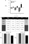
Analysis of phenotype of GP83 deletion virus in infected guinea pigs. (A) Patterns of weight gain or loss following inoculation of wild-type GPCMV and GP83 deletion virus in immunocompromised strain 2 guinea pigs. Cumulative weight gain or loss is indicated over the course of the study (mean ± standard error of the mean). Attenuation of the GP83 deletion virus was evident, based on weight gain in these animals compared to weight loss in wild-type-inoculated animals (P < 0.05, vAM409 versus control and vAM403, one-way ANOVA with Bonferroni correction). (B) Viral culture data. Animals were sacrificed at day 10 or day 21 postviral inoculation, and organ homogenates were prepared for culture. The dilution at which ≥50% of the wells were positive for viral CPE was determined (50% tissue culture infective dose). *, P < 0.05 versus ATCC, Kruskal-Wallis test; **, P < 0.05 versus ATCC and vAM403, Kruskal-Wallis test; , P < 0.05 versus vAM403, Kruskal-Wallis test. (C) qcPCR was performed on liver and spleen homogenates at 21 days postinoculation to compare animals inoculated with wild-type (gray bars), vAM403 (striped bars), and vAM409 (black bars). Tissues from vAM409-infected animals had significantly less viral DNA (P < 0.05, one-way ANOVA with Bonferroni correction), indicating impairment of replication of this mutant in vivo.

Similar articles

Cited by

References

    1. Ace, C. I., T. A. McKee, J. M. Ryan, J. M. Cameron, and C. M. Preston. 1989. Construction and characterization of a herpes simplex virus type 1 mutant unable to transinduce immediate-early gene expression. J. Virol. 63:2260-2269. - PMC - PubMed
    1. Allen, S. S., and D. N. McMurray. 2003. Coordinate cytokine gene expression in vivo following induction of tuberculous pleurisy in guinea pigs. Infect. Immun. 71:7035-7042. - PMC - PubMed
    1. Baldick, C. J., Jr., A. Marchini, C. E. Patterson, and T. Shenk. 1997. Human cytomegalovirus tegument protein pp71 (ppUL82) enhances the infectivity of viral DNA and accelerates the infectious cycle. J. Virol. 71:4400-4408. - PMC - PubMed
    1. Berencsi, K., Z. Gyulai, E. Gonczol, S. Pincus, W. I. Cox, S. Michelson, L. Kari, C. Meric, M. Cadoz, J. Zahradnik, S. Starr, and S. Plotkin. 2001. A canarypox vector-expressing cytomegalovirus (CMV) phosphoprotein 65 induces long-lasting cytotoxic T cell responses in human CMV-seronegative subjects. J. Infect. Dis. 183:1171-1179. - PubMed
    1. Bia, F. J., B. P. Griffith, C. K. Fong, and G. D. Hsiung. 1983. Cytomegaloviral infections in the guinea pig: experimental models for human disease. Rev. Infect. Dis. 5:177-195. - PubMed

Publication types

MeSH terms

LinkOut - more resources