. 2004 Aug 23:4:33.
doi: 10.1186/1471-2180-4-33.

Flagellin acting via TLR5 is the major activator of key signaling pathways leading to NF-kappa B and proinflammatory gene program activation in intestinal epithelial cells

Affiliations

Flagellin acting via TLR5 is the major activator of key signaling pathways leading to NF-kappa B and proinflammatory gene program activation in intestinal epithelial cells

Thomas Tallant et al. BMC Microbiol. .

Abstract

Background: Infection of intestinal epithelial cells by pathogenic Salmonella leads to activation of signaling cascades that ultimately initiate the proinflammatory gene program. The transcription factor NF-kappa B is a key regulator/activator of this gene program and is potently activated. We explored the mechanism by which Salmonella activates NF-kappa B during infection of cultured intestinal epithelial cells and found that flagellin produced by the bacteria and contained on them leads to NF-kappa B activation in all the cells; invasion of cells by the bacteria is not required to activate NF-kappa B.

Results: Purified flagellin activated the mitogen activated protein kinase (MAPK), stress-activated protein kinase (SAPK) and I kappa B kinase (IKK) signaling pathways that lead to expression of the proinflammatory gene program in a temporal fashion nearly identical to that of infection of intestinal epithelial cells by Salmonella. Flagellin expression was required for Salmonella invasion of host cells and it activated NF-kappa B via toll-like receptor 5 (TLR5). Surprisingly, a number of cell lines found to be unresponsive to flagellin express TLR5 and expression of exogenous TLR5 in these cells induces NF-kappa B activity in response to flagellin challenge although not robustly. Conversely, overexpression of dominant-negative TLR5 alleles only partially blocks NF-kappa B activation by flagellin. These observations are consistent with the possibility of either a very stable TLR5 signaling complex, the existence of a low abundance flagellin co-receptor or required adapter, or both.

Conclusion: These collective results provide the evidence that flagellin acts as the main determinant of Salmonella mediated NF-kappa B and proinflammatory signaling and gene activation by this flagellated pathogen. In addition, expression of the fli C gene appears to play an important role in the proper functioning of the TTSS since mutants that fail to express fli C are defective in expressing a subset of Sip proteins and fail to invade host cells. Flagellin added in trans cannot restore the ability of the fli C mutant bacteria to invade intestinal epithelial cells. Lastly, TLR5 expression in weak and non-responding cells indicates that additional factors may be required for efficient signal propagation in response to flagellin recognition.


Figures
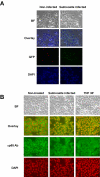
Figure 1
Figure 1
Salmonella infection leads to NF-κB nuclear localization even in non-infected cells. HT29 cells were grown on glass coverslips and either mock-infected, left untreated, infected with Salmonella typhimurium, or treated with TNFα (10 ng/ml). Cells fixed after 30 min (TNF) and 1 h (Salmonella) as described in Experimental Procedures and Salmonella that had invaded HT29 cells were detected by direct fluorescence microscopy of GFP expression, p65(RelA) localization was monitored by indirect immunoflourescence of rabbit anti-p65 antibody detected with FITC-conjugated donkey anti-rabbit antibody. DAPI was used to stain nuclei. A, HT29 cells were mock-infected or infected at an MOI of 50 with Salmonella typhimurium strain SJW1103G which expresses GFP from the ssaH promoter that is only active inside infected host cells [10,34]. Cells were photographed using bright field microscopy (BF), and immunoflourescence to detect GFP or DAPI staining as indicated. Images were merged (overlay) to reveal cells that were infected. B, HT29 cells were left untreated, infected with Salmonella typhimurium strain 1103 or treated with TNFα. NF-κB p65(RelA) localization under various conditions as indicated was monitored by indirect immunofluorescence. Cells were visualized by bright field microscopy (BF), cell nuclei were stained with DAPI and p65(RelA) was visualized with FITC. DAPI staining was falsely colored red to make visualization of the merge (overlay) easier to distinguish.
Figure 2
Figure 2
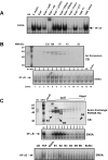
Protein factor in Salmonella culture broth leads to NF-κB activation. A, Salmonella dublin culture broth concentrated 100-fold was treated as indicated or infectious bacteria, as indicated was used to challenge HT29 cells. NF-κB DNA binding activity was assayed by EMSA from whole cell extracts prepared 45 min after treatment. Authenticity of the NF-κB DNA:protein complex was determined using p65(RelA)-specific and p50-specific antibody supershifts. B, Concentrated Salmonella dublin culture broth (IN) was chromatographed by gel permeation on a Superose 12 column. Eluted protein fractions were analyzed by fractionation on 10% SDS-PAGE and visualized by Coomassie blue (CB) staining. Molecular weight markers for chromatography and on the gels are indicated. Aliquots of each fraction as indicated was used to stimulate HT29 cells and resultant WCEs were analyzed by EMSA for NF-κB DNA binding activity. C, Concentrated Salmonella dublin culture broth (IN) was chromatographed by anion exchange chromatography on POROS HQ matrix. Proteins were eluted with an increasing NaCl gradient as indicated and analyzed on 10% SDS-PAGE and visualized by Coomassie blue (CB) staining. Input and aliquots of each fraction as indicated was used to stimulate HT29 cells and resultant WCEs were analyzed by EMSA for NF-κB DNA binding activity. Eluted material corresponding to protein bands B1-B6, a blank portion of the gel was isolated from a duplicate 10% SDS-PAGE gel as described in Experimental Procedures along with buffer samples from the beginning and end NaCl buffer gradient and used to stimulate HT29 cells and resultant WCEs were analyzed by EMSA for NF-κB DNA binding activity.
Figure 3
Figure 3
Identifcation by mass spectrometry of flagellin as the NF-κB activating factor in Salmonella culture broth. Microcapillary HPLC tandem mass spectrometry of Band 2 digested by trypsin. Peaks corresponding to Salmonella peptides are numbered and identified with the corresponding numbered peptide sequence to the right.
Figure 4
Figure 4
Flagellin mutants fail to activate NF-κB. EMSAs assaying for NF-κB DNA binding activity in WCEs prepared 45 min from non-infected cells (UN) and after direct infection of HT29 cells with wild-type E. coli DH5α, wild-type Salmonella dublin or SopE- mutant, SopB- mutant, the SopE-/SopB- double mutant, wild-type Salmonella typhimurium strain 1103, the fliC- mutant (fliC::Tn10), the fliC-/fljB- double mutant as indicated at an MOI of 50. B, EMSAs assaying for NF-κB DNA binding activity in WCEs prepared 45 min after challenge of HT29 cells from non-infected cells (UN) or with sterile-filtered concentrated culture broths from wild-type and mutant bacteria as indicated.
Figure 5
Figure 5
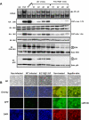
Flagellin is required for activating multiple signaling pathways during Salmonella infection and leads to nuclear localization of NF-κB. HT29 cells were left untreated, stimulated with TNFα (10 ng/ml) or a cocktail of anisomycin [An] (20 μg/ml)/PMA (12.5 ng/ml) for 15 min, or infected with either wild-type (WT) Salmonella typhimurium strain 1103 or the Salmonella typhimurium double fliC-/fljB- mutant strain 134 as indicated. WCE were prepared at the indicated times or at 10 min for TNF-treated cells or 15 min for anisomycin/PMA treated cells and used in EMSAs to analyze NF-κB DNA binding activity, or in immuno-kinase assays (KA) using anti-IKK or anti-JNK antibodies to measure IKK and JNK kinase activity on their respective substrates GST-IκBα 1–54 and GST-cJun 1–79 (as indicated). Immunoblot (IB) analysis of equivalent amounts (40 μg) of protein from each extract was fractionated on SDS-PAGE gels and transferred to PVDF membranes and probed with the indicated antibodies to detect bulk IKK, JNK, ERK and p38 as indicated. Immunoblot analysis using phospho-specific antibodies for ERK and p38 to detect activated ERK and p38 are indicated. B, Immunofluorescence demonstrating that flagellin mutant Salmonella fail to infect HT29 cells and that purified flagellin stimulation of HT29 cells leads to NF-κB nuclear p65 (RelA) localization as determined by indirect immunofluorescence. Imaging of the treatment indicated HT29 cells grown on coverslips was essentially the same as in Fig. 1A & 1B. False coloring of the DAPI stain was used to enhance the visualization of both DAPI stained nuclei and p65 nuclear localization.
Figure 6
Figure 6
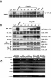
Purified flagellin activates signaling pathways and proinflammatory gene expression in intestinal epithelial cells mimicking that of wildtype a wild-type Salmonella infection. HT29 cells were left untreated or treated with TNFα (10 ng/ml) or a cocktail of anisomycin [An] (20 μg/ml)/PMA (12.5 ng/ml) for 10 min, or with flagellin (1 μg/ml) for the indicated times. WCE were prepared and analyzed by EMSA for NF-κB DNA binding activity, immuno-kinase assays (KA) or immunoblot analysis using phospho-specific antibodies for ERK or p38 to detect activation and with kinase-specific antibodies as described in Fig. 5A to detect bulk kinase abundance as indicated. A, EMSA to detect NF-κB DNA binding activity. Authenticity of the NF-κB bandshift was tested with supershift of the complex with p65(RelA)-specific antibody (α p65), normal rabbit serum (NRS) served as an irrelevant antibody control. B, immunoblot and kinase assays to detect IKK, JNK, ERK and p38 kinase activities and protein abundance as in Fig. 5A. C, semi-quantitative RT-PCR of proinflammatory gene expression of non-treated, wild-type and flagellin double mutant Salmonella typhimurium infected, TNFα (10 ng/ml) or flagellin (1 μg/ml) stimulated cells. HT29 cells were harvested at the indicated times after the indicated treatments and isolated RNA was used to make first strand cDNA that subsequently used in RT-PCR reactions (as described in Experimental Procedures) using gene-specific primers for IL1α, IL1β, IL-8, TNFα, MCP1 and β-actin. β-actin was used as a standard for normalizing expression patterns. Resulting PCR products were fractionated on 2% agarose gels and visualized by eithidium bromide staining.
Figure 7
Figure 7
Flagellin-mediated activation of NF-κB is MyD88 dependent. Infectious wild-type Salmonella Dublin (MOI of 100), IL-1 (20 ng/ml), purified flagellin (1 μg/ml) (as indicated), sterile-filtered and concentrated 100 kDa filter retentate supernatant (spt) from wild-type Salmonella dublin and SopE-/SopB- double mutant Salmonella dublin strain SE1SB2 (S2, as indicated) was used to challenge wild-type, MyD88-/- knockout or TLR2-/-/TLR4-/- double knockout MEFs as indicated. WCEs were prepared 45 min after treatments and examined by EMSA to analyze NF-κB DNA binding activity. IL-1 (20 ng/ml) was used as a positive control to monitor MyD88 function.
Figure 9
Figure 9
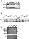
TLR5 is expressed in numerous cell types and has variable responses to flagellin. A, whole cell extracts were prepared from non-stimulated T84, HT29, A549, HeLa, 293T and T98G cells and fractionated on a 8% SDS-PAGE gel, proteins were transferred to PVDF membrane and probed with anti-TLR5 antibody for immunoblot analysis (IB). Protein loading was examined by probing with anti-actin antibody. B, HT29, A549, HeLa, 293T and T98G cells were left untreated (--), treated with flagellin (F) or TNFα (T) and WCEs were prepared after 45 min and used in EMSA to monitor NF-κB DNA binding activity. Authenticity of the NF-κB bandshift was tested with supershift of the complex with p65(RelA)-specific antibody (αp65), normal rabbit serum (NRS) served as an irrelevant antibody control. C, HT29, A549, HeLa, 293T and T98G cells WCEs (50 μg) were fractionated on a 8% SDS-PAGE gel, proteins transferred to Immobilon P and immunoprobed with anti-muc1 (1:450, Santa Cruz). Size markers are listed and muc1 position is indicated with an arrow.
Figure 8
Figure 8
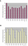
TLR5 inhibits flagellin-mediated NF-κB reporter gene activity. HT29 cells were transfected in triplicate in 6-well dishes using the indicated DN-TLR mammalian expression vectors or antisense TLR5 (AS TLR5) (2 μg/well), 2× NF-κB Luc reporter gene (100 ng/well), pRL-TK Renilla luciferase for normalization (50 ng/well) adjusted to 4 μg total DNA/well with empty vector pCDNA3.1 DNA. A, Fold-induction of 2× NF-κB Luc reporter gene in non-stimulated cells (light shading) and in TNFα (10 ng/ml) treated cells (dark shading). Lysates were prepared 12 h after stimulation. Results of a representative experiment are shown. B, HT29 cells transfected as in A were treated with flagellin (1 μg/ml) and cell lysates were prepared and analyzed as in Fig. 8A. Results of a representative experiment are shown.

Similar articles

Cited by

References

    1. Kagnoff MF, Eckmann L. Epithelial cells as sensors for microbial infection. J Clin Invest. 1997;100:6–10. - PMC - PubMed
    1. Eckmann L, Kagnoff MF, Fierer J. Intestinal epithelial cells as watchdogs for the natural immune system. Trends Microbiol. 1995;3:118–120. doi: 10.1016/S0966-842X(00)88894-0. - DOI - PubMed
    1. Elewaut D, DiDonato JA, Kim JM, Truong F, Eckmann L, Kagnoff MF. NF-kappa B is a central regulator of the intestinal epithelial cell innate immune response induced by infection with enteroinvasive bacteria. J Immunol. 1999;163:1457–1466. - PubMed
    1. Witthoft T, Eckmann L, Kim JM, Kagnoff MF. Enteroinvasive bacteria directly activate expression of iNOS and NO production in human colon epithelial cells. Am J Physiol. 1998;275:G564–G571. - PubMed
    1. Eckmann L, Stenson WF, Savidge TC, Lowe DC, Barrett KE, Fierer J, Smith JR, Kagnoff MF. Role of intestinal epithelial cells in the host secretory response to infection by invasive bacteria. Bacterial entry induces epithelial prostaglandin h synthase-2 expression and prostaglandin E2 and F2 alpha production. J Clin Invest. 1997;100:296–309. - PMC - PubMed

Publication types

MeSH terms

Substances

LinkOut - more resources