. 2004 Oct;15(10):4556-67.
doi: 10.1091/mbc.e04-06-0480. Epub 2004 Aug 10.

Conformational defects slow Golgi exit, block oligomerization, and reduce raft affinity of caveolin-1 mutant proteins

Affiliations

Conformational defects slow Golgi exit, block oligomerization, and reduce raft affinity of caveolin-1 mutant proteins

Xiaoyan Ren et al. Mol Biol Cell. 2004 Oct.

Abstract

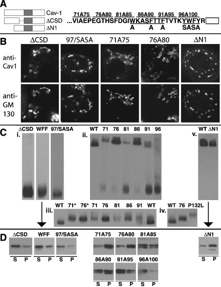
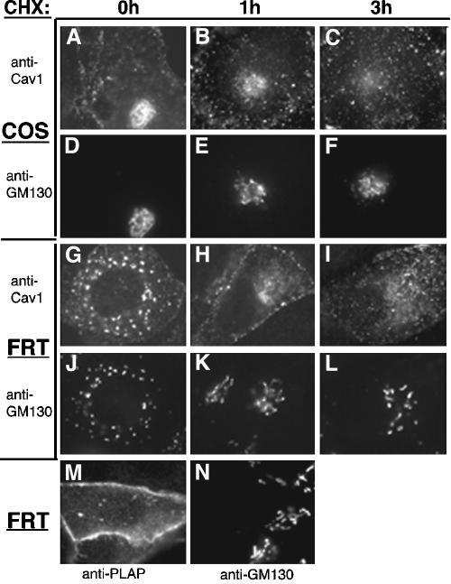
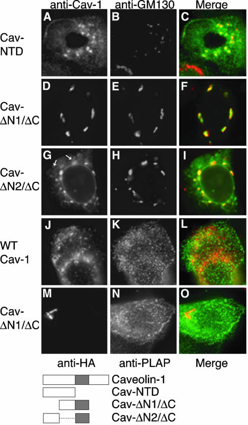
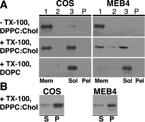
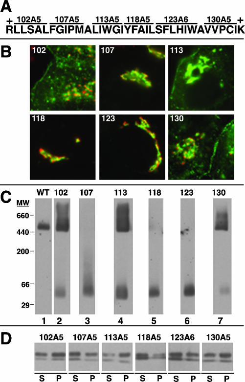
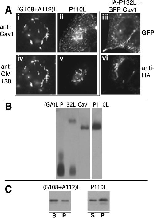
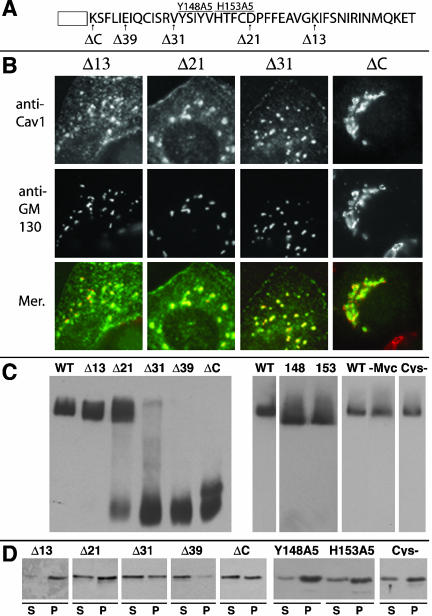
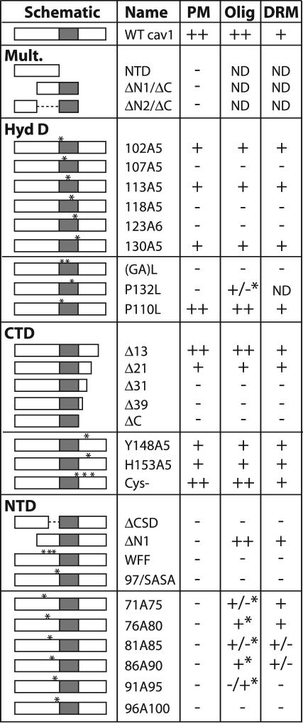
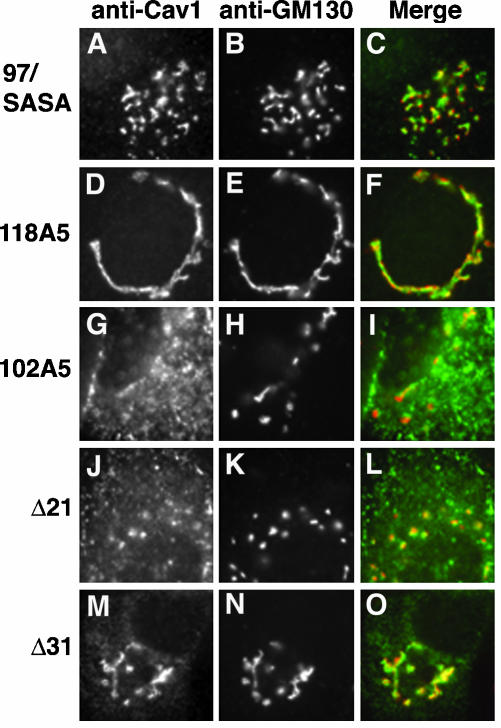
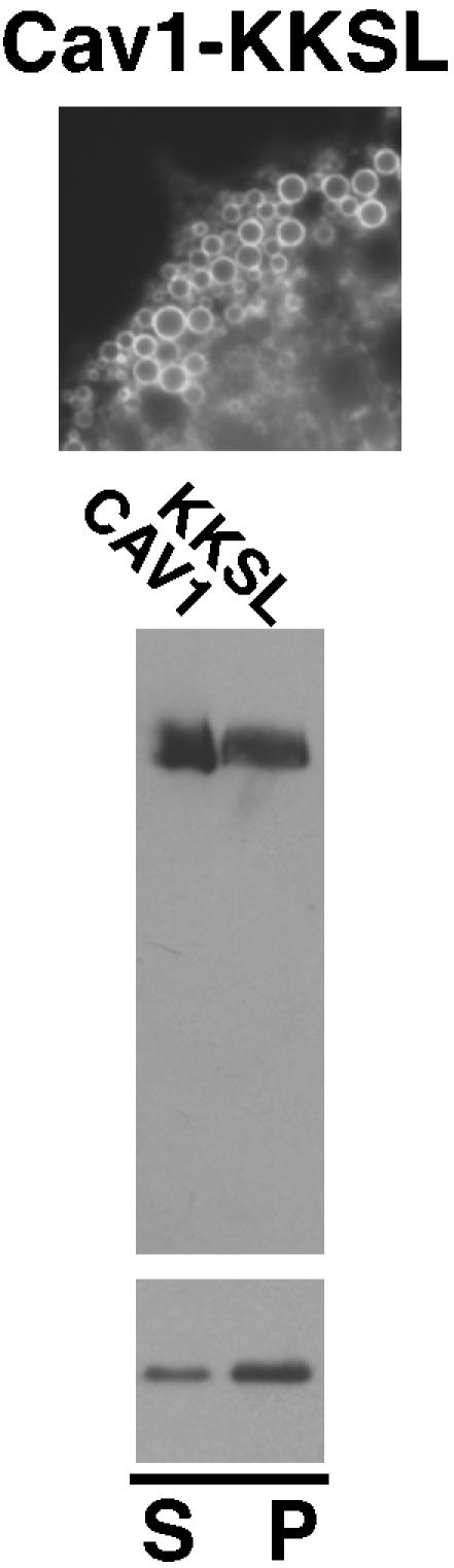
Caveolin-1, a structural protein of caveolae, is cleared unusually slowly from the Golgi apparatus during biosynthetic transport. Furthermore, several caveolin-1 mutant proteins accumulate in the Golgi apparatus. We examined this behavior further in this mutant study. Golgi accumulation probably resulted from loss of Golgi exit information, not exposure of cryptic retention signals, because several deletion mutants accumulated in the Golgi apparatus. Alterations throughout the protein caused Golgi accumulation. Thus, most probably acted indirectly, by affecting overall conformation, rather than by disrupting specific Golgi exit motifs. Consistent with this idea, almost all the Golgi-localized mutant proteins failed to oligomerize normally (even with an intact oligomerization domain), and they showed reduced raft affinity in an in vitro detergent-insolubility assay. A few mutant proteins formed unstable oligomers that migrated unusually slowly on blue native gels. Only one mutant protein, which lacked the first half of the N-terminal hydrophilic domain, accumulated in the Golgi apparatus despite normal oligomerization and raft association. These results suggested that transport of caveolin-1 through the Golgi apparatus is unusually difficult. The conformation of caveolin-1 may be optimized to overcome this difficulty, but remain very sensitive to mutation. Disrupting conformation can coordinately affect oligomerization, raft affinity, and Golgi exit of caveolin-1.


Figures

Figure 7.
Figure 7.
Localization, oligomerization, and raft association of caveolin-1 N-terminal domain mutant proteins. (A) Left, schematic diagram of two deletion mutant proteins, ΔCSD (lacking F81-Y100) and ΔN1 (lacking G3-E48). (A) Right, sequence of the last 31 residues of the N-terminal domain is shown, with the start of the hydrophobic domain shown as an open box. The alterations in two substitution mutant proteins are indicated below the N-terminal domain sequence. In WFF/AAA, Ala is substituted for each of W85, F89, and F92. In 97/SASA, the sequence SASA is substituted for residues Y97-Y100. The alterations in five mutant proteins in which sequential groups of five residues were changed to Ala are shown above the wild-type sequence (Machleidt et al., 2000). These proteins are named for the first and last altered residues (i.e., 71A75 contains Ala substituted for each of residues 71–75). In Figure 7C, these proteins are indicated by the first changed residue (i.e., 71A75 is called 71). (B) Localization of the indicated proteins and GM130 in transfected FRT cells is shown. (C) Oligomerization was determined on blue native gels as in Figure 4C. Only the oligomer bands are shown in iii and iv. 71* and 76*, substitution of Ala for residues 71–75 or 76–80, respectively, in the myc and HA-tagged wild-type caveolin-1 shown on the same blot. In iii, the amounts of lysate loaded in each lane were varied to obtain more uniform oligomer bands. (D) TX100-insolubility of the indicated mutant proteins in raft-containing liposomes was determined as in Figure 4D.
Figure 1.
Figure 1.
Selective clearance of caveolin-1 from the Golgi apparatus in cycloheximide-treated cells. COS (A–F) or FRT cells expressing caveolin-1 (G–L) or PLAP-HA (M and N) were left untreated (A, D, G, J, M, and N) or treated with cycloheximide (CHX) for 1 h (B, E, H, and K) or 3 h (C, F, I, and L) before fixation, permeabilization, and detection of caveolin-1 (A–C and G–I) or PLAP-HA (M) and GM130 in the same cells (D–F, J–L, and N) by IF.
Figure 2.
Figure 2.
Cellular localization of caveolin-1 deletion mutant proteins. Cav-NTD (A), Cav-ΔN1/ΔC (D), or Cav-ΔN2/ΔC (G) were detected together with endogenous GM130 (B, E, and H) in transfected FRT cells by IF. Merged images are shown in C, F, and I. In G, arrows; lipid droplets. In J–O, PLAP was coexpressed with wild-type caveolin-1 (J–L) or with ΔN1/ΔC (M–O) in FRT cells. Caveolin-1 (J) or ΔN1/ΔC (M) was detected with mouse anti-HA antibodies (red), and PLAP in the same cells (K and N, respectively) with anti-PLAP antibodies (green). (L and O); merged images. Schematic diagrams of caveolin-1 and the mutant proteins, indicating the N- and C-terminal hydrophilic domains as open boxes, and the central hydrophobic domain as a shaded box, are shown. The dashed line in the diagram of Cav-ΔN2/ΔC indicates a deleted region.
Figure 3.
Figure 3.
TX100 insolubility of caveolin-1 in mixed proteoliposomes containing cellular proteins and lipids and artificial lipids. OG lysates of COS (left) or transfected MEB-4 cells (right) were mixed with excess OG-solubilized DPPC:cholesterol (2:1) or DOPC as indicated, and proteoliposomes were formed by dialysis. (A) Proteoliposomes were subjected to sucrose density gradient centrifugation (top) or were extracted with TX100 before applying to sucrose gradients (middle and bottom). Gradients were harvested in three fractions from the top. In each case, fraction 1 contained floating membranes (Mem) and fraction 3 contained soluble and detergent-solubilized proteins (Sol). Gradient pellets (Pel) contained any aggregated proteins. (B) Proteoliposomes were extracted with TX100 and subjected to ultracentrifugation, and the TX100-soluble supernatant (S) and TX100-insoluble pellet (P) fractions were separated. (A and B) After resuspension and solubilization of pelleted material, proteins in each fraction were separated by SDS-PAGE and transferred to PVDF membranes. Caveolin-1 was detected by Western blotting.
Figure 4.
Figure 4.
Localization, oligomerization, and raft association of caveolin-1 hydrophobic domain Ala-scan mutant proteins. (A) The sequence of the hydrophobic domain of caveolin-1, bounded by basic residues, is shown. Sequential groups of five or six residues, together spanning the hydrophobic domain, were changed to Ala as indicated. Each mutant protein was named for the position of the first altered residue and the number of altered residues (i.e., 102A5) or simply by the position of the first altered residue (i.e., 102). (B) Localization of the indicated mutant proteins (green) and GM130 (red) in transfected FRT cells is shown. (C) Proteins in lysates of MEB-4 cells expressing wild-type caveolin-1 or the indicated mutant protein were separated by blue native gel electrophoresis and transferred to PVDF membranes. Caveolin-1 and the mutant proteins were detected by Western blotting. Positions of molecular weight standards (MW), detected by Coomassie Blue staining of the membrane, are shown. Exact molecular weights of membrane proteins cannot be determined by native gel analysis. (D) Mixed proteoliposomes formed from lysates of MEB-4 cells expressing the indicated mutant protein and excess DPPC:cholesterol (2:1) were subjected to TX100 extraction and centrifugation. Supernatant (S) and pellet (P) fractions were analyzed for the presence of the mutants by SDS-PAGE and Western blotting.
Figure 5.
Figure 5.
Localization, oligomerization, and raft association of caveolin-1 hydrophobic domain mutant proteins. (A) FRT cells expressing (G108+A112)L (i and iv), P110L (ii and v), or both myc/GFP-tagged caveolin-1 and HA-tagged P132L (iii and vi) were processed for IF. i, ii, iv, and v, (G018+A112)L (i) and P110L (ii) were detected together with GM130 (iv and v, respectively). iii and vi, caveolin-1 was detected by GFP fluorescence (iii), whereas P132L was detected with rabbit anti-HA and Texas Red goat anti-rabbit antibodies (vi). (B) Oligomerization of the indicated proteins [(G108+A112)L, labeled (GA)L; P132L; wild-type caveolin-1; and P110, respectively] was determined on blue native gels as in Figure 4C. The smear migrating more slowly than the (G108+A112)L monomer band was variable. (C) TX100-insolubility of the indicated mutant proteins in raft-containing liposomes was determined as in Figure 4D.
Figure 6.
Figure 6.
Localization, oligomerization, and raft association of caveolin-1 C-terminal domain mutant proteins. (A) Sequence of the C-terminal domain of caveolin-1, with the end of the hydrophobic domain indicated as an open box, is shown. Arrows indicate the last residue in each of a series of mutant proteins. All proteins (except ΔC, which lacked the entire C-terminal domain except K135) are named for the number of deleted residues (i.e., Δ13 lacked the last 13 residues of caveolin-1, and ended with K165). Two mutant proteins, Y148A5 and H153A5 (labeled 148 and 153, respectively, in Figure 6C), contained five Ala residues in place of the indicated residues. Of these proteins, only Δ39 contained a C-terminal myc tag. (B) The localization of the indicated mutant proteins and GM130 in transfected FRT cells is shown. (C) Oligomerization was determined on blue native gels as in Figure 4C. -Myc, wild-type caveolin-1 lacking the Myc tag. Cys-, a nonpalmitoylated mutant protein (Dietzen et al., 1995). (D) TX100-insolubility of the indicated mutant proteins in raft-containing liposomes was determined as in Figure 4D.
Figure 8.
Figure 8.
Summary of localization, oligomerization, and TX100-insolubility results. The caveolin-1 mutant proteins are diagrammed schematically as in Figure 2A. Mutants are grouped according to the domain altered. Mult., deletions in two domains. HydD, CTD, and NTD, changes in the hydrophobic, C-terminal, or N-terminal domains, respectively. Asterisks indicate substitutions. Four or more substitutions are indicated by a single larger asterisk. Column 3 (PM); efficiency of Golgi-to-plasma membrane transport. ++, wild-type localization; +, slightly enhanced Golgi localization; -, Golgi accumulation. None of these proteins except Cav-ΔN2/ΔC accumulated predominantly in the ER, except in highly expressing cells. Column 4 (Olig); oligomerization on blue native gels. ++, wild-type oligomerization, with very little monomer; +, predominantly oligomeric, but some monomer; +/-, substantial oligomer and monomer; -/+, mostly monomer; -, no oligomer detected; *, oligomer migrated more slowly than wild-type caveolin-1. Column 5 (DRM); association with TX100-resistant membranes after extraction of raft-containing mixed proteoliposomes. +, similar to wild-type caveolin-1 (∼70% insoluble); +/-, about equal amounts soluble and insoluble; -, poor DRM association (∼30% insoluble). ND, not determined.
Figure 9.
Figure 9.
Persistence of caveolin-1 mutant proteins in the Golgi apparatus after cycloheximide treatment. The localization of the indicated proteins (A, D, G, J, and M) and GM130 (B, E, H, J, and N) in transfected FRT cells after cycloheximide treatment for 3 h (except cells expressing 118A5, treated for 5 h) is shown. C, F, I, L, and O; merged anti-caveolin-1 (green) and anti-GM130 (red) images.
Figure 10.
Figure 10.
Localization, oligomerization, and raft association of Cav1-KKSL. Top, localization of Cav1-KKSL in FRT cells by IF is shown. Round structures are lipid droplets, as reported previously (Ostermeyer et al., 2001). Middle, oligomerization of wild-type caveolin-1 and Cav-KKSL was determined on blue native gels as in Figure 4C. Bottom, TX100-insolubility of Cav-KKSL in raft-containing liposomes was determined as in Figure 4D.

Similar articles

Cited by

References

    1. Arbuzova, A., Wang, L., Wang, J., Hangyas-Mihalyne, G., Murray, D., Honig, B., and McLaughlin, S. (2000). Membrane binding of peptides containing both basic and aromatic residues. Experimental studies with peptides corresponding to the scaffolding region of caveolin and the effector region of MARCKS. Biochemistry 39, 10330-10339. - PubMed
    1. Arreaza, G., and Brown, D.A. (1995). Sorting and intracellular trafficking of a glycosylphosphatidylinositol-anchored protein and two hybrid proteins with the same ectodomain in MDCK kidney epithelial cells. J. Biol. Chem. 270, 23641-23647. - PubMed
    1. Bretscher, M.S., and Munro, S. (1993). Cholesterol and the Golgi apparatus. Science 261, 1280-1281. - PubMed
    1. Breuza, L., Corby, S., Arsanto, J.-P., Delgrossi, M.-H., Scheiffele, P., and Le Bivic, A. (2002). The scaffolding domain of caveolin 2 is responsible for its Golgi localization in Caco-2 cells. J. Cell Sci. 115, 4457-4467. - PubMed
    1. Brown, D.A., and London, E. (2000). Structure and function of sphingolipid- and cholesterol-rich membrane rafts. J. Biol. Chem. 275, 17221-17224. - PubMed

Publication types

LinkOut - more resources