Comparative Study
. 2004 Aug;114(3):370-8.
doi: 10.1172/JCI20174.

Cancer cachexia is regulated by selective targeting of skeletal muscle gene products

Affiliations
Comparative Study

Cancer cachexia is regulated by selective targeting of skeletal muscle gene products

Swarnali Acharyya et al. J Clin Invest. 2004 Aug.

Abstract

Cachexia is a syndrome characterized by wasting of skeletal muscle and contributes to nearly one-third of all cancer deaths. Cytokines and tumor factors mediate wasting by suppressing muscle gene products, but exactly which products are targeted by these cachectic factors is not well understood. Because of their functional relevance to muscle architecture, such targets are presumed to represent myofibrillar proteins, but whether these proteins are regulated in a general or a selective manner is also unclear. Here we demonstrate, using in vitro and in vivo models of muscle wasting, that cachectic factors are remarkably selective in targeting myosin heavy chain. In myotubes and mouse muscles, TNF-alpha plus IFN-gamma strongly reduced myosin expression through an RNA-dependent mechanism. Likewise, colon-26 tumors in mice caused the selective reduction of this myofibrillar protein, and this reduction correlated with wasting. Under these conditions, however, loss of myosin was associated with the ubiquitin-dependent proteasome pathway, which suggests that mechanisms used to regulate the expression of muscle proteins may be cachectic factor specific. These results shed new light on cancer cachexia by revealing that wasting does not result from a general downregulation of muscle proteins but rather is highly selective as to which proteins are targeted during the wasting state.


Figures
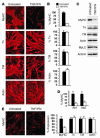
Figure 1
Figure 1
MyHC is selectively targeted by TNF/IFN signaling. C2C12 myoblasts were differentiated in DM for 3 days and subsequently switched to medium alone or containing TNF (10 ng/ml) and IFN (100 U/ml) for 48 hours. (A) Immunofluorescence to detect expression of myofibrillar proteins (MyHC, fast twitch; troponin T [Tn]; tropomyosin, α and β [TM]; actin, α skeletal). Images are shown at ×20 magnification. (B) Immunostained expression levels of myofibrillar proteins were quantitated using Zeiss AxioVision software (Carl Zeiss Inc., Thornwood, New York, USA). The data were calculated from a minimum of ten randomly chosen fields of cells at ×4 magnification. (C) Whole-cell extracts were prepared from untreated or TNF/IFN–treated myocytes, and 50 μg total protein was used in Western analyses to probe for myofibrillar proteins (troponin; α- and β-tropomyosin; MyLC, myosin light chain). (D) Immunofluorescence of untreated (black bars) or TNF/IFN–treated (hatched bars) C2C12 myotubes, probing for MyHC (Ab1, MyHC antibody MY-32; Ab2, MyHC antibody MF20). (E) Primary myotubes were either untreated (black bars) or treated with TNF and IFN (hatched bars) for 48 hours. Cells were stained by immunofluorescence for myofibrillar proteins, and the level of protein expression was quantitated by digital capture and AxioVision software. For histograms, each bar represents mean ± SEM from three independent experiments.
Figure 2
Figure 2
Chronic TNF/IFN signaling is required to induce the temporal downregulation of MyHC mRNA. (A) Myotubes were treated with DM containing increasing doses of TNF (5 ng/ml, lane 1; 10 ng/ml, lane 2), IFN (50 U/ml, lane 3; 100 U/ml, lane 4), or TNF plus IFN (5 ng/ml + 50 U/ml, lane 5; 10 ng/ml + 100 U/ml, lane 6). After 48 hours, RNA was prepared and RT-PCR was performed (MyHC IIb; Tn3, troponin T3; α-TM, α-tropomyosin; α-actin). (B) Myotubes were either untreated or treated with TNF (5 ng/ml), IFN (50 U/ml), or TNF plus IFN (5 ng/ml + 50 U/ml) for 48 hours. RNA was prepared, and Northern blots probing for MyHC IIa and IIb were performed. GAPDH was used as a loading control. Numbers are densitometric values normalized to GAPDH. (C) Myotubes were treated with TNF/IFN, and at the indicated times RNA was prepared and Northern blots probing for MyHC type IIa and IIb were performed. (D) Myotubes were treated with TNF/IFN for 24 hours, at which time cells continued to be treated with cytokines or were washed with PBS and switched to DM (Wash). RNA was prepared at the indicated time points, and Northern blots probing for MyHC IIb were performed. Similar results were obtained from two additional independent experiments.
Figure 3
Figure 3
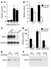
TNF/IFN inhibits MyHC transcription through the concomitant reduction of MyoD. (A) C2C12 cells were transfected in triplicate with a MyHC IIb–luciferase reporter plasmid as described in Methods. The next day, cells were differentiated and at the indicated times were harvested for luciferase assays. (B) Myoblasts were transfected with MyHC IIb–luciferase and subsequently differentiated for 48 hours, at which time cells were left untreated or treated with TNF/IFN. At the indicated times, cells were harvested and luciferase levels determined. (C) Myotubes were either untreated or treated with TNF (5 ng/ml), IFN (50 U/ml), or TNF plus IFN for 48 hours. Northern blots probing for MyoD were performed (25), and GAPDH was used as a loading control. (D) Myoblasts were transfected under conditions similar to those described in B with a MyHC IIb–luciferase reporter along with empty vector or MyoD wild-type or mutant (mut) expression plasmids (50 ng each). Cells were differentiated for 48 hours and subsequently left untreated or treated with TNF and IFN for an additional 24 hours, at which time cell extracts were prepared and luciferase activities determined. (E) Myotubes were either untreated or treated with TNF and IFN in methionine/cysteine–free DM, then pulsed for 1 hour with [35S]-methionine/cysteine prior to collection of cells. Labeled MyHC was detected by immunoprecipitation. (F) Myotubes were pulsed with DM containing [35S]-methionine/cysteine, then chased with fresh DM with or without TNF/IFN. For histograms, the data are representative of experiments performed a minimum of three times, plotted as mean ± SD.
Figure 4
Figure 4
Expression of TNF and IFN in vivo causes the selective downregulation of MyHC. Saline used as a control (100 μl) or CHO cells expressing TNF and IFN (1 × 10⁷ cells per 0.1 ml) were injected into gastrocnemius muscles of nude mice (n = 4 per group). At 6 and 12 days after injection, muscles were harvested, divided into equal sections, and homogenized to prepare either total protein or RNA. Core myofibrillar gene products were examined by Western blotting (A) or Northern analyses (B) (MyHC IIb, α-tropomyosin, troponin T3, α-actin).
Figure 5
Figure 5
C-26 tumors induce severe muscle wasting associated with a selective loss of MyHC protein. (A) Saline used as control (100 μl) or C-26 adenocarcinoma cells (1 × 10⁶ cells per 0.1 ml) were injected subcutaneously into the right flank of CD2F1 male mice. Body weights of saline- or C-26–injected mice (n = 4 per group) were recorded every 48 hours from day 8 to day 22 postinjection. (B and C) Mice (n = 6) were injected in the right flank with either saline (control) or C-26 cells. At the indicated days after injection, tibialis anterior muscles from the right hind limb were collected and divided into two sections. From one section, tissue homogenates were prepared, and Western blot analyses probing for myofibrillar proteins were performed (B). From the remaining section, total RNA was prepared, and Northern blot analyses were undertaken (C). (D) Extracts of soleus muscles from mice described in B and C were prepared and used for Western blot analysis for expression of MyHC type I and α-sarcomeric actin.
Figure 6
Figure 6
C-26 tumors enhance ubiquitin/E3 ligase expression and MyHC ubiquitination products. Mice were injected in the right flank with either control (n = 3) or C-26 cells (n = 3), and at days 23–25 after injection, tibialis muscles were isolated and prepared for RNA or protein analyses. (A) Northern blots probing for ubiquitin (Ub), MuRF1, and atrogin-1/MAFbx in muscles from control versus C-26–injected mice. (B and C) Homogenates were prepared from mouse muscles or C2C12 myotubes, and protein complexes were subsequently immunoprecipitated overnight with an anti-ubiquitin antibody (IP). Immunoprecipitates were fractionated by SDS-PAGE, and immunoblot (IB) analyses probing for MyHC (upper panels) or tropomyosin (TM; lower panels) were performed. The Western blot for MyHC in B was purposely overexposed to accentuate differences in MyHC protein products coupled to ubiquitin (bracketed). Asterisks in the tropomyosin Western blot denote nonspecific bands. (D and E) Homogenates described in B and C were immunoprecipitated with an α-sarcomeric actin antibody (IP), and complexes were fractionated by SDS-PAGE and immunoblotted (IB) for MyHC and α-actin.

Similar articles

Cited by

References

    1. Tisdale MJ. Biology of cachexia. J. Natl. Cancer Inst. 1997;89:1763–1773. - PubMed
    1. Argiles JM, Moore-Carrasco R, Fuster G, Busquets S, Lopez-Soriano FJ. Cancer cachexia: the molecular mechanisms. Int. J. Biochem. Cell Biol. 2003;35:405–409. - PubMed
    1. Tisdale MJ. Cachexia in cancer patients. Nat. Rev. Cancer. 2002;2:862–871. - PubMed
    1. Body JJ. The syndrome of anorexia-cachexia. Curr. Opin. Oncol. 1999;11:255–260. - PubMed
    1. Evans WK, et al. Limited impact of total parenteral nutrition on nutritional status during treatment for small cell lung cancer. Cancer Res. 1985;45:3347–3353. - PubMed

Publication types

MeSH terms