. 2004 Aug;78(16):8552-64.
doi: 10.1128/JVI.78.16.8552-8564.2004.

Dissecting the requirement for subgenomic promoter sequences by RNA recombination of brome mosaic virus in vivo: evidence for functional separation of transcription and recombination

Affiliations

Dissecting the requirement for subgenomic promoter sequences by RNA recombination of brome mosaic virus in vivo: evidence for functional separation of transcription and recombination

Rafal Wierzchoslawski et al. J Virol. 2004 Aug.

Abstract

Previously, we and others mapped an increased homologous recombination activity within the subgenomic promoter (sgp) region in brome mosaic virus (BMV) RNA3. In order to correlate sgp-mediated recombination and transcription, in the present work we used BMV RNA3 constructs that carried altered sgp repeats. We observed that the removal or extension of the poly(U) tract reduced or increased recombination, respectively. Deletion of the sgp core hairpin or its replacement by a different stem-loop structure inhibited recombination activity. Nucleotide substitutions at the +1 or +2 transcription initiation position reduced recombination. The sgp core alone supported only basal recombination activity. The sites of crossovers mapped to the poly(U) region and to the core hairpin. The observed effects on recombination did not parallel those observed for transcription. To explain how both activities operate within the sgp sequence, we propose a dual mechanism whereby recombination is primed at the poly(U) tract by the predetached nascent plus strand, whereas transcription is initiated de novo at the sgp core.


Figures
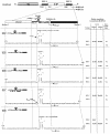
FIG. 1.
FIG. 1.
BMV RNA3 constructs (ID RNA3s) used in these studies. A schematic of the genetic organization of BMV RNA3 is shown at the top. Thin lines, noncoding regions; solid rectangles show the locations of the sgp sequences. Shaded and open rectangles, ORFs for the movement protein (3a) and CP, respectively. Arrows mark the regions of the sg RNA4 and RNA4′ transcriptions. EcoRI and XhoI marker restriction sites are shown at INT-1. Below the RNA3 schematic, the elements of the sgp insert of INT-2 [the enhancer, the poly(U) tract, the hairpin, the core region, the initiation site, and a downstream sequence] are shown. Below the INT-2 schematic, the nucleotide sequences of constructs ID2 to ID13 are given, with structures represented on the left and locations of marker restriction sites in the two variants of each construct tabulated on the right. ID2 carries the wt sequence; ID5 and ID6 carry mutations at positions +1 and +2, respectively; ID7 carries nine single marker mutations (indicated by italicized capital letters); ID8 carries hairpin D; ID9 has no poly(U) tract; ID10 has no enhancer and no poly(U) elements, so the core sequence forms a new hairpin B structure by pairing with the adjacent upstream region; ID11 carries a replacement of hairpin A with the 3′ hairpin C; ID12 carries an extended poly(U) tract; and ID13 has no hairpin structure. Nucleotide positions of junction sites are given according to the numbering of the wt sequence, as counted from the 5′ end. Thin V-shaped lines represent deleted elements. For the ID7 RNA3, only one variant was created (ID7-1), and it was tested for recombination with ID1-1 (described previously by Wierzchoslawski et al. [54]).
FIG. 1.
FIG. 1.
BMV RNA3 constructs (ID RNA3s) used in these studies. A schematic of the genetic organization of BMV RNA3 is shown at the top. Thin lines, noncoding regions; solid rectangles show the locations of the sgp sequences. Shaded and open rectangles, ORFs for the movement protein (3a) and CP, respectively. Arrows mark the regions of the sg RNA4 and RNA4′ transcriptions. EcoRI and XhoI marker restriction sites are shown at INT-1. Below the RNA3 schematic, the elements of the sgp insert of INT-2 [the enhancer, the poly(U) tract, the hairpin, the core region, the initiation site, and a downstream sequence] are shown. Below the INT-2 schematic, the nucleotide sequences of constructs ID2 to ID13 are given, with structures represented on the left and locations of marker restriction sites in the two variants of each construct tabulated on the right. ID2 carries the wt sequence; ID5 and ID6 carry mutations at positions +1 and +2, respectively; ID7 carries nine single marker mutations (indicated by italicized capital letters); ID8 carries hairpin D; ID9 has no poly(U) tract; ID10 has no enhancer and no poly(U) elements, so the core sequence forms a new hairpin B structure by pairing with the adjacent upstream region; ID11 carries a replacement of hairpin A with the 3′ hairpin C; ID12 carries an extended poly(U) tract; and ID13 has no hairpin structure. Nucleotide positions of junction sites are given according to the numbering of the wt sequence, as counted from the 5′ end. Thin V-shaped lines represent deleted elements. For the ID7 RNA3, only one variant was created (ID7-1), and it was tested for recombination with ID1-1 (described previously by Wierzchoslawski et al. [54]).
FIG. 2.
FIG. 2.
General illustration of homologous crossovers occurring between two coinoculated variants of ID RNA3s. At the top, the regions of sgp-mediated crossovers at the INT-2 locus, the INT-1 locus, and the control CP ORF are shown schematically (not to scale). The elements of the RNA3 molecule are represented as in Fig. 1; arrows indicate homologous crossovers. The types of ID RNA3s carrying specific restriction sites at four restriction marker positions (indicated by circles, triangles, diamonds, and inverted triangles) are given in the balloons (see also the restriction marker table in Fig. 1, right side). Below the crossover schematic, the molecules of two parental ID RNA3 variants [designated generally as ID RNA3(1) and ID RNA3(2)] are shown on the left, whereas the predicted types of progeny RNA3-RNA3 recombinants that arise due to crossovers within the recombination regions (see also Table 1) are shown on the right. Solid rectangles, INT-1 and INT-2 regions; shaded and open rectangles, ORFs.
FIG. 3.
FIG. 3.
(A) Northern blot analysis of the accumulation of BMV RNA3 ID constructs. Leaves were coinoculated mechanically with in vitro-transcribed wt RNA1 and RNA2 and with RNA3 ID mutants (as indicated above the gel). Total (tot.) RNA was extracted from the infected tissue, separated electrophoretically in a 1% agarose-formamide-formaldehyde gel, and blotted to a nylon membrane (HybondN+; Amersham), and BMV RNA sequences were detected by using a 32P-labeled RNA probe that was complementary to an 83-nt fragment common to the 3′ ends of all four BMV RNAs (nt 1870 to 1953 on the BMV RNA3 positive strand). Lanes 2 to 11, RNAs extracted from the local lesion tissues of C. quinoa. Lane 1 (control), RNA from systemically infected barley seedlings. The positions of BMV RNA components are indicated on the right. (B) Histogram showing the quantification of the accumulation of RNA3 and sg RNA4 and RNA4′. The Northern blot autoradiogram shown in panel A was scanned in the PhosphorImager and subjected to computer-based densitometry by using the ImageQuant (version 5.0) program from Amersham Biosciences. Bars represent the percentages which the band intensities for RNA3, RNA4, and RNA4′ contributed to the total intensity of all BMV RNA components. The numbers below show the actual results of quantification and the observed recombination frequencies at INT-2 (as a reference; repeated from Table 1).
FIG. 3.
FIG. 3.
(A) Northern blot analysis of the accumulation of BMV RNA3 ID constructs. Leaves were coinoculated mechanically with in vitro-transcribed wt RNA1 and RNA2 and with RNA3 ID mutants (as indicated above the gel). Total (tot.) RNA was extracted from the infected tissue, separated electrophoretically in a 1% agarose-formamide-formaldehyde gel, and blotted to a nylon membrane (HybondN+; Amersham), and BMV RNA sequences were detected by using a 32P-labeled RNA probe that was complementary to an 83-nt fragment common to the 3′ ends of all four BMV RNAs (nt 1870 to 1953 on the BMV RNA3 positive strand). Lanes 2 to 11, RNAs extracted from the local lesion tissues of C. quinoa. Lane 1 (control), RNA from systemically infected barley seedlings. The positions of BMV RNA components are indicated on the right. (B) Histogram showing the quantification of the accumulation of RNA3 and sg RNA4 and RNA4′. The Northern blot autoradiogram shown in panel A was scanned in the PhosphorImager and subjected to computer-based densitometry by using the ImageQuant (version 5.0) program from Amersham Biosciences. Bars represent the percentages which the band intensities for RNA3, RNA4, and RNA4′ contributed to the total intensity of all BMV RNA components. The numbers below show the actual results of quantification and the observed recombination frequencies at INT-2 (as a reference; repeated from Table 1).
FIG. 4.
FIG. 4.
Results of sequencing of RT-PCR-generated cDNA clones of progeny BMV RNA3 extracted from ID8-1 × ID8-2-coinfected leaves. Schematic representations of ID8 RNA3, and of the sgp INT-2 insert with the modified hairpin D (see Fig. 1), are shown at the top. Elements are represented as in Fig. 1. Below the schematics are representations of deletion variants. Thin bent lines, deleted sequences. Nucleotide positions (according to wt RNA3 numbering) are shown on either side. The number of clones (recombinant clones) representing each particular sequence is given on the right.
FIG. 5.
FIG. 5.
Mapping of the crossover sites to the poly(U) tract based on ID1 × ID7 coinfection. (Top) Diagram showing the structures of the two ID RNA3 constructs that carry either the nonmutated sgp insert (ID1 RNA3) or the sgp insert with multiple single-nucleotide mutations (ID7 RNA3) at INT-2. All elements are as described in the legend to Fig. 1. The construction of ID1 RNA3 has been described previously (54). Two thick arrows flank the hot spot region. (Bottom) Table shows the presence (+) or absence (−) of marker mutations within the INT-2 repeats of nine RT-PCR-generated cDNA clones (numbered from 1 through 9) derived from the recombinant RNA3 progeny. Marker nucleotides are italicized, and their positions are given below the ID7 RNA3 sequence.
FIG. 6.
FIG. 6.
Diagrammatic representation of the model of crossovers at the sgp region. Thick lines, minus strands of BMV RNA3; dashed lines, progeny plus strands. Arrows indicate the direction of migration of the BMV replicase enzyme (solid circle). (Top) The RdRp (solid circle) approaches the sgp region (thick line), detaches at the sgp region [e.g., hairpin or poly(U) tract], hybridizes to another RNA molecule, and reinitiates by primer extension with the reannealed nascent plus strand (thin line). (Bottom) The poly(U) area in the minus strand of the sgp. The detached poly(U) region reanneals near the sgp core and extends RNA synthesis. Dotted box, replicase binding region (as determined in vitro).

Similar articles

Cited by

References

    1. Adkins, S., and C. C. Kao. 1998. Subgenomic RNA promoters dictate the mode of recognition by bromoviral RNA-dependent RNA polymerases. Virology 252:1-8. - PubMed
    1. Adkins, S., R. W. Siegel, J. H. Sun, and C. C. Kao. 1997. Minimal templates directing accurate initiation of subgenomic RNA synthesis in vitro by the brome mosaic virus RNA-dependent RNA polymerase. RNA 3:634-647. - PMC - PubMed
    1. Adkins, S., S. S. Stawicki, G. Faurote, R. W. Siegel, and C. C. Kao. 1998. Mechanistic analysis of RNA synthesis by RNA-dependent RNA polymerase from two promoters reveals similarities to DNA-dependent RNA polymerase. RNA 4:455-470. - PMC - PubMed
    1. Ahlquist, P. 1992. Bromovirus RNA replication and transcription. Curr. Opin. Genet. Dev. 2:71-76. - PubMed
    1. Ahlquist, P., V. Luckow, and P. Kaesberg. 1981. Complete nucleotide sequence of brome mosaic virus RNA3. J. Mol. Biol. 153:23-38. - PubMed

Publication types

LinkOut - more resources