. 2004 Oct;15(10):4426-43.
doi: 10.1091/mbc.e03-12-0872. Epub 2004 Jul 21.

Autoantigen Golgin-97, an effector of Arl1 GTPase, participates in traffic from the endosome to the trans-golgi network

Affiliations

Autoantigen Golgin-97, an effector of Arl1 GTPase, participates in traffic from the endosome to the trans-golgi network

Lei Lu et al. Mol Biol Cell. 2004 Oct.

Abstract

The precise cellular function of Arl1 and its effectors, the GRIP domain Golgins, is not resolved, despite our recent understanding that Arl1 regulates the membrane recruitment of these Golgins. In this report, we describe our functional study of Golgin-97. Using a Shiga toxin B fragment (STxB)-based in vitro transport assay, we demonstrated that Golgin-97 plays a role in transport from the endosome to the trans-Golgi network (TGN). The recombinant GRIP domain of Golgin-97 as well as antibodies against Golgin-97 inhibited the transport of STxB in vitro. Membrane-associated Golgin-97, but not its cytosolic pool, was required in the in vitro transport assay. The kinetic characterization of inhibition by anti-Golgin-97 antibody in comparison with anti-Syntaxin 16 antibody established that Golgin-97 acts before Syntaxin 16 in endosome-to-TGN transport. Knock down of Golgin-97 or Arl1 by their respective small interference RNAs (siRNAs) also significantly inhibited the transport of STxB to the Golgi in vivo. In siRNA-treated cells with reduced levels of Arl1, internalized STxB was instead distributed peripherally. Microinjection of Golgin-97 antibody led to the fragmentation of Golgi apparatus and the arrested transport to the Golgi of internalized Cholera toxin B fragment. We suggest that Golgin-97 may function as a tethering molecule in endosome-to-TGN retrograde traffic.


Figures
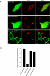
Figure 6.
Figure 6.
Overexpression of EGFP-GRIP domain prevented the perinuclear accumulation of STxB in HeLa cells. (A) HeLa cells were transiently transfected to overexpress EGFP-GRIP (a–c), EGFP-GRIP/Y697A (d–f), or Myc-Golgin-97CC (g–i). Cells were subsequently allowed to continuously internalize STxB for 3 h and processed to reveal STxB by IF (b, e, and h). Bars, 10 μm. (B) Statistical analysis of the morphology relating the ratio of cells with perinuclear to cells with only peripheral distribution of STxB under different transfection conditions. The ratio in nontransfected cells is defined as 100%. In each case, ∼100 cells, which contain both the exogenously over expressed protein and internalized STxB, were randomly chosen for scoring the distribution of STxB.
Figure 9.
Figure 9.
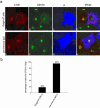
The retrograde trafficking of CTxB was arrested before the Golgi when Golgin-97's functions were interfered by its microinjected pAb. (a) HeLa cells were microinjected with Golgin-97 pAb (A–D) or preimmune rabbit IgG (E–H) and then allowed to internalize Alexa Fluor 555-conjugated CTxB for 30 min and followed by 60-min chase. Cells were subsequently triple labeled to reveal CTxB (A and E), GM130 (B and F), and injected rabbit IgG (C and G). In D and H, the intensity of blue color was reduced fivefold for clearer observation of red and green signals. Bars, 10 μm. (b) For those injected cells, which also internalized CTxB, the distribution of CTxB was analyzed and the percentage of cells with CTxB in the Golgi region was shown in the bar chart. About 50 cells microinjected with Golgin-97 pAb or control rabbit IgG were examined and scored.
Figure 1.
Figure 1.
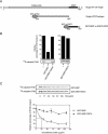
The recombinant GRIP domain of Golgin-97 inhibited in vitro EE/RE–TGN transport. (A) Schematic representation of the GRIP domain and the central coiled-coil region (Golgin-97CC) used in this study. (B) Relative efficiencies of STxB transport to the TGN as measured by the extents of its sulfation under various conditions as indicated. Lanes 1 and 4 are positive controls, and the transports were set as 100%. In lane 2, the assay contains 133 μg/ml GST-GRIP. In lane 3, GST-GRIP (133 μg/ml) was preboiled before adding to the assay. In lane 5, the reaction includes 133 μg/ml GST-GRIP/Y697A mutant. Error bars represent the standard deviations of two independent experiments. Bottom, typical gel autographic images. (C) Inhibition of EE/RE-TGN transport by GST-GRIP is dose dependent with a half-maximal inhibition at concentration of 25 μg/ml. Top, typical autoradiographs of 35S-sulfated STxB. Bottom shows the correspondent quantification curves. Points with deviation error bars indicate that these experiments were independently repeated twice. Transport in the absence of the recombinant protein is defined as 100%.
Figure 2.
Figure 2.
Generation and characterization of Golgin-97 pAb. (A) Golgin-97CC (Figure 1A) was used to raise rabbit antibodies and the purified Golgin-97 pAb recognized an endogenous protein of ∼97 kDa in human (lane 1) but not rat (lane 2) cell lysate. HeLa or NRK cell lysate (30 μg) was resolved by SDS-PAGE and analyzed by immunoblotting by using Golgin-97 pAb. (B) Golgin-97 pAb, but not the control rabbit IgG, efficiently immunoprecipitated (IP) endogenous human Golgin-97 from 293T cell lysate, which was detected by a commercial Golgin-97 mAb. Lane 1, 5% IP input; lane 2, IP using control rabbit IgG; lane 3, IP using Golgin-97 pAb.
Figure 3.
Figure 3.
Golgin-97 pAB inhibited in vitro EE/RE–TGN transport of STxB. Top, quantification of bottom panels, which are typical autoradiographs of 35S-sulfated STxB. (A) Golgin-97 pAb, but not control rabbit IgG, inhibited STxB transport in a dose-dependent manner. The STxB transport was reduced to 19% with the increasing amount of Golgin-97 pAb added to a concentration of 133 μg/ml (black bars), whereas the transport was still 85% when control rabbit IgG was increased to the same concentration (gray bars). Error bars represent the standard deviations of two independent experiments. Bottom, typical autoradiograph of 35S-sulfated STxB in the presence of increasing amount of Golgin-97 pAb or control rabbit IgG. Lane 1, transport assay in the absence of cytosol, serving as a negative control. The transport assays conducted in lanes 2–5 contained the indicated amount of Golgin-97 pAb or control rabbit IgG. (B) Inhibition of Golgin-97 pAb in STxB transport was specific as it could be relieved by preincubation with antigen. Lane 1, transport reaction in the absence of cytosol serving as a negative control. Lane 2, complete reaction, as positive control and the STxB transport was set as 100%. Lane 3, 33 μg/ml Golgin-97 pAb was added. Lane 4, 33 μg/ml Golgin-97 pAb was first neutralized by 67 μg/ml antigen (Ag, GST-Golgin-97CC) before addition to the assay. In lane 5, 67 μg/ml Ag was added to the reaction. Lane 6 contains 33 μg/ml Golgin-97 pAb preincubated with 67 μg/ml GST protein. In lane 7, 67 μg/ml GST protein was added to the reaction. Error bars represent the standard deviations of two independent experiments.
Figure 4.
Figure 4.
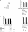
The membrane pool of Golgin-97 was responsible for in vitro EE/RE-TGN transport of STxB. (A) HeLa cytosol used in the STxB transport assay were mock depleted by control rabbit IgG (lane 1) or Golgin-97 pAb (lane 2). Top, Golgin-97 levels in two treated cytosols. Bottom, equal loading of two cytosols as assessed by blotting with antibodies against β-tubulin. About 90% of Golgin-97 was depleted in Golgin-97 pAb-treated cytosol. In B–D, top shows the relative percentage of STxB transport in bar charts. A typical set of autoradiographs of 35S-sulfated STxB is shown in the bottom panels for each experiment. Black bars are positive controls for adjacent gray bars and their STxB transports are set as 100%. Error bars represent standard deviations of two independent experiments. (B) Transport of STxB was reduced only slightly using Golgin-97 depleted HeLa cytosol. Lane 1, reaction without cytosol. Lanes 2 and 4, assays with 1 and 2 mg/ml control depleted cytosol, respectively. Lanes 3 and 5, assays with 1 and 2 mg/ml Golgin-97 pAb-depleted cytosol, respectively. (C) Separate treatment of membrane and cytosolic pools of Golgin-97 affected STxB transport differently. Lane 1, reaction without cytosol, as negative control. Lane 2, complete reaction (cytosol and semiintact cells), as positive control, and the transport is set as 100%. Lane 3, Golgin-97 pAb was mixed with cytosol and semiintact cells. In lanes 4 and 5, the cytosol was incubated without (lane 4, control for lane 5) or with Golgin-97 pAb (lane 5) for 1 h on ice (step 1), the excess amount of Golgin-97 pAb was then neutralized by twice amount of antigen (step 2). The resulting cytosol in lane 5 supported the transport of STxB with efficiency similar to control in lane 4 after addition of semiintact cells (step 3). In lanes 6 and 7, semiintact cells (containing membrane pool of Golgin-97) were incubated without (lane 6, as positive control for lane 7) or with Golgin-97 pAb (lane 7) on ice for 1 h (step 1). After antigen neutralization (step 2), the resulting semiintact cells were mixed with cytosol to complete the reaction (step 3). In lane 7, the STxB transport was reduced to ∼50% compared with lane 6. (D) Transport of STxB in HeLa semiintact cells was equally inhibited by human Golgin-97 specific pAb in the presence of either rat liver (which provided rat Golgin-97 that is not reactive with the antibody) or HeLa cytosol. Lane 1, without cytosol, as negative control. Lane 2, rat liver cytosol as positive control for lane 3. Lane 3, rat liver cytosol with 133 μg/ml Golgin-97 pAb. Lane 4, HeLa cell cytosol as positive control for lane 5. Lane 6, HeLa cell cytosol with 133 μg/ml Golgin-97 pAb.
Figure 7.
Figure 7.
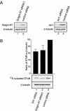
Knockdown of Arl1 or Golgin-97 inhibited in vivo trafficking of STxB to the TGN. (A) HeLa cells were transfected with indicated siRNAs. After 48 h, the total cell lysate was subjected to immunoblotting analysis to detect Golgin-97 (lanes 1 and 2 in the top panel), Arl1 (lanes 3 and 4 of the top panel), and β-tubulin (bottom). (B) The relative transport of STxB to Golgi in siRNA-treated HeLa cells. siRNA-treated HeLa cells were allowed to internalize STxB for 90 min in the presence of 35S-sulfate. The 35S-sulfated STxB was revealed by autoradiograph (middle panel shows a typical result). The loading of each lane was normalized by immunoblotting with antibody against β-tubulin (bottom). For each lane, the relative ratio of sulfated STxB to β-tubulin densities was quantified as bar chart in the top panel. Error bars represent standard deviations of four independent experiments. Knock down of Golgin-97 or Arl1 significantly reduced the sulfation of STxB compared with controls (p <7E-5 and 0.006, respectively).
Figure 5.
Figure 5.
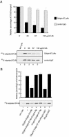
Golgin-97 was required before SNARE-mediated docking/fusion step in EE/RE–TGN transport in vitro. (A) Set of typical autoradiographs of 35S-sulfated STxB derived from the in vitro transport assays showed the temporal sensitivities to antibodies against Golgin-97 and Syntaxin 16. The complete transport assays were assembled and conducted for various time points as per normal. The Golgin-97 (top) or Syntaxin 16 (bottom) pAb was subsequently added at these time points, and the transport reactions were allowed to proceed in the presence of antibody for a total of 90 min. (B) Quantification of relative percentage of inhibition. The inhibition of STxB transport in which Golgin-97 and Syntaxin16 was added at 10-min time point is set as 100% and 90-min time point is defined as 0%. Error bars represent the standard deviations of two independent experiments.
Figure 10.
Figure 10.
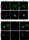
Internalized STxB was arrested at a stage before the Golgi in Arl1 knocked down HeLa cells. Arl1 siRNA (A–C and G–I) and control siRNA (D–F and J–L)-treated HeLa cells were allowed to internalized STxB for 3 h and subsequently double labeled to reveal internalized STxB (A, D, G, and J) and Ar1l (B and E) or GT (H and K). Bars, 10 μm.
Figure 12.
Figure 12.
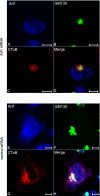
Internalized CTxB was similarly arrested at a stage before the Golgi in Ar1l knocked-down HeLa cells. Arl1 siRNA (A–D) and control siRNA (E–H)-treated HeLa cells were allowed to internalize Alexa Fluor 555-conjugated CTxB for 3 h and subsequently triple labeled to reveal Arl1 (A and E), GM130 (B and F) and internalized CTxB (C and G). Bars, 10 μm.
Figure 8.
Figure 8.
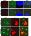
Microinjection Golgin-97 pAb-fragmented Golgi apparatus. HeLa cells were microinjected with Golgin-97 pAb (A–D and I–K) or preimmune rabbit IgG (E–H and L–N) and subsequently triple labeled to reveal Golgin-245 (A and E), TGN46 (B and F) and injected rabbit IgG (C and G) or double labeled to reveal γ-adaptin (I and L) and injected rabbit IgG (J and M). In D and H, the intensity of blue color was reduced fivefold for clearer observation of red and green signals. Bars, 10 μm.
Figure 11.
Figure 11.
The trafficking of STxB to Golgi was inhibited in Arl1-Q71L expressing cells. HeLa cells were transiently transfected with Arl1-Q71L and then allowed to continuously internalize STxB for 3 h. The coverslips were then processed for immunofluorescence microscopy to reveal over expressed Arl1-Q71L (by a limited amount of Arl1 antibody; A), Golgin-97 (in nontransfected cells; D), and STxB (B and E). Bar, 10 μm.

Similar articles

Cited by

References

    1. Allan, B.B., Moyer, B.D., and Balch, W.E. (2000). Rab1 recruitment of p115 into a cis-SNARE complex: programming budding COPII vesicles for fusion. Science 289, 444-448. - PubMed
    1. Antonny, B., and Schekman, R. (2001). ER export: public transportation by the COPII coach. Curr. Opin. Cell Biol. 13, 438-443. - PubMed
    1. Barr, F.A. (1999). A novel Rab6-interacting domain defines a family of Golgi-targeted coiled-coil proteins. Curr. Biol. 9, 381-384. - PubMed
    1. Barr, F.A., Puype, M., Vandekerckhove, J., and Warren, G. (1997). GRASP65, a protein involved in the stacking of Golgi cisternae. Cell 91, 253-262. - PubMed
    1. Barr, F.A., and Short, B. (2003). Golgins in the structure and dynamics of the Golgi apparatus. Curr. Opin. Cell Biol. 15, 405-413. - PubMed

Publication types

MeSH terms

LinkOut - more resources