. 2004 Jul 19;166(2):237-48.
doi: 10.1083/jcb.200401078.

Endocytosis of E-cadherin regulated by Rac and Cdc42 small G proteins through IQGAP1 and actin filaments

Affiliations

Endocytosis of E-cadherin regulated by Rac and Cdc42 small G proteins through IQGAP1 and actin filaments

Genkichi Izumi et al. J Cell Biol. .

Abstract

E-cadherin is a key cell-cell adhesion molecule at adherens junctions (AJs) and undergoes endocytosis when AJs are disrupted by the action of extracellular signals. To elucidate the mechanism of this endocytosis, we developed here a new cell-free assay system for this reaction using the AJ-enriched fraction from rat liver. We found here that non-trans-interacting, but not trans-interacting, E-cadherin underwent endocytosis in a clathrin-dependent manner. The endocytosis of trans-interacting E-cadherin was inhibited by Rac and Cdc42 small G proteins, which were activated by trans-interacting E-cadherin or trans-interacting nectins, which are known to induce the formation of AJs in cooperation with E-cadherin. This inhibition was mediated by reorganization of the actin cytoskeleton by Rac and Cdc42 through IQGAP1, an actin filament-binding protein and a downstream target of Rac and Cdc42. These results indicate the important role of the Rac/Cdc42-IQGAP1 system in the dynamic organization and maintenance of the E-cadherin-based AJs.


Figures

Figure 1.
Figure 1.
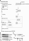
A cell-free assay system for the endocytosis of E-cadherin. (A) Flow diagram of the steps and schematic morphologies of the AJ plasma membranes. (B) Time-dependent endocytosis of E-cadherin. At the indicated times, the reactions were stopped by chilling each tube on ice and the MSP (16,000 g) and the HSP (100,000 g) were prepared by differential centrifugation as described in A. The amount of the MSP and the HSP (20 and 70% of total SDS solubilized membranes, respectively) were quantitated by immunoblotting with the anti–E-cadherin mAb, the anti-nectin-2 pAb, and the anti-clathrin HC mAb. (C) Requirement of the cytosol, ATP, and GTP for the endocytosis of E-cadherin. (Ca) The AJ plasma membranes were incubated in the presence of the indicated concentrations of the rat brain cytosol (RBC) or the boiled rat brain cytosol and assayed for the endocytosis of E-cadherin. (Cb) The AJ plasma membranes were incubated in the presence of 1 mM ATP and 100 μM GTP (control), 20U/ml of apyrase, 1 mM ATP, and 100 μM GTP (ATP depletion), or 1 mM ATP and 100 μM GTPγS (GTPγS), and assayed for the endocytosis of E-cadherin. All the reaction mixtures contained the rat brain cytosol. The results shown in all panels are representative of three independent experiments.
Figure 2.
Figure 2.
Characteristics of the endocytosed vesicles of E-cadherin. (A) Specific endocytosis of E-cadherin. The relative amount of the MSP and the HSP (15 μg of protein in each fraction) were analyzed by quantitative immunoblotting with various antibodies against the indicated proteins. The results were obtained from the same experiments and the same gels. (B) Isolation of endocytosed vesicles of E-cadherin. (Ba) Electron microscopic morphology of the HSP. The reaction for the formation of vesicles was performed as shown in Fig. 1 A. Bar, 100 nm. (Bb) Immunoelectron microscopic morphology of endocytosed vesicles of E-cadherin. The reaction for the formation of vesicles was performed as shown in Fig. 1 A. The vesicles were isolated from the MSS using the anti– β-catenin mAb-coated magnetic beads. The accumulated vesicles on the beads were processed for electron microscopy. The membranes bound to the beads consisted largely of a homogenous population of 60–80-nm vesicles (arrowheads). (inset) Immunogold labeling for E-cadherin. The immunoisolated beads were stained with the anti-pan-cadherin (cytoplasmic portion) pAb, followed by 10 nm gold-conjugated secondary antibody (arrows). Bars, 100 nm. (Bc) Composition of the endocytosed vesicles of E-cadherin. The vesicles were immunoisolated with the anti–β-catenin mAb or the anti-mouse IgG (control IgG)-coated magnetic beads from the MSS as shown in Bb. The bound proteins were analyzed by immunoblotting with various antibodies against the indicated proteins. The results were obtained from the same experiments and the same gels. The results shown in all panels are representative of three independent experiments.
Figure 3.
Figure 3.
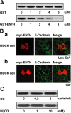
Clathrin-dependent endocytosis of E-cadherin. (A) Epsin-dependent endocytosis of E-cadherin in the cell-free assay system. The AJ plasma membranes were incubated in the presence of recombinant GST-ENTH or control GST at the indicated concentrations and assayed for the endocytosis of E-cadherin. (B) Epsin- dependent endocytosis of E-cadherin in MDCK cells. (Ba) MDCK cells were transfected with pEFBOS-myc-ENTH and cultured for 24 h. After the culture, the cells were incubated at 2 μM Ca2+ for 30 min. The cells were then fixed and stained for myc-ENTH and E-cadherin with the anti-myc pAb and the anti-E-cadherin mAb, respectively. Bar, 10 μm. (Bb) MDCK cells were transfected with pEFBOS-myc-ENTH and cultured for 24 h. After the culture, the cells were incubated with 10 ng/ml HGF for 3 h. The cells were then fixed and stained for myc-ENTH and E-cadherin with the anti-myc pAb and the anti-E-cadherin mAb, respectively. Bar, 10 μm. (C) Caveolin- independent endocytosis of E-cadherin. The AJ plasma membranes were incubated in the presence of cholesterol oxidase (CO) and cyclic heptasaccharide MβCD (MβCD) at the indicated concentrations and assayed for the endocytosis of E-cadherin. The results shown in all panels are representative of three independent experiments.
Figure 4.
Figure 4.
Regulation of endocytosis of E-cadherin by trans interaction of E-cadherin. (A) Enhancement of the endocytosis of E-cadherin by the E-cadherin–blocking antibody. The AJ plasma membranes were first assayed for the endocytosis of E-cadherin. The resulting membranes were incubated with the indicated concentrations of the E-cadherin–blocking antibody and assayed for the endocytosis of E-cadherin. (B) Inhibition of the endocytosis of non-trans-interacting E-cadherin by Cef. (Ba) After the preincubation at various concentrations of Ca2+, the membranes were collected and assayed for the endocytosis of E-cadherin. (Bb) The AJ plasma membranes were incubated with the indicated concentrations of Cef in the presence of 2 μM Ca2+ and assayed for the endocytosis of E-cadherin. (Bc) The AJ plasma membranes were incubated with the indicated concentrations of Cef in the presence of 2 mM Ca2+ and assayed for the endocytosis of E-cadherin. In all panels, the quantification of immunoblot is shown as the mean ± SD of duplicate assays in the bottom panel. The results shown in all panels are representative of three independent experiments.
Figure 5.
Figure 5.
Regulation of endocytosis of E-cadherin through Rac and Cdc42 activated by trans-interacting E-cadherin. (A) Activation of Rac and Cdc42 by Cef. The AJ plasma membranes were incubated with [35S]GTPγS and the GDP-bound form of recombinant Rac, Cdc42, or Rho in the presence or absence of Cef for the indicated periods of time. The bound [35S]GTPγS to Rac, Cdc42, or Rho was scintillation counted and plotted. The mean ± SD of duplicate assays is shown. (B) Enhancement of the endocytosis of E-cadherin by Rho GDI. The AJ plasma membranes were incubated with the indicated concentrations of GST-Rho GDI. The membranes were collected and assayed for the endocytosis of E-cadherin. The amount of remaining Rac on the AJ plasma membranes (10% of total) or the extent of the endocytosis of E-cadherin was determined by quantitative immunoblotting with the anti-Rac mAb or the anti–E-cadherin mAb, respectively. The mean ± SD of duplicate assays is shown. (C) Inhibition of the Rho GDI-enhanced endocytosis of E-cadherin by Cef. (Ca) The AJ plasma membranes were first incubated with Cef in the presence of 2 mM Ca2+ and then incubated with GST-Rho GDI. The membranes were collected and assayed for the endocytosis of E-cadherin. The numbers 1 and 2 indicate the sequence of the incubation. (Cb) Conversely, the AJ plasma membranes were first incubated with GST-Rho GDI and then incubated with Cef in the presence of 2 mM Ca2+. The membranes were collected and assayed for the endocytosis of E-cadherin. The numbers 1 and 2 indicate the sequence of the incubation. (D) Inhibition of the Rho GDI-enhanced endocytosis by Rac and Cdc42, but not by Rho. (Da) The AJ plasma membranes were first incubated with GST-Rho GDI and collected. The collected membranes were restored by adding the GTPγS-bound form of recombinant Rac, Cdc42, or Rho at the indicated concentrations and assayed for the endocytosis of E-cadherin. The numbers 1 and 2 indicate the sequence of the incubation. (Db) The membrane bound amount of added GTPγS-bound form of recombinant Rac in Da. (D, a and b) The quantification of immunoblot is shown as the mean ± SD of duplicate assays in the bottom panel. The numbers 1 and 2 indicate the sequence of the incubation. The results shown in all panels are representative of three independent experiments.
Figure 6.
Figure 6.
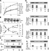
Involvement of IQGAP1 in the inhibitory effect of Rac and Cdc42 on the endocytosis of E-cadherin. (A) Inhibition of the endocytosis of E-cadherin by IQGAP1. (Aa) The AJ plasma membranes were incubated with the indicated concentrations of recombinant His-IQGAP1 and assayed for the endocytosis of E-cadherin. (Ab) The AJ plasma membranes were first incubated with GST-Rho GDI and collected. The collected membranes were incubated with recombinant His-IQGAP1 at the indicated concentrations and assayed for the endocytosis of E-cadherin. The numbers 1 and 2 indicate the sequence of the incubation. (Ac) The AJ plasma membranes were incubated with the GTPγS-bound form of recombinant Rac and recombinant His-IQGAP1 at the indicated concentrations and assayed for the endocytosis of E-cadherin. (Ad) The AJ plasma membranes were incubated with the GTPγS-bound form of recombinant Cdc42 and recombinant His-IQGAP1 at the indicated concentrations and assayed for the endocytosis of E-cadherin. (Ae) The AJ plasma membranes were incubated with the indicated concentrations of recombinant His-ΔCHD-IQGAP1 and assayed for the endocytosis of E-cadherin. (Af) The AJ plasma membranes were incubated with the GTPγS-bound form of recombinant Rac and recombinant His-ΔCHD-IQGAP1 at the indicated concentrations and assayed for the endocytosis of E-cadherin. (A, a–f) The quantification of immunoblot is shown as the mean ± SD of duplicate assays in the bottom panel. (B) Endocytosis efficiency (%) is expressed relative to the controls in each experiment in A (a–d). The results shown in all panels are representative of three independent experiments.
Figure 7.
Figure 7.
Involvement of the actin cytoskeleton in the inhibitory effect of IQGAP1 on the endocytosis of E-cadherin. (A) Prevention of the inhibitory effect of IQGAP1 on the endocytosis of E-cadherin by Lat-A. (Aa) The AJ plasma membranes were incubated with Lat-A or nocodazole and assayed for the endocytosis of E-cadherin. (Ab) The AJ plasma membranes were first incubated with Lat-A and collected. The collected membranes were incubated with the GTPγS-bound form of recombinant Rac and assayed for the endocytosis of E-cadherin. The numbers 1 and 2 indicate the sequence of the incubation. (Ac) The AJ plasma membranes were first incubated with Lat-A and collected. The collected membranes were incubated with recombinant His-IQGAP1 and assayed for the endocytosis of E-cadherin. (A, a–c) The quantification of immunoblot is shown as the mean ± SD of duplicate assays in the bottom panel. The numbers 1 and 2 indicate the sequence of the incubation. (B) F-Actin levels in the AJ plasma membranes. Increase in total F-actin in the AJ plasma membranes treated with the indicated factors was expressed as fold increase to the amount of F-actin in the nontreated control AJ plasma membranes. The mean ± SD of duplicate assays is shown. (C) Effect of the Arp2/3 complex on the endocytosis of E-cadherin. (Ca) The AJ plasma membranes were incubated with the indicated concentrations of the Arp2/3 complex and assayed for the endocytosis of E-cadherin. (Cb) The AJ plasma membranes were assayed for the endocytosis of E-cadherin in the presence of either control cytosol or Arp2/3-depleted cytosol. The results shown in all panels are representative of at least three independent experiments.
Figure 8.
Figure 8.
Inhibition of the endocytosis of E-cadherin by trans-interacting E-cadherin or trans-interacting nectin-1 in intact cells. (A) Inhibition of the constitutive endocytosis of E-cadherin by trans-interacting E-cadherin in intact EL cells. (Aa) EL cells were incubated in the medium with 400 nM Cef or 400 nM human IgG (control IgG) for 60 min. The cells were surface-biotinylated on ice and cultured at 18°C for the indicated periods of time to allow the endocytosis of E-cadherin. Biotinylated proteins on the plasma membrane were then stripped off by glutathione treatment, and biotinylated proteins inside the cells were recovered on streptavidin beads. The bound proteins were analyzed by immunoblotting with the anti-E-cadherin mAb. The relative amounts of endocytosed E-cadherin were expressed as percentage of total biotinylated E-cadherin in the bottom panel. The mean ± SD of duplicate assays is shown. (Ab) EL cells pretreated with the 400 nM Cef or 400 nM control IgG were incubated at 18°C for 20 min to allow the endocytosis of E-cadherin. EL cells overexpressing GFP, GFP-N17Rac1, or GFP-N-WASP-CRIB-CAAX were pretreated with the 400 nM Cef or 400 nM control IgG and then incubated at 18°C for 20 min to allow the endocytosis of E-cadherin. The cells were then fixed and immunostained with the anti–E-cadherin mAb and the anti-IQGAP1 pAb. Bars, 10 μm. (B) Inhibition of the constitutive endocytosis of E-cadherin by trans-interacting nectin-1 in intact nectin-1-EL cells. (Ba) Nectin-1-EL (N1-EL) cells were incubated in the medium with 400 nM Nef-3 or 400 nM control IgG for 60 min. The cells were surface-biotinylated on ice and cultured at 18°C for the indicated time to allow the endocytosis of E-cadherin. Biotinylated proteins on the plasma membranes were then stripped off by glutathione treatment, and biotinylated proteins inside the cells were recovered on streptavidin beads. The bound proteins were analyzed by immunoblotting with the anti–E-cadherin mAb. The relative amounts of endocytosed E-cadherin were expressed as percentage of total biotinylated E-cadherin in the bottom panel. The mean ± SD of duplicate assays is shown. (Bb) N1-EL cells pretreated with 400 nM Nef-3 or 400 nM control IgG were incubated at 18°C for 20 min to allow the endocytosis of E-cadherin. N1-EL cells overexpressing GFP, GFP-N17Rac1, or GFP-N-WASP-CRIB-CAAX were pretreated with the 400 nM Nef-3 or 400 nM control IgG and then incubated at 18°C for 20 min to allow the endocytosis of E-cadherin. The cells were then fixed and immunostained with the anti–E-cadherin mAb and the anti-IQGAP1 pAb. Bars, 10 μm. The results shown in all panels are representative of at least three independent experiments.

Similar articles

Cited by

References

    1. Adams, C.L., and W.J. Nelson. 1998. Cytomechanics of cadherin-mediated cell-cell adhesion. Curr. Opin. Cell Biol. 10:572–577. - PubMed
    1. Adams, C.L., Y.T. Chen, S.J. Smith, and W.J. Nelson. 1998. Mechanisms of epithelial cell-cell adhesion and cell compaction revealed by high-resolution tracking of E-cadherin-green fluorescent protein. J. Cell Biol. 142:1105–1119. - PMC - PubMed
    1. Akhtar, N., and N.A. Hotchin. 2001. RAC1 regulates adherens junctions through endocytosis of E-cadherin. Mol. Biol. Cell. 12:847–862. - PMC - PubMed
    1. Anastasiadis, P.Z., and A.B. Reynolds. 2000. The p120 catenin family: complex roles in adhesion, signaling and cancer. J. Cell Sci. 113:1319–1334. - PubMed
    1. Arribas, J., and A. Borroto. 2002. Protein ectodomain shedding. Chem. Rev. 102:4627–4638. - PubMed

Publication types