. 2004 Jun 2;24(22):5193-201.
doi: 10.1523/JNEUROSCI.0854-04.2004.

Cytoplasmic polyadenylation element binding protein-dependent protein synthesis is regulated by calcium/calmodulin-dependent protein kinase II

Affiliations

Cytoplasmic polyadenylation element binding protein-dependent protein synthesis is regulated by calcium/calmodulin-dependent protein kinase II

Coleen M Atkins et al. J Neurosci. .

Abstract

Phosphorylation of cytoplasmic polyadenylation element binding protein (CPEB) regulates protein synthesis in hippocampal dendrites. CPEB binds the 3' untranslated region (UTR) of cytoplasmic mRNAs and, when phosphorylated, initiates mRNA polyadenylation and translation. We report that, of the protein kinases activated in the hippocampus during synaptic plasticity, calcium/calmodulin-dependent protein kinase II (CaMKII) robustly phosphorylated the regulatory site (threonine 171) in CPEB in vitro. In postsynaptic density fractions or hippocampal neurons, CPEB phosphorylation increased when CaMKII was activated. These increases in CPEB phosphorylation were attenuated by a specific peptide inhibitor of CaMKII and by the general CaM-kinase inhibitor KN-93. Inhibitors of protein phosphatase 1 increased basal CPEB phosphorylation in neurons; this was also attenuated by a CaM-kinase inhibitor. To determine whether CaM-kinase activity regulates CPEB-dependent mRNA translation, hippocampal neurons were transfected with luciferase fused to a 3' UTR containing CPE-binding elements. Depolarization of neurons stimulated synthesis of luciferase; this was abrogated by inhibitors of protein synthesis, mRNA polyadenylation, and CaMKII. These results demonstrate that CPEB phosphorylation and translation are regulated by CaMKII activity and provide a possible mechanism for how dendritic protein synthesis in the hippocampus may be stimulated during synaptic plasticity.


Figures
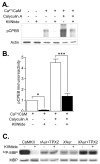
Figure 2.
Figure 2.
Endogenous CPEB is phosphorylated by CaMKII in the PSD. A, PSD fractions were phosphorylated with or without 1.4 mm Ca2+ and 5 μm CaM, 50 nm CA, or the specific CaMKII inhibitor CaMKIINtide (KIINtide; 10 μm) and then Western blotted for pCPEB. Basal phosphorylation levels were negligible. Ca2+/CaM and CA increased pCPEB levels. Protein levels were equivalent as shown by reprobing with an actin antibody. B, Densitized results, normalized to the Ca2+/CaM stimulation condition (n = 3 for each). CA increased CPEB phosphorylation significantly above the increase seen with Ca2+/CaM (n = 3; p < 0.001). The selective, potent peptide inhibitor of CaMKII, CaMKIINtide, significantly blocked pCPEB in PSD fractions treated with Ca2+/CaM (n = 3; p < 0.05) or CA (n = 3; p < 0.001). C, To test whether another CPEB kinase found in the PSD, Aurora, could be attenuated by CaMKIINtide, phosphorylation of an Aurora kinase substrate, MBP, was assayed. Shown is a representative phosphor-imaging blot for 32P-MBP and the corresponding Coomassie Blue stained gel (MBP). MBP was phosphorylated with CaMKII, rat Aurora kinase (rAur), or Xenopus Aurora kinase (XAur). CaMKIINtide (KIINtide, 10 μm) abolished CaMKII activity. Rat Aurora kinase stimulated by the Aurora kinase chaperone, TPX2, and basal and stimulated Xenopus Aurora A kinase were not affected by CaMKIINtide. Thus, another CPEB kinase found in the PSD, Aurora kinase, was not inhibited by CaMKIINtide.
Figure 3.
Figure 3.
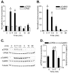
Time course of CPEB phosphorylation in neurons stimulated by depolarization. A, Representative Western blots of pCPEB and pCaMKII from neurons depolarized with 90 mm KCl for the indicated time points. For the 10 and 60 min time points, neurons were depolarized for 5 min and then incubated in control solution to assess whether phosphorylation levels were prolonged beyond the initial depolarization. B, Densitized results of the fold increase in pCPEB. pCPEB levels were significantly elevated at 10 sec (0.17 min; n = 18; p < 0.001) and 0.5 min (n = 10; p < 0.001) but not at 5 min (n = 6), 10 min (n = 6), or 60 min (n = 6). There was no significant change in CPEB total levels. C, Densitized fold increase in pCaMKII levels. Similar to pCPEB, increases in pCaMKII were significant at 10 sec (0.17 min; n = 18; p < 0.001) and 0.5 min (n = 10; p < 0.001). D, Densitized results of the fold increase in pCPEB (left y-axis) and pCaMKII (right y-axis) when neurons were stimulated with glutamate (100 μm) for the indicated times. Both pCPEB (n = 3) and pCaMKII (n = 3) increased at 10 sec (0.17 min; p < 0.001) but returned to baseline levels after 5 min.
Figure 5.
Figure 5.
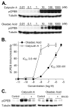
Identification of the protein phosphatase acting on CPEB. A, Representative Western blots of pCPEB and corresponding control blots for total protein (Tubulin). Hippocampal neurons were treated (60 min) with increasing concentrations of the protein phosphatase 1 and 2a inhibitors, calyculin A or okadaic acid, respectively. B, Densitized changes in pCPEB in response to calyculin A and okadaic acid (n = 6 for all concentrations except for 5 μm; n = 4). Lower concentrations of calyculin A (IC50 3.81 nm) were needed to increase pCPEB levels compared with okadaic acid (IC50 301 nm). C, Representative Western blots of pCPEB and total protein (Tubulin). Hippocampal neurons were treated with calyculin A (2.5 nm) or okadaic acid (5 nm) for 60 min. The CaM-kinase inhibitor KN-93 (10 μm) attenuated the increase in pCPEB with calyculin A or okadaic acid. Results are representative of experiments repeated in triplicate.
Figure 1.
Figure 1.
Phosphorylation of CPEB by CaMKII in vitro. A, Purified CPEB-GST fusion protein or a ubiquitous kinase substrate, MBP, was phosphorylated in vitro with [γ-32P]ATP and the indicated protein kinases. Representative phosphor-imaging blots for 32P-CPEB and 32P-MBP are shown with the corresponding Coomassie Blue stained gels (CPEB and MBP). All protein kinases were active as demonstrated by 32P-incorporation into MBP, but only CaMKII robustly phosphorylated CPEB. B, Densitized results of 32P-incorporation into WT CPEB versus Thr171Ala CPEB, normalizing phosphorylation of WT CPEB to 100% for each protein kinase. CaMKII phosphorylation of CPEB was significantly affected by Thr171Ala mutation (KII, 50 nm; n = 10; p < 0.001), but PKC (0.0125 U; n = 4), PKA (50 nm; n = 10), MAPK (20 nm; n = 7), or CaMKI (KI, 50 nm; n = 4) and CaMKIV (KIV, 50 nm; n = 12), both activated by CaMKK, were unaffected by the mutation. GST phosphorylation was negligible. C, Purified CPEB-GST fusion proteins, WT or Thr171Ala (A), were phosphorylated in vitro and then Western blotted with a pCPEB monoclonal antibody or a total CPEB antibody. D, Densitized results, normalizing the phosphorylation level of WT CPEB by CaMKII to 100%. Phosphorylation by CaMKII, but not activated CaMKI or CaMKIV, was significantly affected by Thr171Ala mutation (n = 3 for each; p < 0.001).
Figure 4.
Figure 4.
CPEB phosphorylation in depolarized neurons requires extracellular calcium, calmodulin, and voltage-gated calcium channels; all of which lead to activation of CaMKII. A, Densitized results of the fold increase in pCPEB levels (left y-axis) and pCaMKII levels (right y-axis). The increases in pCPEB (KCl; n = 24; p < 0.001) and pCaMKII (KCl; n = 24; p < 0.001) during neuronal depolarization were blocked when calcium was removed from the extracellular solution and 1 mm EGTA was included (0 Ca2+ pCPEB, n = 8; pCaMKII, n = 8) or by 5 μm CZ (pCPEB, n = 9; pCaMKII, n = 10). A voltage-gated calcium channel blocker, 300 μm CdCl2, also blocked the increases in pCPEB (n = 10) and partially attenuated the increases in pCaMKII (n = 10; p < 0.001), but an NMDA receptor antagonist, APV (50 μm), did not affect pCPEB levels (n = 8; p < 0.001) or pCaMKII levels (n = 8; p < 0.001). B, Application of the CaMKK inhibitor STO-609 (STO; 5 μm; n = 11), the PKA inhibitor H-89 (5 μm; n = 6), the PKC inhibitor chelerythrine (Chele; 1 μm; n = 4), or the MAPK inhibitor U0126 (5 μm; n = 6) had no effect on CPEB phosphorylation. C, The CaM-kinase inhibitor, KN-93 (10 μm), attenuated CPEB phosphorylation when neurons were depolarized (n = 16; p < 0.01). This attenuation was comparable with the attenuation in CaMKII activation by KN-93 (n = 16; p < 0.001). The inactive analog KN-92 had no significant effect on either pCPEB or pCaMKII. D, Correlation analysis of the inhibition of pCPEB and pCaMKII with increasing concentrations of KN-93 (0, 2.5, 5, and 10 μm). When hippocampal neurons were depolarized, the increases in pCPEB and pCaMKII were inhibited to a similar extent by KN-93 (R = 0.901).
Figure 6.
Figure 6.
Regulation of CPE-mediated protein synthesis by CaMKII in neurons. A, Cultured hippocampal neurons were transfected with plasmids expressing firefly luciferase fused to a portion of CaMKII 3′ UTR that contained two WT CPE domains or mutated CPE domains (MT). Neurons were stimulated with depolarization by 90 mm KCl for 5 min, incubated for 3 hr in control saline, and then assayed for luciferase levels. Luciferase expression significantly increased in depolarized neurons only when the CPE-binding domains were intact (n = 100; p < 0.001). Treatment with 0.8 μm anisomycin (n = 15) or 80 μm cordycepin (n = 17) decreased basal translation levels by 40–50% (data not shown) and blocked the increases in luciferase expression when neurons were depolarized. B, CPE-mediated luciferase translation was blocked by a specific inhibitor of CaMKII (KIIN). Cotransfection of neurons with luciferase fused to the 3′ UTR of CaMKII and empty vector elicited significant increases in luciferase when the CPE domains were not mutated (control, n = 24; p < 0.001). However, cotransfection of CaMKIIN, an endogenous protein inhibitor of CaMKII, blocked the increases in luciferase (n = 22), whereas the PKA inhibitor 5 μm H-89 (n = 11; p < 0.01) or the MAPK inhibitor 5 μm U0126 (n = 16; p < 0.001) did not block the increases in luciferase translation.

Similar articles

Cited by

References

    1. Aakalu G, Smith WB, Nguyen N, Jiang C, Schuman EM (2001) Dynamic visualization of local protein synthesis in hippocampal neurons. Neuron 30: 489–502. - PubMed
    1. Blitzer RD, Connor JH, Brown GP, Wong T, Shenolikar S, Iyengar R, Landau EM (1998) Gating of CaMKII by cAMP-regulated protein phosphatase activity during LTP. Science 280: 1940–1942. - PubMed
    1. Brewer GJ (1997) Isolation and culture of adult rat hippocampal neurons. J Neurosci Methods 71: 143–155. - PubMed
    1. Brown GP, Blitzer RD, Connor JH, Wong T, Shenolikar S, Iyengar R, Landau EM (2000) Long-term potentiation induced by theta frequency stimulation is regulated by a protein phosphatase-1-operated gate. J Neurosci 20: 7880–7887. - PMC - PubMed
    1. Cao Q, Richter JD (2002) Dissolution of the maskin-eIF4E complex by cytoplasmic polyadenylation and poly(A)-binding protein controls cyclin B1 mRNA translation and oocyte maturation. EMBO J 21: 3852–3862. - PMC - PubMed

Publication types

MeSH terms