Dissection of the mammalian midbody proteome reveals conserved cytokinesis mechanisms
- PMID: 15166316
- PMCID: PMC3679889
- DOI: 10.1126/science.1097931
Dissection of the mammalian midbody proteome reveals conserved cytokinesis mechanisms
Abstract
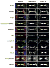
Cytokinesis is the essential process that partitions cellular contents into daughter cells. To identify and characterize cytokinesis proteins rapidly, we used a functional proteomic and comparative genomic strategy. Midbodies were isolated from mammalian cells, proteins were identified by multidimensional protein identification technology (MudPIT), and protein function was assessed in Caenorhabditis elegans. Of 172 homologs disrupted by RNA interference, 58% displayed defects in cleavage furrow formation or completion, or germline cytokinesis. Functional dissection of the midbody demonstrated the importance of lipid rafts and vesicle trafficking pathways in cytokinesis, and the utilization of common membrane cytoskeletal components in diverse morphogenetic events in the cleavage furrow, the germline, and neurons.
Figures


Similar articles
-
Mitotic spindle proteomics in Chinese hamster ovary cells.PLoS One. 2011;6(5):e20489. doi: 10.1371/journal.pone.0020489. Epub 2011 May 27. PLoS One. 2011. PMID: 21647379 Free PMC article.
-
Profiling of the mammalian mitotic spindle proteome reveals an ER protein, OSTD-1, as being necessary for cell division and ER morphology.PLoS One. 2013 Oct 10;8(10):e77051. doi: 10.1371/journal.pone.0077051. eCollection 2013. PLoS One. 2013. PMID: 24130834 Free PMC article.
-
Determination of the cleavage plane in early C. elegans embryos.Annu Rev Genet. 2008;42:389-411. doi: 10.1146/annurev.genet.40.110405.090523. Annu Rev Genet. 2008. PMID: 18710303 Review.
-
The large GTPase dynamin associates with the spindle midzone and is required for cytokinesis.Curr Biol. 2002 Dec 23;12(24):2111-7. doi: 10.1016/s0960-9822(02)01390-8. Curr Biol. 2002. PMID: 12498685 Free PMC article.
-
Proteomics of regulated secretory organelles.Mass Spectrom Rev. 2009 Sep-Oct;28(5):844-67. doi: 10.1002/mas.20211. Mass Spectrom Rev. 2009. PMID: 19301366 Review.
Cited by
-
Novel role for the midbody in primary ciliogenesis by polarized epithelial cells.J Cell Biol. 2016 Aug 1;214(3):259-73. doi: 10.1083/jcb.201601020. Epub 2016 Jul 25. J Cell Biol. 2016. PMID: 27458130 Free PMC article.
-
The Exocyst Complex in Health and Disease.Front Cell Dev Biol. 2016 Apr 12;4:24. doi: 10.3389/fcell.2016.00024. eCollection 2016. Front Cell Dev Biol. 2016. PMID: 27148529 Free PMC article. Review.
-
Klotho Exerts an Emerging Role in Cytokinesis.Genes (Basel). 2020 Sep 4;11(9):1048. doi: 10.3390/genes11091048. Genes (Basel). 2020. PMID: 32899868 Free PMC article.
-
Profilin-mediated competition between capping protein and formin Cdc12p during cytokinesis in fission yeast.Mol Biol Cell. 2005 May;16(5):2313-24. doi: 10.1091/mbc.e04-09-0781. Epub 2005 Mar 2. Mol Biol Cell. 2005. PMID: 15743909 Free PMC article.
-
Balance of actively generated contractile and resistive forces controls cytokinesis dynamics.Proc Natl Acad Sci U S A. 2005 May 17;102(20):7186-91. doi: 10.1073/pnas.0502545102. Epub 2005 May 3. Proc Natl Acad Sci U S A. 2005. PMID: 15870188 Free PMC article.
References
Publication types
MeSH terms
Substances
Grants and funding
LinkOut - more resources
Full Text Sources
Other Literature Sources
Molecular Biology Databases

