. 2004 Jun;78(12):6621-35.
doi: 10.1128/JVI.78.12.6621-6635.2004.

Short duration of elevated vIRF-1 expression during lytic replication of human herpesvirus 8 limits its ability to block antiviral responses induced by alpha interferon in BCBL-1 cells

Affiliations

Short duration of elevated vIRF-1 expression during lytic replication of human herpesvirus 8 limits its ability to block antiviral responses induced by alpha interferon in BCBL-1 cells

Veronika P Pozharskaya et al. J Virol. 2004 Jun.

Abstract

Human herpesvirus 8 (HHV-8) encodes multiple proteins that disrupt the host antiviral response, including viral interferon (IFN) regulatory factor 1 (vIRF-1). The product of the vIRF-1 gene blocks responses to IFN when overexpressed by transfection, but the functional consequence of vIRF-1 that is expressed during infection with HHV-8 is not known. These studies demonstrate that BCBL-1 cells that were latently infected with HHV-8 expressed low levels of vIRF-1 that were associated with PML bodies, whereas much higher levels of vIRF-1 were transiently expressed during the lytic phase of HHV-8 replication. The low levels of vIRF-1 that were associated with PML bodies were insufficient to block alpha IFN (IFN-alpha)-induced alterations in gene expression, whereas cells that expressed high levels of vIRF-1 were resistant to some changes induced by IFN-alpha, including the expression of the double-stranded-RNA-activated protein kinase. High levels of vIRF-1 were expressed for only a short period during the lytic cascade, so many cells with HHV-8 in the lytic phase responded to IFN-alpha with increased expression of antiviral genes and enhanced apoptosis. Furthermore, the production of infectious virus was severely compromised when IFN-alpha was present early during the lytic cascade. These studies indicate that the transient expression of high levels of vIRF-1 is inadequate to subvert many of the antiviral effects of IFN-alpha so that IFN-alpha can effectively induce apoptosis and block production of infectious virus when present early in the lytic cascade of HHV-8.


Figures
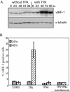
FIG. 1.
FIG. 1.
Changes in expression of vIRF-1. (A) Total cellular extracts (50 μg/lane) were prepared from BCBL-1 cells that were cultured in the absence or the presence of TPA (20 ng/ml) for the indicated durations. Levels of vIRF-1 and α-tubulin in these extracts were determined by Western blot analysis with the antibodies described in Materials and Methods. (B) The percentages of BCBL-1 cells that expressed high levels of vIRF-1 after incubation for 24 and 48 h with the indicated agents were assessed by IFA. BCBL-1 cells from nine random fields were photographed at a magnification of ×200 for each experimental condition in duplicate. The number of intensely vIRF-1-positive cells within each field was determined with Image Pro Plus software and was compared to the total number of cells. Error bars indicate standard deviations.
FIG. 2.
FIG. 2.
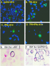
Morphology of BCBL-1 cells that express vIRF-1. (A to D) BCBL-1 cells were incubated for 48 h in standard medium (A) or in medium supplemented with TPA (20 ng/ml) (B), with IFN-α (1,000 U/ml) (C), or with both TPA and IFN-α simultaneously (D). IFA was done with a polyclonal antibody to vIRF-1 and a secondary antibody conjugated with Alexa Fluor 488 (green). The nuclei of all cells were labeled with DAPI (blue). (E and F) In situ hybridization was performed on uninduced BCBL-1 cells by using digoxigenin-labeled antisense riboprobes to vIRF-1 (E) and to GAPDH (F). The specificity of the riboprobes was demonstrated by using sense vIRF-1 (data not shown) and sense GAPDH (inset in panel F), both of which were nonreactive. Magnifications, ×200 (A to D) and ×400 (E and F).
FIG. 3.
FIG. 3.
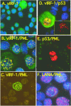
Colocalization of vIRF-1 and PML in latently infected BCBL-1 cells. BCBL-1 cells were incubated for 48 h in standard medium. Single and double immunofluorescent staining was done with vIRF-1 polyclonal antibody (A to D), p53 monoclonal antibody (D and E), LANA monoclonal antibody (F), and PML monoclonal antibody (B, C, E, and F), with secondary antibodies conjugated to Alexa Fluor 488 (green) and Alexa Fluor 568 (red). For all images, vIRF-1 was secondarily labeled with Alexa Fluor 488 (green), and p53 and LANA were secondarily labeled with Alexa Fluor 568 (red). PML was labeled with Alexa Fluor 568 (red) in panels B, C, and F and with Alexa Fluor 488 (green) in panel E. Cellular nuclei were counterstained with DAPI (blue). Imaging was performed with a Nikon Eclipse E-800 microscope equipped with an Optronics MagnaFire S99800 digital camera (A, B, D, and F) or with a Zeiss axioplasm laser-scanning confocal microscope with a Zeiss X100 1.3 oil emersion objective (C and E). Magnifications, ×400 (A) and ×1,000 (B to F).
FIG. 4.
FIG. 4.
Multinucleated BCBL-1 cells and deficient centrosomes in cells expressing elevated levels of vIRF-1. (A) Double immunofluorescent staining was done with vIRF-1 polyclonal antibody and γ-tubulin monoclonal antibody to detect centrosomes. Secondary antibodies were conjugated to Alexa Fluor 488 (green) and Alexa Fluor 568 (red) for vIRF-1 and γ-tubulin, respectively. Cellular nuclei were counterstained with DAPI. Magnification, ×400. (B) Immunofluorescent staining with vIRF-1 antibody detected by Alexa Fluor 488 (green) followed by FISH with a probe for the centromere region of chromosome 8 conjugated to SpectrumOrange (red). Cellular nuclei were counterstained with DAPI. Magnification, ×1,000.
FIG.5.
FIG.5.
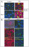
Expression of vIRF-1 and PPF. (A) BCBL-1 cells were incubated in standard medium (control) or in medium supplemented with TPA (20 ng/ml) for 2 and 5 days, and expression of vIRF-1 (green) and PPF (red) was analyzed by IFA. Colocalization of vIRF-1 and PPF appears yellow. The nuclei of all cells were counterstained with DAPI (blue). Magnification, ×200. (B) BCBL-1 cells were incubated in standard medium (control) or in medium supplemented with TPA (20 ng/ml) for 2 days. The medium was then changed, cells were maintained in fresh medium for 24 h, and expression of vIRF-1 (green) and PPF (red) was individually determined by IFA. The nuclei of all cells were counterstained with DAPI (blue). Magnification, ×200.
FIG. 6.
FIG. 6.
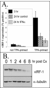
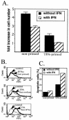
Duration of vIRF-1 expression. (A) BCBL-1 cells were primed with TPA (20 ng/ml) for 48 h or were not primed. The medium was then changed, and cells were maintained in fresh medium or medium supplemented with IFN-α (1,000 U/ml) for 24 h. The percentage of cells that expressed high levels of vIRF-1 was determined by IFA with anti-vIRF-1 antibody. BCBL-1 cells from nine random fields were photographed at a magnification of ×200 for each of duplicate samples prepared per experimental condition. The number of vIRF-1-positive cells within each field was determined with Image Pro Plus software. Error bars indicate standard deviations. (B) BCBL-1 cells were incubated with TPA for 48 h. Protein synthesis was inhibited by incubation with cycloheximide (Cx) (10 μg/ml) for the indicated durations. Total cellular protein (50 μg) was size fractionated by SDS-polyacrylamide gel electrophoresis and analyzed by Western blot analysis for expression of vIRF-1 and α-tubulin.
FIG. 7.
FIG. 7.
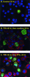
Effect of vIRF-1 on IFN-α-induced PKR expression. BCBL-1 cells were incubated in the absence (A) or presence (B and C) of TPA for 48 h. After the medium was changed, cells were incubated for 24 h in standard medium (A and B) or in the presence of IFN-α (1,000 U/ml) for 24 h (C). Expression of vIRF-1 was detected by using vIRF-1 polyclonal antibody followed by Alexa Fluor 488-conjugated antibody (green). Expression of PKR was determined by using PKR monoclonal antibody followed by Alexa Fluor 568-cojugated antibody (red). Cellular DNA was counterstained with DAPI. Magnification, ×400.
FIG. 8.
FIG. 8.
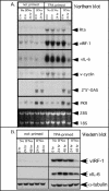
Effect of TPA priming on gene induction by IFN-α. BCBL-1 cells at a density of 5 × 10⁵/ml were incubated in the absence or presence of TPA (20 ng/ml) for 48 h and then switched at time zero to standard medium or medium supplemented with IFN-α (1,000 U/ml). Cells were harvested at the indicated time points. (A) Total cellular RNA (15 μg) was analyzed by Northern blotting with the indicated probes. Ethidium bromide staining was carried out to assess RNA loading (bottom panel). (B) Whole-cell extracts (50 μg) were analyzed by Western blotting with antibodies to vIRF-1, vIL-6, and α-tubulin.
FIG. 9.
FIG. 9.
Effect of TPA priming and incubation with IFN-α on cell number and apoptosis. Cells were incubated in control medium (nonprimed) or medium supplemented with 20 ng of TPA per ml (primed) for 48 h. At time zero, the cell number was adjusted to 5 × 10⁵/ml, and the medium was then replaced with fresh medium with no supplements (control) or supplemented with IFN-α (1,000 U/ml) for 24 and 48 h. (A) BCBL-1 cell number and viability were measured by direct counting of cells that excluded trypan blue by using a hemacytometer as described in the text. The fold increase was determined by dividing the number of cells after a 48-h incubation by the number at time zero. Error bars indicate standard deviations for triplicate experiments. (B) Apoptosis in nonprimed and TPA-primed cells was assessed at 24 h following stimulation by using the TUNEL assay. For each sample, 10,000 events were acquired. The percentage of apoptotic cells was determined by using a gate that contained 90% of the peak from the nonprimed control but lacked the shoulder to the right of the peak. A shift to the right is indicative of increased TUNEL labeling, indicating increased apoptosis. FITC, fluorescein isothiocyanate. (C) The percentage of cells to the right of the gate for each of the experimental conditions was determined and plotted as percent apoptotic cells.
FIG. 10.
FIG. 10.
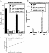
Impact of IFN-α on cell number and production of infectious HHV-8 in TPA-stimulated cells. BCBL-1 cells were incubated in the absence of TPA (control) or with TPA (20 ng/ml), and IFN-α (1,000 U/ml) was added at the same time as TPA (simultaneous) or 24 or 48 h after TPA stimulation. The amount of infectious virus in the cultured medium and the number of surviving cells were examined 4 and 5 days after TPA stimulation. (A) The number of cells was determined by direct counting of cells that excluded trypan blue by using a hemacytometer. The data represent means and standard deviations from triplicate determinations. (B) The amount of infectious virus in 200 μl of the medium was determined by using the T1H6 reporter cell line and is shown as relative luminescence units. (C) A standard curve was generated by using aliquots of the infectious virus from BCBL-1 cells.

Similar articles

Cited by

References

    1. Barber, G. N. 2001. Host defense, viruses and apoptosis. Cell Death Differ. 8:113-126. - PubMed
    1. Benech, P., Y. Mory, M. Revel, and J. Chebath. 1985. Structure of two forms of the interferon-induced (2′5′)-oligo A synthetase of human cells based on cDNAs and gene sequences. EMBO J. 4:2249-2256. - PMC - PubMed
    1. Burysek, L., and P. M. Pitha. 2001. Latently expressed human herpesvirus 8-encoded interferon regulatory factor 2 inhibits double-stranded RNA-activated protein kinase. J. Virol. 75:2345-2352. - PMC - PubMed
    1. Burysek, L., W. S. Yeow, B. Lubyova, M. Kellum, S. L. Schafer, Y. Q. Huang, and P. M. Pitha. 1999. Functional analysis of human herpesvirus 8-encoded viral interferon regulatory factor 1 and its association with cellular interferon regulatory factors and p300. J. Virol. 73:7334-7342. - PMC - PubMed
    1. Burysek, L., W. S. Yeow, and P. M. Pitha. 1999. Unique properties of a second human herpesvirus 8-encoded interferon regulatory factor (vIRF-2). J. Hum. Virol. 2:19-32. - PubMed

Publication types

LinkOut - more resources