. 2004 Jun;78(12):6527-42.
doi: 10.1128/JVI.78.12.6527-6542.2004.

Ability of the human cytomegalovirus IE1 protein to modulate sumoylation of PML correlates with its functional activities in transcriptional regulation and infectivity in cultured fibroblast cells

Affiliations

Ability of the human cytomegalovirus IE1 protein to modulate sumoylation of PML correlates with its functional activities in transcriptional regulation and infectivity in cultured fibroblast cells

Hye-Ra Lee et al. J Virol. 2004 Jun.

Abstract

In one of the earliest events in human cytomegalovirus (HCMV)-infected cells, the major immediate-early (IE) protein IE1 initially targets to and then disrupts the nuclear structures known as PML oncogenic domains (PODs) or nuclear domain 10. Recent studies have suggested that modification of PML by SUMO is essential to form PODs and that IE1 both binds to PML and may disrupt PODs by preventing or removing SUMO adducts on PML. In this study, we showed that in contrast to herpes simplex virus type 1 (HSV-1) IE110 (ICP0), the loss of sumoylated forms of PML by cotransfected IE1 was resistant to the proteasome inhibitor MG132 and that IE1 did not reduce the level of unmodified PML. Reduced sumoylation of PML was also observed in U373 cells after infection with wild-type HCMV and proved to require IE1 protein expression. Mutational analysis revealed that the central hydrophobic domain of IE1, including Leu174, is required for both PML binding and loss of PML sumoylation and confirmed that all IE1 mutants tested that were deficient in these functions also failed both to target to PODs and to disrupt PODs. These same mutants were also inactive in several reporter gene transactivation assays and in inhibition of PML-mediated repression. Importantly, a viral DNA genome containing an IE1 gene with a deletion [IE1(Delta290-320)] that was defective in these activities was not infectious when transfected into permissive fibroblast cells, but the mutant IE1(K450R), which is defective in IE1 sumoylation, remained infectious. Our mutational analysis strengthens the idea that interference by IE1 with both the sumoylation of PML and its repressor activity requires a physical interaction with PML that also leads to disruption of PODs. These activities of IE1 also correlate with several unusual transcriptional transactivation functions of IE1 and may be requirements for efficient initiation of the lytic cycle in vivo.


Figures

FIG. 1.
FIG. 1.
Characterization of the desumoylation activity of IE1 in transient-DNA-transfection assays. (A) Comparison of the activities of HCMV IE1 and HSV-1 IE110 in abolishing sumoylation of PML. 293T cells were transfected with 1 μg of plasmid encoding HA-PML VI alone or cotransfected with 1 μg of plasmid encoding HCMV IE1, HSV-1 IE110, or HSV-1 IE68. At 48 h after transfection, extracts were prepared as described in Materials and Methods. Equal amounts of cell extracts were separated on an SDS-8% polyacrylamide gel, and immunoblot analysis was performed with a rat anti-HA MAb. The results of two separate experiments are shown to ensure that the levels of unmodified forms of PML are significantly reduced in cells that received IE110, but not IE1. (B) Effects of proteasome inhibitor MG132 on interference with sumoylation of PML by IE1 or IE110. 293T cells were transfected with 1 μg of plasmid encoding HA-PML VI alone or cotransfected with 0.5 μg of plasmid encoding either IE1 or IE110. At 17 h after infection, culture media were replaced with fresh media and cells were either further incubated for 22 h or incubated for 0, 5, 10, and 22 h with media containing 5 μM proteasome inhibitor MG132 before harvesting, extract preparation, and immunoblotting as described above. The unmodified form of PML is indicated by arrowheads, and sumoylated forms of PML are also indicated as PML-S.
FIG. 2.
FIG. 2.
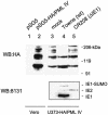
Effect of HCMV infection on sumoylation in the U373 cell line overexpressing PML. Lanes 1 and 2, control Vero cells were transfected with either vector (pSG5) or plasmid encoding HA-PML IV. At 48 h after transfection, the cells were harvested and total extracts were prepared as described for Fig. 1. Lanes 3 to 5, the U373 pooled cell line overexpressing HA-PML IV was either mock infected or infected with wild-type (wt) HCMV(Towne) or IE1 deletion HCMV(CR208) virus at an MOI of 2. Cells were harvested at 6 h after infection, and total extracts were prepared as described for Fig. 1. Equal amounts of cell extracts were separated on an SDS-8% polyacrylamide gel, and immunoblot analysis (WB) was performed with rat anti-HA MAb conjugated with peroxidase (top panel). The same filter membrane for lanes 3, 4, and 5 was stripped, and a second immunoblot analysis was carried out with MAb 8131, which detects both IE1 and IE2 (bottom panel).
FIG. 3.
FIG. 3.
Domains of IE1 required for desumoylation of PML and disruption of PODs. (A) PML sumoylation assays with the deletion mutant IE1 proteins. 293T cells were transfected with 1 μg of plasmid (pUS112) encoding HA-PML alone or cotransfected with 1 μg of plasmid encoding wild-type (wt) IE1, IE1(1-420), IE1(1-346), IE1(1-231), IE1(Δ132-274), or IE1(Δ290-320). At 48 h after transfection, a PML sumoylation assay was carried out as described for Fig. 1. (B) Expression levels of the mutant IE1 proteins. The same cell extracts described for panel A were separated on an SDS-4 to 20% gradient polyacrylamide gel, and immunoblot analysis (WB) was carried out with mouse MAb 8131 against IE1. (C) 293T cells were transfected with a plasmid encoding HA-PML or cotransfected with various amounts of plasmid encoding wild-type IE1 (0.1, 0.5, or 1.0 μg), IE1(K450R) (0.1, 0.5, or 1.0 μg), or IE1(L174P) (1.0 μg) protein. At 48 h after transfection, cell extracts were prepared and immunoblot analysis was carried out with anti-HA MAb as described for panel A. Unmodified and sumoylated (PML-S) forms of PML are indicated. (D) Structures of the IE1 proteins used in domain mapping experiments. 1-491 (wild-type IE1 in pJHA303), 1-420 (pJHA423), 1-346 (pJHA304), 1-231 (pJHA307), Δ132-274 (pJHA308), Δ290-320 (pJHA346), and Δ1-85 (pJHA305) are illustrated. Coding regions corresponding to each exon of IE1 are shown at the top. Closed bars represent exons 2 and 3, and gray bars represent the C-terminal acidic domains. The estimated map locations for epitopes recognized by MAbs 8131 and 6E1 are shown at the bottom. (E) Localization patterns of the mutant IE1 proteins and their effects on distribution of the endogenous PML proteins. Vero cells were transfected with plasmids encoding wild-type IE1, IE1(1-420), IE1(1-346), IE1(1-231), IE1(Δ132-274), IE1(Δ290-320), IE1(Δ1-85), IE1(L174P), or IE1(K450R) and fixed in methanol at 48 h after transfection, followed by double-label IFA for IE1 and PML. IE1 was detected with mouse MAb 8131 or 6E1 and fluorescein isothiocyanate-labeled anti-mouse immunoglobulin G. PML was detected with rabbit polyclonal antibody PML(C) and rhodamine-coupled anti-rabbit immunoglobulin G.
FIG. 3.
FIG. 3.
Domains of IE1 required for desumoylation of PML and disruption of PODs. (A) PML sumoylation assays with the deletion mutant IE1 proteins. 293T cells were transfected with 1 μg of plasmid (pUS112) encoding HA-PML alone or cotransfected with 1 μg of plasmid encoding wild-type (wt) IE1, IE1(1-420), IE1(1-346), IE1(1-231), IE1(Δ132-274), or IE1(Δ290-320). At 48 h after transfection, a PML sumoylation assay was carried out as described for Fig. 1. (B) Expression levels of the mutant IE1 proteins. The same cell extracts described for panel A were separated on an SDS-4 to 20% gradient polyacrylamide gel, and immunoblot analysis (WB) was carried out with mouse MAb 8131 against IE1. (C) 293T cells were transfected with a plasmid encoding HA-PML or cotransfected with various amounts of plasmid encoding wild-type IE1 (0.1, 0.5, or 1.0 μg), IE1(K450R) (0.1, 0.5, or 1.0 μg), or IE1(L174P) (1.0 μg) protein. At 48 h after transfection, cell extracts were prepared and immunoblot analysis was carried out with anti-HA MAb as described for panel A. Unmodified and sumoylated (PML-S) forms of PML are indicated. (D) Structures of the IE1 proteins used in domain mapping experiments. 1-491 (wild-type IE1 in pJHA303), 1-420 (pJHA423), 1-346 (pJHA304), 1-231 (pJHA307), Δ132-274 (pJHA308), Δ290-320 (pJHA346), and Δ1-85 (pJHA305) are illustrated. Coding regions corresponding to each exon of IE1 are shown at the top. Closed bars represent exons 2 and 3, and gray bars represent the C-terminal acidic domains. The estimated map locations for epitopes recognized by MAbs 8131 and 6E1 are shown at the bottom. (E) Localization patterns of the mutant IE1 proteins and their effects on distribution of the endogenous PML proteins. Vero cells were transfected with plasmids encoding wild-type IE1, IE1(1-420), IE1(1-346), IE1(1-231), IE1(Δ132-274), IE1(Δ290-320), IE1(Δ1-85), IE1(L174P), or IE1(K450R) and fixed in methanol at 48 h after transfection, followed by double-label IFA for IE1 and PML. IE1 was detected with mouse MAb 8131 or 6E1 and fluorescein isothiocyanate-labeled anti-mouse immunoglobulin G. PML was detected with rabbit polyclonal antibody PML(C) and rhodamine-coupled anti-rabbit immunoglobulin G.
FIG. 4.
FIG. 4.
Yeast two-hybrid interaction assays between PML and mutant IE1 proteins. The yeast Y190 cells were cotransformed with plasmids encoding either GAL4-DB/PML VI or GAL4-DB/EBNA-1 (2) and GAL4-A alone (pACTII), GAL4-A/wild-type IE1, GAL4-A/IE1(1-420), GAL4-A/IE1(Δ290-320), GAL4-A/IE1(L174P), or GAL4-A/IE1(K450R). Transformants were assayed for their β-galactosidase production. The average values and standard errors for β-galactosidase units in duplicated assays are shown. Assays of the interaction between Epstein-Barr virus and IE1 were used as negative interaction controls.
FIG. 5.
FIG. 5.
Effects of PML desumoylation on transactivation and PML repression functions of IE1. (A) Effects of wild-type or mutant IE1 proteins on the transcriptional repressor activity of PML. HeLa cells were transfected with 0.5 μg of reporter plasmid containing a (GAL4)5/TK-LUC gene or cotransfected with 0.4 μg of plasmid encoding GAL4-PML alone or together with effector plasmid encoding wild-type (wt) IE1, IE1(1-420), IE1(1-346), IE1(1-231), IE1(Δ132-274), IE1(Δ290-320), IE1(L174P), or IE1(K450R). At 36 h after transfection, total cell extracts were prepared and assayed for luciferase activity. Shown are mean values with standard errors for the percentage of luciferase activity from three independent assays. (B) Effects of mutant IE1 proteins on transactivation of the cellular DNA Pol α promoter. U373-MG cells were transfected with 0.4 μg of reporter plasmid (pDPALΔ5′) containing a DNA Pol α-luciferase reporter gene and 1.0 μg of effector plasmid encoding wild-type or mutant IE1 proteins as described above. At 48 h after transfection, total cell extracts were prepared and assayed for luciferase activity. Luciferase activities are indicated as fold activation over the basal level of each reporter gene. The results shown are the mean values along with standard errors from three independent experiments. (C) Effects of mutant IE1 proteins on augmentation of transactivation of the HCMV Pol promoter (Pol-LUC) by IE2. U373-MG cells were transfected with 0.3 μg of target reporter plasmid (pLA12) encoding the HCMV UL54 (Pol) promoter-driven Pol-LUC gene and 0.3 μg of effector plasmid encoding wild-type IE1, IE1(L174P), IE1(Δ290-320), or IE1(K450R) either alone or together with 0.3 μg of effector plasmid encoding wild-type IE2 (pJHA124). At 48 h after transfection, total cell extracts were prepared and assayed for luciferase activity as described for panel B. (D) Effects of wild-type or mutant IE1 on transactivation of the MMTV promoter. U373-MG cells were transfected with 0.4 μg of reporter plasmid (pMMTV-LUC) containing MMTV LTR-luciferase reporter gene and 0.2 μg of plasmid encoding wild-type IE1, IE1(L174P), IE1(Δ290-320), or IE1(K450R). At 24 h after transfection, culture media were replaced with medium with or without 1 μM dexamethasone (DEX). After incubation for another 24 h, total cell extracts were prepared and assayed for luciferase activity. Luciferase activities are indicated as fold activation over the basal level of each reporter gene and are shown as averages from duplicated experiments. (E) Expression levels of the mutant IE1 proteins. The same cell extracts described for panel A were separated on an SDS-10% polyacrylamide gel, and immunoblot analysis was carried out with MAb 8131.
FIG. 6.
FIG. 6.
Summary of the activities of wild-type or mutant IE1 proteins. Several activities of the wild-type (wt) or mutant IE1 proteins characterized in this study are summarized at a semiquantitative level, including PML binding in yeast two-hybrid assays, POD targeting by IFA, loss of PML sumoylation in cotransfection assays, POD disruption in IFA, ability to function as a target for sumoylation, and derepression of PML or positive transactivation functions on other promoters in transient reporter gene cotransfection assays. The PML binding activities of mutants IE1(1-346), IE1(1-231), and IE1(Δ132-274) were shown previously (2). Sumoylation of IE1(L174P) was originally shown by Muller and Dejean (60), but we found that the sumoylation level of IE1(L174P) is reduced to only 30% that of the wild-type protein (data not shown). Domain mapping of IE1 for POD disruption was also reported by Wilkinson et al. (79).
FIG.7.
FIG.7.
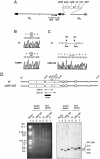
Construction of recombinant HCMV-BAC genomes encoding mutant IE1 proteins. (A) Genome structure of the parent Towne HCMV-BAC plasmid used in this study. The F plasmid sequences containing the replication origin (ori), replication and partitioning functions (repE, parA, and parB), chloramphenicol resistance (Cmr), and the GFP eukaryotic expression cassette, which have been substituted in place of the US1-to-US12 gene region, are indicated as boxes. The locations of UL122 (IE2) and UL123 (IE1) are also indicated. (B and C) Relevant DNA sequences of the wild-type (wt) and recovered mutant IE1 alleles of the HCMV-BAC plasmids. Genomic DNA containing either wild-type IE1(1-491) or the mutant IE1 alleles IE1(K450R) (B) and IE1(Δ290-320) (C) were PCR amplified from BAC DNAs and sequenced with specific primers. The two SpeI sites at amino acid positions 290 and 320 in wild-type IE1 are also indicated (C). (D) Genome structure of the Towne HCMV-BAC containing the deleted IE1(Δ290-320) allele. Top, the 6.6-kb EcoRI-SalI regions encompassing the MIE locus and the locations of the restriction enzyme sites used for deletion (SpeI) and mapping by Southern blot analysis (EcoRV, NcoI, and BamHI) are shown. Bottom, restriction fragment DNA patterns obtained following EcoRV-BamHI (lanes 2, 3, and 4) or NcoI-BamHI (lanes 5, 6, and 7) digestion of three HCMV-BAC DNAs (wild type [lanes 2 and 5], Δ290-320 [lanes 3 and 6], and the revertant [R] for Δ290-320 [lanes 4 and 7]) are shown (left). The 530-bp probe (BglII-BamHI fragment) used for Southern blot analysis (right) is shown in the top panel. The sizes of λ-HindIII/EcoRI are shown in the marker lane.
FIG. 8.
FIG. 8.
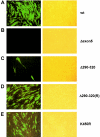
Infectivities of transfected HCMV-BAC DNAs. HF cells were electroporated with T-BAC-wt, T-BAC-Δexon5, T-BAC-Δ290-320, T-BAC-Δ290-320(R), or T-BAC-K450R and monitored for spreading of GFP signals (see text). GFP images (left panels) and their phase-contrast images (right panels) were photographed at 4 weeks after electroporation. Representative images from at least three independent experiments are shown. HF cells that received T-BAC-Δ290-320 usually gave no GFP-positive cells in most microscopic fields. The image shown in panel C was selected to include three GFP-positive cells, demonstrating that the few GFP-positive cells did not spread into the surrounding cells.

Similar articles

Cited by

References

    1. Adamson, A. L., and S. Kenney. 2001. Epstein-Barr virus immediate-early protein BZLF1 is SUMO-1 modified and disrupts promyelocytic leukemia bodies. J. Virol. 75:2388-2399. - PMC - PubMed
    1. Ahn, J. H., E. J. Brignole III, and G. S. Hayward. 1998. Disruption of PML subnuclear domains by the acidic IE1 protein of human cytomegalovirus is mediated through interaction with PML and may modulate a RING finger-dependent cryptic transactivator function of PML. Mol. Cell. Biol 18:4899-4913. - PMC - PubMed
    1. Ahn, J. H., and G. S. Hayward. 2000. Disruption of PML-associated nuclear bodies by IE1 correlates with efficient early stages of viral gene expression and DNA replication in human cytomegalovirus infection. Virology 274:39-55. - PubMed
    1. Ahn, J. H., and G. S. Hayward. 1997. The major immediate-early proteins IE1 and IE2 of human cytomegalovirus colocalize with and disrupt PML-associated nuclear bodies at very early times in infected permissive cells. J. Virol. 71:4599-4613. - PMC - PubMed
    1. Ahn, J. H., W. J. Jang, and G. S. Hayward. 1999. The human cytomegalovirus IE2 and UL112-113 proteins accumulate in viral DNA replication compartments that initiate from the periphery of promyelocytic leukemia protein-associated nuclear bodies (PODs or ND10). J. Virol. 73:10458-10471. - PMC - PubMed

Publication types

MeSH terms

LinkOut - more resources