. 2004 May 24;165(4):575-89.
doi: 10.1083/jcb.200311141.

NG2-expressing cells in the subventricular zone are type C-like cells and contribute to interneuron generation in the postnatal hippocampus

Affiliations

NG2-expressing cells in the subventricular zone are type C-like cells and contribute to interneuron generation in the postnatal hippocampus

Adan A Aguirre et al. J Cell Biol. .

Abstract

The subventricular zone (SVZ) is a source of neural progenitors throughout brain development. The identification and purification of these progenitors and the analysis of their lineage potential are fundamental issues for future brain repair therapies. We demonstrate that early postnatal NG2-expressing (NG2+) progenitor cells located in the SVZ self-renew in vitro and display phenotypic features of transit-amplifier type C-like multipotent cells. NG2+ cells in the SVZ are highly proliferative and express the epidermal growth factor receptor, the transcription factors Dlx, Mash1, and Olig2, and the Lewis X (LeX) antigen. We show that grafted early postnatal NG2+ cells generate hippocampal GABAergic interneurons that propagate action potentials and receive functional glutamatergic synaptic inputs. Our work identifies Dlx+/Mash1+/LeX+/NG2+/GFAP-negative cells of the SVZ as a new class of postnatal multipotent progenitor cells that may represent a specific cellular reservoir for renewal of postnatal and adult inhibitory interneurons in the hippocampus.


Figures
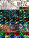
Figure 1.
Figure 1.
NG2 + /EGFP + cells express NSCs markers in the SVZ. Coronal sections of the SVZ at P8. (A1–A3) Anti-NG2 staining (DAB reaction, brown) shows that NG2 cells are found lining the wall of the lateral ventricle (yellow line) and throughout the entire lateral SVZ. The dotted line (A3) indicates the area analyzed in this paper. (A5–A7) Most of the EGFP+ cells (A5, green) were labeled with NG2 antibodies (A6, red), and all NG2+ cells were EGFP+ (A7). (B–G) All micrographs were obtained from the lateral SVZ (laSVZ). (B) All NG2+/EGFP+ cells (blue/green, respectively) express Olig2 (red). (C and D) NG2+/EGFP+ cells proliferate in the SVZ, as shown by BrdU incorporation (C, red) and by Ki67 immunolabeling (D, red). (E and G) A large percentage of NG2+/EGFP+ cells (blue/green, respectively) express the transcription factor Mash1 (E, red), the adult NSC markers LeX antigen (F, red), and EGFR (G, red). Arrows indicate NG2+/EGFP+ cells double-labeled with any of the markers used. NG2+/EGFP+ cells in boxed areas are shown at higher magnification. LV, lateral ventricle; Str, striatum; CC, corpus callosum. Bars: (A1) 500 μm; (A2) 300 μm; (A3) 50 μm; (B–E) 50 μm; (F and G) 100 μm.
Figure 2.
Figure 2.
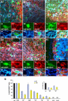
A subpopulation of NG2 + /EGFP + cells displays an immature neuronal phenotype in the SVZ. P8 coronal sections. (A and B) NG2+/EGFP+ cells (blue/green, respectively) in the SVZ are not labeled for the astroglial markers GFAP (A, red) or GLAST (B, red). (C) A large percentage of the NG2+/EGFP+ cells (blue/green, respectively) in the SVZ are labeled with anti-Dlx antibodies (red) for neuronal progenitor cells. (D) A significant percentage of the NG2+/EGFP+ cells (blue/green, respectively) in the SVZ express the early neuronal markers PSA-NCAM (D, red) and βIII- tubulin (E, TUJ1; red). (F) The majority of the EGFP+/TUJ1+ cells (green/blue, respectively) are Dlx+ (red). Arrows indicate double-labeled NG2+/EGFP+ cells. NG2+/EGFP+ cells in boxed areas are shown at higher magnification. Bar, 50 μm. (G) NG2+/EGFP+ cells in the lateral ventricle of the SVZ. Virtually all NG2+/EGFP+ cells expressed Olig2 at P8 and P40, and EGFRs at P8. At P40 the percentage of EGFR+/NG2+ cells decreases by 50%. A similar decrease is also observed for Lex, Dlx, PSA-NCAM, and TUJ1. None of the NG2+/EGFP+ cells are labeled with anti-GFAP or anti-GLAST antibodies. (inset) Percentage of NG2+/EGFP+ cells that incorporated BrdU after 2 h of pulse labeling. No significant differences are detected between P8 and P40. This result was also confirmed by anti-Ki67 immunostaining. Percentages were obtained by counting NG2+/EGFP+ cells located into the SVZ region. Data are means ± SEM (total NG2+/EGFP+ cells counted equals 850 at P8 and 400 at P40; for each age, four to six brain sections from four different brains were used).
Figure 3.
Figure 3.
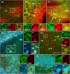
FACS ® -purified NG2 + /EGFP + and LeX + /NG2 + /EGFP + cells self-renew and generate GABAergic interneurons in vitro. FACS®-purified NG2+/EGFP+ (A–C) and LeX+/NG2+/EGFP+ (D–G) cells generate neurospheres. After primary neurosphere formation, cell dissociation to single cell suspension and replating in SCM at clonal dilution generated secondary neurospheres (A1 and F1, insets). (A) A single secondary neurosphere was plated and processed for immunocytochemistry 6 d after plating. All the TUJ1+ cells (red) are labeled with anti–GAD-67 (blue) antibodies. (B and C) The phenotype of the neurons derived from NG2+/EGFP+ cells is GABAergic. Neurons are double labeled with anti-Dlx (red) and anti–GAD-67 (blue) antibodies (B), and anti-parvalbumin (PV, red) and anti–GAD-67 (C, blue). (D) Sorting profile for NG2+/EGFP+ cells (D1; NG2 immunolabeling was scattered with PE-Cy5, R2 box) or MMA+/EGFP+ cells (D2; LeX antigen was scattered with R-phycoerythrin, RPE). To scatter the triple positive (LeX+/NG2+/EGFP+) cells, the NG2+/EGFP+ fraction (R2 box in D1) was double sorted using PE-Cy5 for NG2 and RPE for LeX (D3). Note that a significant percentage of NG2+/EGFP+ cells (D1, R2 window) express the LeX antigen (D3). (E) A typical sample of LeX+/NG2+/EGFP+ cells after FACS® shows high purity. (F) LeX+/NG2+/EGFP+ FACS®-purified cells were assayed for neurosphere formation. LeX+/NG2+/EGFP+ neurospheres displayed similar properties to those derived from NG2+/EGFP+ cells and generated differentiated GABAergic neurons, as shown by immunocytochemistry with anti-NeuN (red) and anti–GAD-67 (blue) antibodies. (G) The GABAergic neuronal fate is confirmed by anti-Dlx (red) and anti–GAD-67 (blue) coexpression. EGFP+ cells (green) in boxed areas are shown at higher magnification. Bars, 50 μm.
Figure 4.
Figure 4.
Grafted NG2 + /EGFP + cells differentiate to neurons in the hippocampus. (A–C) 3 wk after transplantation, all the grafted EGFP+ cells (green) in the hippocampus are labeled with TUJ1 (red) antibodies. EGFP+/TUJ1+ cells (green/red, respectively) are found in the pyramidal layer of the CA1 (A1) and CA3 (B1). Grafted cells are also found in the hilar region of the DG (C1). (D) A large percentage of transplanted EGFP+ cells (green) are also labeled with anti-NeuN (blue) antibodies in the CA3 area and hilar region of the DG (not depicted), confirming the neural fate of the grafted NG2+/EGFP+ cells. The tissue was also stained with anti-GFP antibodies (D3, red), confirming that these neurons are derived from the transplanted NG2+/EGFP+ fraction. (E–G) Neurons derived from grafted NG2+/EGFP+ cells are viable, as determined by TUNEL assay. (E) Positive control (Dnase I-treated tissue). EGFP+/TUJ1+ (F, green/blue, respectively) and EGFP+/NeuN+ (G, green/blue, respectively) grafted cells are TUNEL-negative (red) 6 wk after transplantation. Arrows in A1–D1 indicate EGFP+ neurons derived from grafted cells. Arrows in E1 indicate EGFP+/TUNEL+ cells after DNAse treatment of the tissue (positive control). Arrows in G1 indicate endogenous EGFP-negative TUNEL+ cells in DG. EGFP+ cells in boxed areas are shown at higher magnification. Bars: (A–D , F, and G) 50 μm; (E) 100 μm.
Figure 5.
Figure 5.
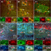
Grafted NG2 + /EGFP + cells give rise to GABAergic interneurons in the hippocampus. (A–C) 3 wk after transplantation, EGFP+/Dlx+ cells were found in the stratum radiatum, stratum oriens, and stratum pyramidale of the CA1 (A) and CA3 (B) and in the hilar region of the DG (C). (D–F) Immunostaining with anti–GAD-67 antibodies 3 wk after transplantation. Note that at this time the vast majority of the grafted EGFP+ cells (green) were labeled with anti–GAD-67 (blue). Immunoreactivity of the graft-derived GAD-67+ neurons for calcium-binding proteins and neuropeptides confirmed the interneuron fate of the grafted EGFP+ cells. (D and E) A proportion of the graft-derived neurons are double labeled with anti–GAD-67 (blue) and parvalbumin (D, PV; red), somatostatin (E, SOM; red), or doublecortin (F, DC; red). Arrows in A1–F1 indicate EGFP+ neurons derived from grafted cells. EGFP+ cells in boxed areas are shown at higher magnification. Bar, 50 μm.
Figure 6.
Figure 6.
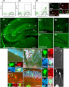
Grafted NG2 + /EYFP + cells give rise to GABAergic interneurons in the hippocampus. (A1–A3) Typical sorting profile for NG2+/EYFP+ cells (NG2 immunolabeling was scattered with RPE-Cy5 [R2 box]). (A4–A6) A typical sample of NG2+/EYFP+ cells after FACS® shows high purity. (B) 1 wk after grafting, EYFP+ cells (green) were found in the hippocampus (arrows) displaying a typical neuronal morphology in the stratum oriens of the CA1 (B3) and CA3 (not depicted), in the stratum pyramidale of the CA1 and CA3 (B2 and B4, respectively), and in the DG (B5). The tissue was also stained with anti-GFP antibodies (C, red), confirming that these neurons are derived from the transplanted NG2+/EYFP+ fraction. Migratory EYFP+ cells acquired immature neuronal markers 1 wk after grafting, as shown by Dlx (D, red) and TUJ1 (not depicted). (E and F) 3 wk after transplantation, all the grafted EYFP+ cells (green) found in the hippocampus are labeled with anti-NeuN antibodies (blue). EYFP+/NeuN+ cells (green/blue) are found in the pyramidal layer of the CA1 (not depicted) and CA3 (E) and in the subgranular layer of the DG (F). EYFP+ cells are also labeled with anti–GAD-67 (red) antibodies in the CA3 area (E) and DG (F), confirming their neural fate. In E6 and F6, EYFP was converted to grayscale. Anti-GFP antibodies recognize all the GFP variants, including EGFP, EYFP, and ECFP. EYFP+ cells are shown at higher magnification in boxed areas. Bars: (A, C, E, and F) 50 μm; (B1) 300 μm; (B2–B5) 100 μm.
Figure 7.
Figure 7.
Grafted NG2 + /EGFP + cells in the hippocampus possess physiological properties of mature inhibitory interneurons. (A) Schematic of hippocampus illustrating the positions of the grafted EGFP+ cells that physiological recordings were attained from. The open circle indicates the cell from which the single example current (B) and voltage clamp (E and F) traces were obtained. (B) Action potential generation in response to a depolarizing current injection (120 pA for 1,000 ms) from a membrane potential of −70 mV. (C) Pooled data of injection current (30–180 pA; step = 30 pA) versus spike frequency during the 1,000-ms depolarizing pulse. (D) Pooled data illustrating spike frequency during the first and last 200-ms epochs (0–200 and 800–1,000 ms) of depolarizing current injections (120, 150, and 180 ms pA). (E) Continuous voltage clamp trace (holding potential −70 mV; 3 min) illustrating the presence of spontaneous inward currents that were abolished by inclusion of 10 μM NBQX (arrow, 2 min; NBQX is present for the remainder of trace). (F) Magnification of part of the trace in E, illustrating the time course of individual spontaneous inward currents (four superimposed consecutive traces of 700 ms each). (G) Average time course (267 events; average amplitude equals 73 pA) of the inward currents, as determined by single exponential fitting of the decay (dotted line). (H and I) Biocytin (brown) images of two cells from which recordings were taken. GCL, granule cell layer; SO, stratum orien; SP, stratum pyramidale; SR, stratum radiatum. Images were converted to grayscale and inverted for clarity. Insets in H illustrate a high magnification of the biocytin-conjugated fluorescence (red; top inset) and the corresponding EGFP+ fluorescence (green; bottom inset). Bar, 50 μm.
Figure 8.
Figure 8.
Endogenous EGFP + cells in the postnatal hippocampus display a GABAergic interneuron phenotype. P8 sagittal sections and CA3 and DG regions of the hippocampus. (A) A proportion of the NG2+/EGFP+ cells (blue/green, respectively) in the DG express PSA-NCAM (red). (B) A proportion of the EGFP+ cells coexpress Dlx (red) and class III β-tubulin (blue). (C) The majority of the EGFP+ cells (green) that express Dlx (red) also express GAD-67 (blue). (D) All the EGFP+/NeuN+ cells (green/blue, respectively) also express Dlx (red). (E and F) A proportion of the EGFP+ cells in CA3 (E) and DG (F) express parvalbumin (E, PV; red), doublecortin (F, DC; red), and GAD-67 (blue). (G) In the hilar region of the DG and in CA3 (not depicted), a significant proportion of the EGFP+ cells express the neuropeptide somatostatin (SOM, red) and GAD-67 (blue). (H) EGFP+ cells that are positive for GAD-67 (blue) are not labeled with antibodies against calretinin (CAL, red). White arrows in A1–H1 indicate EGFP+ cells that are also labeled for all the markers analyzed. In H1, black arrows indicate EGFP-negative CAL+/GAD-67+ cells. Cells in boxed areas are shown at higher magnification. Quantitative analysis of these data is shown in Table II. Bars, 50 μm.

Similar articles

Cited by

References

    1. Alvarez-Buylla, A., and J.M. Garcia-Verdugo. 2002. Neurogenesis in adult subventricular zone. J. Neurosci. 22:629–634. - PMC - PubMed
    1. Belachew, S., R. Chittajallu, A.A. Aguirre, X. Yuan, M. Kirby, S. Anderson, and V. Gallo. 2003. Postnatal NG2 proteoglycan-expressing progenitor cells are intrinsically multipotent and generate functional neurons. J. Cell Biol. 161:169–186. - PMC - PubMed
    1. Capela, A., and S. Temple. 2002. Lex/ssea-1 is expressed by adult mouse CNS stem cells, identifying them as nonependymal. Neuron. 35:865–875. - PubMed
    1. Casaccia-Bonnefil, P., R. Tikoo, H. Kiyokawa, V. Friedrich Jr., M.V. Chao, and A. Koff. 1997. Oligodendrocyte precursor differentiation is perturbed in the absence of the cyclin-dependent kinase inhibitor p27Kip1. Genes Dev. 11:2335–2346. - PMC - PubMed
    1. Dawson, M.R., J.M. Levine, and R. Reynolds. 2000. NG2-expressing cells in the central nervous system: are they oligodendroglial progenitors? J. Neurosci. Res. 61:471–479. - PubMed

Publication types

MeSH terms