. 2004 May 19;24(20):4728-36.
doi: 10.1523/JNEUROSCI.0757-04.2004.

Block of AMPA receptor desensitization by a point mutation outside the ligand-binding domain

Affiliations

Block of AMPA receptor desensitization by a point mutation outside the ligand-binding domain

Maria V Yelshansky et al. J Neurosci. .

Abstract

Desensitization of ionotropic glutamate receptors (GluRs), specifically the AMPA receptor subtype, shapes the postsynaptic response at certain synapses in the brain. All known mechanisms that alter desensitization, either pharmacological or mutational, are associated with the ligand-binding domain. Here we report that substitution of a conserved positively charged arginine (R) with a negatively charged glutamate in the linker between the pore-forming M3 segment and the S2 lobe, a region outside the ligand-binding domain, blocks desensitization in homomeric AMPA receptors composed of GluR-B(i) subunits. A charge-reversing substitution of a glutamate adjacent to this conserved R enhanced desensitization, consistent with these effects attributable to electrostatics. Homologous substitutions of the conserved R in GluR-B(o), GluR-A(i) and the kainate receptor GluR-6 subunits produced comparable but less visible effects on desensitization. Subunit specificity was also apparent for accessibility of substituted cysteines in the M3-S2 linker, suggesting that this part of the channel is not structurally identical in different GluRs. Additionally, reactivity with a sulfhydryl-specific reagent was state dependent, suggesting that the conformations of the nonconducting closed and desensitized states are different at the level of the M3-S2 linker. Our results therefore represent the first identification of elements outside the ligand-binding domain affecting desensitization in non-NMDA receptor channels and suggest that electrostatic interactions involving charged residues in the M3-S2 linker influence channel gating in a subunit- and subtype-specific manner.


Figures
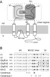
Figure 1.
Figure 1.
M3–S2 linker in GluRs. A, Topology of a GluR subunit. Presumedα-helical regions of the membrane-spanning segments M1–M4 and the pore loop M2 are shown as open cylinders. The S1 (N-terminal to M1) and S2 (between M3 and M4) lobes constitute the ligand-binding core (dashed box), for which a crystal structure exists (Armstrong et al., 1998; Armstrong and Gouaux, 2000). The large N-terminal domain is not shown. The asterisk indicates the approximate location of positions mutated in the present study. B, Amino acid sequence of non-NMDAR subunits within and around the M3–S2 linker. Subunits shown includeδ (δ1, δ2), AMPAR (GluR-A, GluR-B), and low-affinity (GluR-6) and high-affinity (KA-2) KAR. Within AMPAR and low- and high-affinity KAR subunits, these regions are completely conserved. Residues conserved in all non-NMDAR subtypes (Consensus) or in all subtypes except for one (Consensus–1) are shown under the alignment. Charged residues are shown in bold. Positions within the dashed box are resolved in the crystal structure of the GluR-B ligand-binding core (Armstrong et al., 1998; Armstrong and Gouaux, 2000).
Figure 2.
Figure 2.
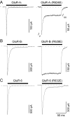
Effect of the R → E charge reversal in the M3–S2 linker on glutamate-activated currents in AMPAR and KAR channels. Glutamate-activated currents in outside-out patches isolated from HEK 293 cells expressing wild-type (left traces) or mutant (right traces) GluR-Ai (A), GluR-Bi (B), and GluR-6 (C) channels are shown. The mutant channels have the positively charged arginine (R) in the M3–S2 linker replaced by a glutamate (E) (R → E substitution). Currents were elicited by a 100 msec application of glutamate (3 mm; filled bar) at a holding potential of –60 mV. Current amplitudes were measured either at the peak (Ip) or near the end of the glutamate applications when they reached steady-state (Iss). Dashed lines show zero current level. The time scale is the same for all panels.
Figure 3.
Figure 3.
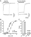
The R → E substitution disrupts desensitization in GluR-Bi channels. A, Superimpositions of glutamate-activated currents for wild-type (left traces) and mutant (R → E; right traces) GluR-Bi channels recorded either in the absence or in the presence of 30 μm CTZ. Currents were elicited by a 100 msec application of glutamate (3 mm; filled bar) at a holding potential of –60 mV. B, Glutamate concentration response curves for wild-type (wt; open symbols) and mutant (R → E; filled symbols) GluR-Bi channels. The solid lines are a fitted Hill equation with an EC50 of 134 ± 3 μm (n = 5) for wild type and 24 ± 2 μm (n = 4) for GluR-Bi (R628E). C, Average extent of desensitization, %des, for wild-type and GluR-Bi (R628E) channels measured in the absence or presence of 100 μm EDTA. Currents were recorded either in our standard external solution containing 1.8 mm Ca2+ and 1 mm Mg2+ or in the same solution but with 100 μm EDTA and no added divalent cations.
Figure 4.
Figure 4.
Desensitization properties of wild-type and mutant GluR-Bi channels. Average time constant of desensitization, τdes (A), the extent of desensitization measured relative to the peak currentin CTZ, %desCTZ (B), and the time constant of recovery from desensitization, τrec (C) measured for wild-type and mutant GluR-Bi channels are shown.τrec was not determined (n.d.) for R628E channels because of a small desensitizing component. In this and all subsequent figures, filled bars indicate values statistically different from those for wild type.
Figure 5.
Figure 5.
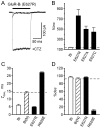
Effect of an E → R substitution on desensitization in GluR-Bi channels. A, Superimposition of glutamate-activated currents for GluR-Bi (E627R) channels (E → R substitution) recorded in the absence or in the presence of 30 μm CTZ. Currents were elicited by a 100 msec application of glutamate (3 mm; filled bar) at the holding potential of –60 mV. B, Average values for %incpeak for wild-type and mutant GluR-Bi channels. C, D, Average values for the rate of desensitization, τdes (C), and the extent of desensitization measured relative to the peak current, %des (D), for wild-type channels (Bi) and mutant channels containing an asparagine at the Q/R site [Bi (N)]. Opposite charge substitution of E and R was made in the Bi (N) background. Values for Bi (N) are significantly different from those in Bi (Q). Filled bars indicate values statistically different from those for GluR-Bi (N).
Figure 6.
Figure 6.
Opposite charge substitutions of both E and R. A, Superimposition of glutamate-activated currents for GluR-Bi (E627R/R628E) channels (ER → RE substitution) recorded in the absence or in the presence of 30 μm CTZ. Currents were elicited by a 100 msec application of glutamate (3 mm; filled bar) at the holding potential of –60 mV. B, Average values for the plateau/peak ratio normalized to wild type (ER channels) in the respective background of either GluR-Bi (Q) or GluR-Bi (N). Mutant channels include RR (E → R substitution), EE (R → E substitution), and RE (ER → RE substituion). Values for GluR-Bi (Q) (RR) could not be determined because the peak current amplitudes were too small.
Figure 7.
Figure 7.
State-dependent effect of a sulfhydryl-specific reagent on glutamate-activated currents in cysteine-substituted AMPAR channels. A, Protocols to measure the state dependence of reactivity. Whole-cell currents recorded from Xenopus oocytes expressing GluR-Bi (top trace), GluR-Bi (E627C) (middle trace), and GluR-Bi (R628C) (bottom trace) subunits are shown. Currents were elicited by applications of glutamate (1 mm; thin lines; typically 15 sec in duration except during the MTSET application). MTSET (2 mm; thick lines) was applied in the presence of glutamate and CTZ (50 μm; open boxes, top trace), in the absence of glutamate and in the presence of the competitive antagonist CNQX (10 μm; shaded box, middle trace), or just in the presence of glutamate (bottom trace). During these different applications, we assume that the predominant channel state is open (O; top trace), closed (C; middle trace), or desensitized (D; bottom trace). Current amplitudes were measured either before (Ipre) or after (Ipost) application of MTSET. B, Changes in the current amplitude measured before (Ipre) and after (Ipost) exposure to MTSET (1 – Ipost/Ipre) according to protocols in A for wild-type (first row) and cysteine-substituted (second, third rows) GluR-Ai (left column) and GluR-Bi (right column) channels. Up- and down-pointing bars indicate inhibition and potentiation of glutamate-activated currents, respectively. Filled bars indicate a significant difference between Ipost and Ipre (n = 3–8).
Figure 8.
Figure 8.
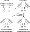
Possible state-dependent electrostatic interactions in GluRs. The diagram illustrates possible electrostatic interactions between E and R in the M3–S2 linker and a complimentary charge in other parts of the protein. This complimentary charge may arise from negative or polar residues in other extracellularly located linkers or from proximal parts of the ligand-binding domain (LBD). Alternatively, it may arise from α-helical dipoles. Because of subunit- and subtype-specific structural differences, which may be quite small, E and R and the putative complimentary charge may show various degrees of proximity in the desensitized state, leading to differences in the degree of desensitization in wild-type channels.

Similar articles

Cited by

References

    1. Armstrong N, Gouaux E (2000) Mechanisms for activation and antagonism of an AMPA-sensitive glutamate receptor: crystal structures of the GluR2 ligand binding core. Neuron 28: 165–181. - PubMed
    1. Armstrong N, Sun Y, Chen GQ, Gouaux E (1998) Structure of a glutamate-receptor ligand-binding core in complex with kainate. Nature 395: 913–917. - PubMed
    1. Banke TG, Greenwood JR, Christensen JK, Liljefors T, Traynelis SF, Schousboe A, Pickering DS (2001) Identification of amino acid residues in GluR1 responsible for ligand binding and desensitization. J Neurosci 21: 3052–3062. - PMC - PubMed
    1. Beck C, Wollmuth LP, Seeburg PH, Sakmann B, Kuner T (1999) NMDAR channel segments forming the extracellular vestibule inferred from the accessibility of substituted cysteines. Neuron 22: 559–570. - PubMed
    1. Bowie D, Lange GD, Mayer ML (1998) Activity-dependent modulation of glutamate receptors by polyamines. J Neurosci 18: 8175–8185. - PMC - PubMed

Publication types

MeSH terms

LinkOut - more resources