Fluorescence spectral properties of labeled thiolated oligonucleotides bound to silver particles
- PMID: 15150802
- PMCID: PMC2763907
- DOI: 10.1002/bip.20071
Fluorescence spectral properties of labeled thiolated oligonucleotides bound to silver particles
Abstract
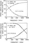
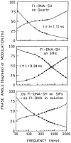
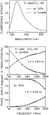
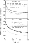
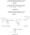
We examined the fluorescent spectral properties of fluorescein-labeled DNA oligomers when directly bound to metallic silver particles via a terminal sulfhydryl group. We found a 12-fold increase in fluorescence intensity and 25-fold decrease in lifetime for a fluorescein residue positioned 23 nucleotides from the silver surface compared to labeled oligomers in free solution. Similar results were found for a 23-mer labeled with five fluorescein residues. The absence of long lifetime components in the intensity decays suggests that all labeled oligomers are bound to silver and affected similarly by the metallic surfaces. These results provide the basic knowledge needed to begin use of metal-enhanced fluorescence for the detection of target sequences in simple formats potentially without a washing separation step. The use of metal-enhanced fluorescence provides a generic approach to obtaining a hybridization-dependent increase in fluorescence with most, if not all, commonly used fluorophores.
Copyright 2004 Wiley Periodicals, Inc. Biopolymers, 2004
Figures





Similar articles
-
Fluorescence spectral properties of cyanine dye-labeled DNA oligomers on surfaces coated with silver particles.Anal Biochem. 2003 Jun 15;317(2):136-46. doi: 10.1016/s0003-2697(03)00005-8. Anal Biochem. 2003. PMID: 12758251 Free PMC article.
-
DNA hybridization assays using metal-enhanced fluorescence.Biochem Biophys Res Commun. 2003 Jun 20;306(1):213-8. doi: 10.1016/s0006-291x(03)00935-5. Biochem Biophys Res Commun. 2003. PMID: 12788090 Free PMC article.
-
Fluorescence lifetime correlation spectroscopic study of fluorophore-labeled silver nanoparticles.Anal Chem. 2008 Oct 1;80(19):7313-8. doi: 10.1021/ac8009356. Epub 2008 Sep 5. Anal Chem. 2008. PMID: 18771274 Free PMC article.
-
Fluorescence spectral properties of cyanine dye labeled DNA near metallic silver particles.Biopolymers. 2003;72(2):96-104. doi: 10.1002/bip.10301. Biopolymers. 2003. PMID: 12583012
-
DNA-templated fluorescent silver nanoclusters.Anal Bioanal Chem. 2012 Jan;402(1):129-38. doi: 10.1007/s00216-011-5307-6. Epub 2011 Aug 21. Anal Bioanal Chem. 2012. PMID: 21858647 Review.
Cited by
-
Fluorescence enhancement of fluorophores tethered to different sized silver colloids deposited on glass substrate.Biopolymers. 2005 Jan;77(1):31-7. doi: 10.1002/bip.20179. Biopolymers. 2005. PMID: 15578680 Free PMC article.
-
Metal particle-enhanced fluorescent immunoassays on metal mirrors.Anal Biochem. 2007 Apr 15;363(2):239-45. doi: 10.1016/j.ab.2007.01.030. Epub 2007 Jan 26. Anal Biochem. 2007. PMID: 17316540 Free PMC article.
-
Molecular fluorescence, phosphorescence, and chemiluminescence spectrometry.Anal Chem. 2006 Jun 15;78(12):4047-68. doi: 10.1021/ac060683m. Anal Chem. 2006. PMID: 16771540 Free PMC article. No abstract available.
-
Fluorescence Enhancement Using Bimetal Surface Plasmon-Coupled Emission from 5-Carboxyfluorescein (FAM).Micromachines (Basel). 2018 Sep 12;9(9):460. doi: 10.3390/mi9090460. Micromachines (Basel). 2018. PMID: 30424393 Free PMC article.
-
Increasing the sensitivity of DNA microarrays by metal-enhanced fluorescence using surface-bound silver nanoparticles.Nucleic Acids Res. 2007;35(2):e13. doi: 10.1093/nar/gkl1054. Epub 2006 Dec 14. Nucleic Acids Res. 2007. PMID: 17169999 Free PMC article.
References
Publication types
MeSH terms
Substances
Grants and funding
LinkOut - more resources
Full Text Sources
Miscellaneous

