. 2004 Jun;24(11):4968-78.
doi: 10.1128/MCB.24.11.4968-4978.2004.

Tyrosine phosphorylation of Jak2 in the JH2 domain inhibits cytokine signaling

Affiliations

Tyrosine phosphorylation of Jak2 in the JH2 domain inhibits cytokine signaling

Edward P Feener et al. Mol Cell Biol. 2004 Jun.

Abstract

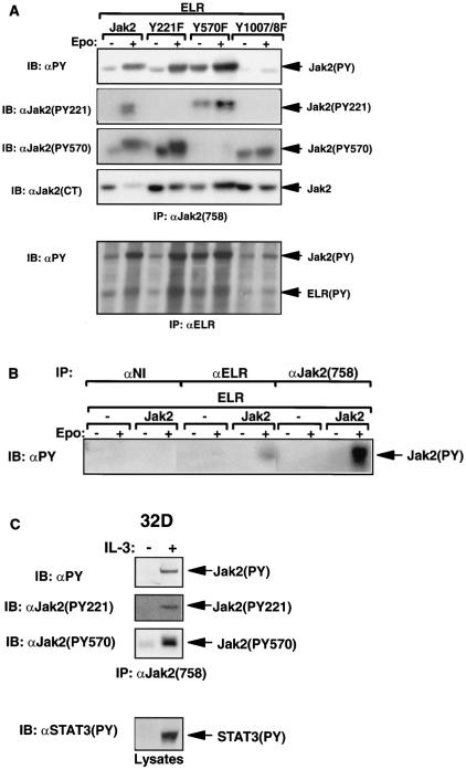
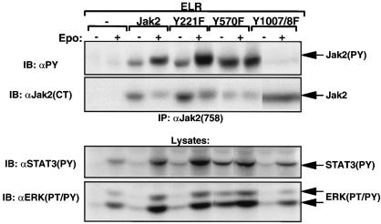
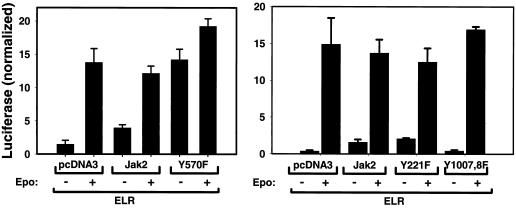
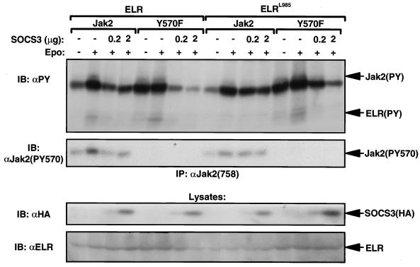
Jak family tyrosine kinases mediate signaling by cytokine receptors to regulate diverse biological processes. Although Jak2 and other Jak kinase family members are phosphorylated on numerous sites during cytokine signaling, the identity and function of most of these sites remains unknown. Using tandem mass spectroscopic analysis of activated Jak2 protein from intact cells, we identified Tyr(221) and Tyr(570) as novel sites of Jak2 phosphorylation. Phosphorylation of both sites was stimulated by cytokine treatment of cultured cells, and this stimulation required Jak2 kinase activity. While we observed no gross alteration of signaling upon mutation of Tyr(221), Tyr(570) lies within the inhibitory JH2 domain of Jak2, and mutation of this site (Jak2(Y570F)) results in constitutive Jak2-dependent signaling in the absence of cytokine stimulation and enhances and prolongs Jak2 activation during cytokine stimulation. Mutation of Tyr(570) does not alter the ability of SOCS3 to bind or inhibit Jak2, however. Thus, the phosphorylation of Tyr(570) in vivo inhibits Jak2-dependent signaling independently of SOCS3-mediated inhibition. This Tyr(570)-dependent mechanism of Jak2 inhibition likely represents an important mechanism by which cytokine function is regulated.


Figures

FIG. 1.
FIG. 1.
Purification and analysis of Jak2 protein. (A) HEK 293 cells were left untransfected or were transfected with the cDNAs for ELR and Jak2, were made quiescent, and were then incubated a further 15 min in the absence (−) or presence (+) of EPO (10 U/ml). Cells were lysed, and lysates were immunoprecipitated with α-Jak2(758). Immunoprecipitated proteins were resolved by SDS-PAGE and detected by staining with Coomassie blue; the relevant region of a representative gel is shown in the panel. The migration of Jak2 protein is indicated to the right of the gel. (B) MS/MS spectra for the Jak2 tryptic peptide phosphorylated at Tyr221 (m/z 620.43; top) and corresponding synthetic peptide, IQDY(P)HILTR (m/z 620.25; bottom). (C) MS/MS spectra for the Jak2 tryptic peptide phosphorylated at Tyr570 (m/z 691.25; top) and corresponding synthetic peptide, REVGDY(P)GQLHK (m/z 691.64; bottom). Sequest assignments of y+ and b+ ions are shown in red and blue, respectively. Fragment ions corresponding to the neutral loss of 40 m/z from these doubly charged precursors are shown in green.
FIG. 1.
FIG. 1.
Purification and analysis of Jak2 protein. (A) HEK 293 cells were left untransfected or were transfected with the cDNAs for ELR and Jak2, were made quiescent, and were then incubated a further 15 min in the absence (−) or presence (+) of EPO (10 U/ml). Cells were lysed, and lysates were immunoprecipitated with α-Jak2(758). Immunoprecipitated proteins were resolved by SDS-PAGE and detected by staining with Coomassie blue; the relevant region of a representative gel is shown in the panel. The migration of Jak2 protein is indicated to the right of the gel. (B) MS/MS spectra for the Jak2 tryptic peptide phosphorylated at Tyr221 (m/z 620.43; top) and corresponding synthetic peptide, IQDY(P)HILTR (m/z 620.25; bottom). (C) MS/MS spectra for the Jak2 tryptic peptide phosphorylated at Tyr570 (m/z 691.25; top) and corresponding synthetic peptide, REVGDY(P)GQLHK (m/z 691.64; bottom). Sequest assignments of y+ and b+ ions are shown in red and blue, respectively. Fragment ions corresponding to the neutral loss of 40 m/z from these doubly charged precursors are shown in green.
FIG. 2.
FIG. 2.
Phosphorylation of Tyr221 and Tyr570 in HEK 293 and 32D cells. (A and B) HEK 293 cells were transfected with ELR and the indicated Jak2 isoform, made quiescent, and incubated in the absence (−) or presence (+) of EPO (10 U/ml) for an additional 15 min before being lysed. (C) Quiescent 32D cells were incubated in the absence (−) or presence (+) of IL-3 (10 U/ml) for 15 min and lysed. In all panels, lysates were immunoprecipitated with the indicated antibodies. Lysates or immunoprecipitated proteins (as indicated) were resolved by SDS-PAGE and transferred to nitrocellulose for immunoblotting with the indicated antibody. The migration of detected signaling proteins is noted to the right of the panels.
FIG. 3.
FIG. 3.
Role of phosphorylation of Jak2 Tyr221 and Tyr570 in ELR signaling. HEK 293 cells were transfected with ELR and the indicated Jak2 isoform, made quiescent, and incubated in the absence (−) or presence (+) of EPO (10 U/ml) for 15 min before lysis. Lysates were subjected to immunoprecipitation with the indicated antibody or directly resolved by SDS-PAGE before transfer to nitrocellulose for immunoblotting with the indicated antibodies. The migration of detected signaling proteins is noted to the right of the panels.
FIG. 4.
FIG. 4.
Activation of Jak2Y570F in the absence and presence of ELR. HEK 293 cells were transfected with ELR plus control plasmid or the indicated Jak2 isoform, made quiescent, and incubated in the absence (−) or presence (+) of EPO (10 U/ml) for an additional 15 min before lysis. Lysates were subjected to immunoprecipitation with the indicated antibody or directly resolved by SDS-PAGE before transfer to nitrocellulose for immunoblotting with the indicated antibodies. The migration of detected signaling proteins is noted to the right of the panels.
FIG. 5.
FIG. 5.
Enhanced STAT3 signaling mediated by Jak2Y570F. HEK 293 cells were transfected with the cDNA for ELR plus control plasmid or the indicated isoform of Jak2, along with the STAT3-responsive GAS-luciferase and control pRL-TK (constitutively expressing Renilla luciferase) plasmids (4). After transfection, cells were placed in serum free medium (−) or the same medium containing 10 U of EPO/ml (+) for 12 h before being lysed and subjected to a dual luciferase assay. Firefly luciferase activity from the GAS-luciferase construct (normalized to Renilla luciferase activity) is shown in the graph. Left and right panels represent independent experiments, each representative of at least two similar experiments. Numbers represent the means of triplicate determinations ± the standard error of the mean for the single experiment shown.
FIG. 6.
FIG. 6.
Prolonged activation of Jak2Y570F. HEK 293 cells were transfected with ELR and the indicated Jak2 isoform, made quiescent, and incubated in the absence (−) or presence of EPO (10 U/ml) for the indicated time before lysis. Lysates were immunoprecipitated with α-Jak2(758), and immunoprecipitated proteins were resolved by SDS-PAGE and transferred to nitrocellulose before immunoblotting with the indicated antibody. The migration of signaling proteins is indicated to the right of the panels. This figure is representative of multiple similar experiments.
FIG. 7.
FIG. 7.
Role of Jak2 Tyr570 in SOCS3-mediated inhibition. HEK 293 cells were transiently transfected with ELR or ELRL985, Jak2 or Jak2Y570F plus empty vector, and/or the indicated amount of pcDNA3SOCS3 (0.2 or 2 μg; vector plus pcDNA3 totaled 2 μg in each case). Cells were incubated in the absence or presence of EPO for 5 min before lysis and immunoprecipitation of Jak2. Washed immune complexes or lysates (as indicated) were resolved by SDS-PAGE and immunoblotted with the indicated antibodies. The migration of signaling proteins is indicated to the right of the panels. This figure is representative of two independent experiments.

Similar articles

Cited by

References

    1. Argetsinger, L. S., J.-L. Kouadio, H. Steen, A. Stensballe, O. N. Jensen, and C. Carter-Su. 2004. Autophosphorylation of JAK2 on tyrosines 221 and 570. Mol. Cell. Biol. 24:4955-4967. - PMC - PubMed
    1. Banks, A. S., S. M. Davis, S. H. Bates, and M. G. Myers, Jr. 2000. Activation of downstream signals by the long form of the leptin receptor. J. Biol. Chem. 275:14563-14572. - PubMed
    1. Bjorbaek, C., K. El Haschimi, J. D. Frantz, and J. S. Flier. 1999. The role of SOCS-3 in leptin signaling and leptin resistance. J. Biol. Chem. 274:30059-30065. - PubMed
    1. Bjorbak, C., H. J. Lavery, S. H. Bates, R. K. Olson, S. M. Davis, J. S. Flier, and M. G. Myers, Jr. 2000. SOCS3 mediates feedback inhibition of the leptin receptor via Tyr985. J. Biol. Chem. 275:40649-40657. - PubMed
    1. Carpino, N., R. Kobayashi, H. Zang, Y. Takahashi, S. T. Jou, J. Feng, H. Nakajima, and J. N. Ihle. 2002. Identification, cDNA cloning, and targeted deletion of p70, a novel, ubiquitously expressed SH3 domain-containing protein. Mol. Cell. Biol. 22:7491-7500. - PMC - PubMed

Publication types