. 2004 May 1;18(9):1072-87.
doi: 10.1101/gad.1171104.

Interactions between Sox9 and beta-catenin control chondrocyte differentiation

Affiliations

Interactions between Sox9 and beta-catenin control chondrocyte differentiation

Haruhiko Akiyama et al. Genes Dev. .

Abstract

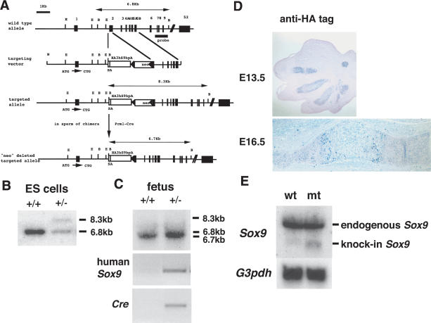
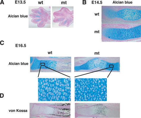
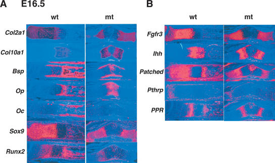
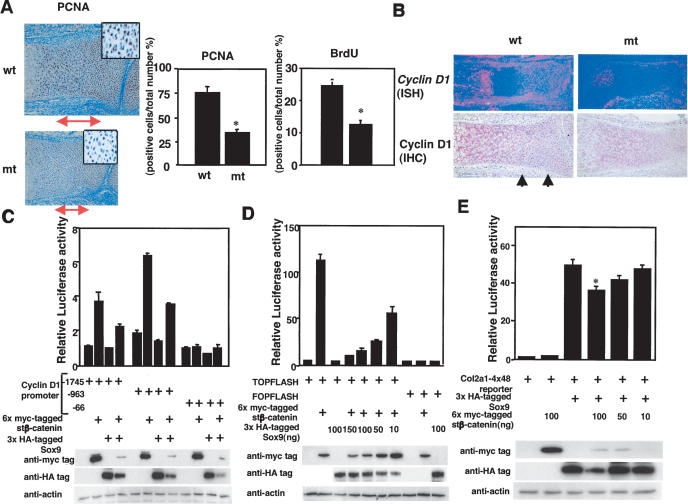
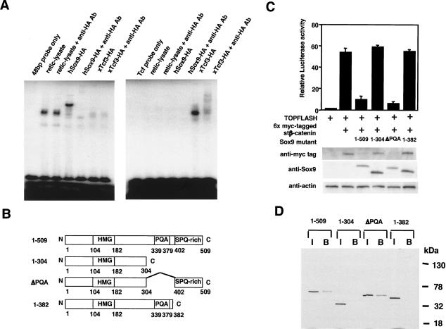
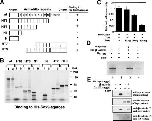
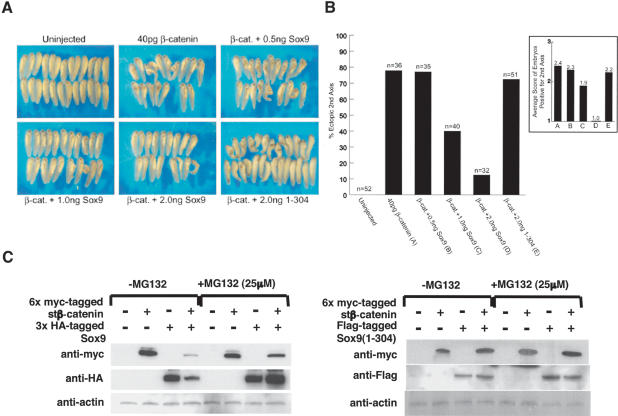
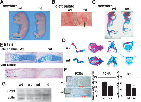
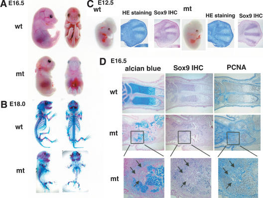
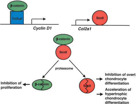
Chondrogenesis is a multistep process that is essential for endochondral bone formation. Previous results have indicated a role for beta-catenin and Wnt signaling in this pathway. Here we show the existence of physical and functional interactions between beta-catenin and Sox9, a transcription factor that is required in successive steps of chondrogenesis. In vivo, either overexpression of Sox9 or inactivation of beta-catenin in chondrocytes of mouse embryos produces a similar phenotype of dwarfism with decreased chondrocyte proliferation, delayed hypertrophic chondrocyte differentiation, and endochondral bone formation. Furthermore, either inactivation of Sox9 or stabilization of beta-catenin in chondrocytes also produces a similar phenotype of severe chondrodysplasia. Sox9 markedly inhibits activation of beta-catenin-dependent promoters and stimulates degradation of beta-catenin by the ubiquitination/proteasome pathway. Likewise, Sox9 inhibits beta-catenin-mediated secondary axis induction in Xenopus embryos. Beta-catenin physically interacts through its Armadillo repeats with the C-terminal transactivation domain of Sox9. We hypothesize that the inhibitory activity of Sox9 is caused by its ability to compete with Tcf/Lef for binding to beta-catenin, followed by degradation of beta-catenin. Our results strongly suggest that chondrogenesis is controlled by interactions between Sox9 and the Wnt/beta-catenin signaling pathway.


Figures

Figure 1.
Figure 1.
Targeting strategy for insertion of Sox9 cDNA in the Col2a1 locus. (A) Structure of the genomic Col2a1 locus, targeting vector, and targeted allele. Exons are depicted as closed boxes, and intronic sequences are shown as solid lines. DNA fragments revealed in Southern blot analysis are indicated by double arrows. (B) BamHI; (E) EcoRI; (N) NotI. (B,C) Southern blot analysis of ES cells and fetal genomic DNA. Genomic DNA isolated from ES cells or the skin of wild-type and Col2a1/Sox9 knock-in mutants is digested with BamHI and then hybridized with the 3′ probe. The wild-type, mutant, and mutant alleles without neo are detected as 6.8-kb, 8.3-kb, and 6.7-kb fragments, respectively. (D) Expression of 3xHA-tagged Sox9 protein in limb buds of E13.5 and E16.5 Col2a1/Sox9 knock-in mutants. (E) Expression of 3xHA-tagged Sox9 mRNA in cartilage of wild-type and Col2a1/Sox9 knock-in mutant newborn mice using a mouse Sox9 cDNA probe. The same result occurs with a human Sox9 cDNA probe (data not shown).
Figure 2.
Figure 2.
Analysis of skeletal phenotypes in Col2a1/Sox9 knock-in mutant embryos. (A) Gross appearance of E13.5 and E16.5 embryos and newborn mice. (B) A cleft secondary palate in mutant newborn mice (the arrow). (C) Skeletons of newborn mice stained by alcian blue followed by alizarin red. Cartilage and endochondral bones in the mutant are hypoplastic.
Figure 5.
Figure 5.
Rescue of Sox9 heterozygous mice with Col2a1/Sox9 knock-in mutant mice. (A) Appearance of 14-day-old mice. (B) Skeletons of newborn mice stained with alcian blue followed by alizarin red. In Sox9wt/-; Sox9 knock-in (KI) double mutants, the chondrodysplasia is partially corrected. Alcian blue staining of humerus shows the reduction of width of the hypertrophic zone (the bars) in Sox9wt/-; Sox9 knock-in (KI) double mutants, compared with that in Sox9wt/-.
Figure 3.
Figure 3.
Histological analysis of limbs in Col2a1/Sox9 knock-in mutant embryos. (A) Alcian blue staining of limb buds in E13.5 embryos. Chondrogenic mesenchymal condensations normally occur in the mutants. (B) Alcian blue staining of radius in E14.5 embryos. No hypertrophic chondrocytes are seen in mutant embryos. (C) Alcian blue staining of radius in E16.5 embryos shows delay of endochondral bone formation in the mutants. Boxed regions are shown at a higher magnification. (D) Staining by von Kossa's method visualizes mineral deposition of radius in E16.5 wild-type and mutant embryos.
Figure 4.
Figure 4.
Expression of chondrocyte and osteoblast markers and signaling molecules in Col2a1/Sox9 knock-in mutant embryos. Sections of limb buds of E16.5 wild-type and mutant embryos are hybridized with Col2a1, Col10a1, Bsp, Op, Oc, Sox9, and Runx2 probes (A); and Fgfr3, Ihh, Patched, Pthrp, and PPR probes (B).
Figure 6.
Figure 6.
Inhibition of cell proliferation and Cyclin D1 expression in Col2a1/Sox9 knock-in mutant embryos, and negative functional interactions between Sox9 and β-catenin in vitro. (A) PCNA staining shows a decrease in PCNA-positive cells (brown nuclei) in the radius of E16.5 mutant embryos. The double arrows indicate the zone of proliferating chondrocytes. Boxed regions show a higher magnification of proliferating chondrocytes. BrdU incorporation also decreases in the radius of E16.5 mutant embryos. Statistical significance is assessed by one-way analysis of variance and unpaired Student's t-test. (*) Statistically significant difference between wild-type and mutant embryos at p < 0.001. (B) Expression of Cyclin D1 mRNA (in situ hybridization; ISH) and protein (immunohistochemistry; IHC) in the radius of E16.5 wild-type and mutant embryos. The arrows indicate Cyclin D1-positive proliferating chondrocytes. (C) Activation of the Cyclin D1 promoter by stβ-catenin is inhibited by Sox9 in 293 cells. Cells are cotransfected with 6x myc-tagged stβ-catenin and 3xHA-tagged Sox9. Activity of the Cyclin D1 promoter is measured by luciferase assay. Cell extracts are fractionated by electrophoresis and blotted with anti-myc and anti-HA antibodies. Cotransfection of Sox9 and β-catenin results in a reduction of the levels of both proteins. (D) Activation of TOPFLASH by stβ-catenin is inhibited by Sox9. TOPFLASH and FOPFLASH activities are measured by luciferase assay. Cotransfection of 6x myc-tagged stβ-catenin and 3xHA-tagged Sox9 in 293 cells results in a reduction of the levels of these proteins in a dose-dependent manner. (E) Activation of the Col2a1/4 × 48-bp enhancer reporter construct was inhibited by stβ-catenin in C3H10T1/2 cells. Cotransfection of 6x myc-tagged stβ-catenin and 3xHA-tagged Sox9 results in a reduction of the levels of these proteins in a dose-dependent manner. Statistical significance is assessed by one-way analysis of variance and unpaired Student's t-test. (*) Statistically significant difference between Sox9 transfection and Sox9 and stβ-catenin cotransfection at p < 0.001.
Figure 7.
Figure 7.
Functional and structural analysis of the interactions between Sox9 and β-catenin. (A) EMSAs performed using in vitro translated 3xHA-tagged Sox9 and 3xHA-tagged xTcf-3 proteins show that Sox9 does not bind to Tcf/Lef DNA-binding sites and that Tcf does not bind to Sox9 consensus DNA-binding sites. (B) Schematic representation of the Sox9 deletion mutants. (C) Activation of TOPFLASH by stβ-catenin is inhibited by Sox9 mutants containing the C-terminal transactivation domain. Cotransfection of 6x myc-tagged stβ-catenin and Sox9 mutants containing the C-terminal transactivation domain results in a reduction of the levels of these proteins. (D) In vitro binding of Sox9 deletion mutants to 6xHis-tagged β-catenin bound to a nickel-resin. (I) Input; (B) bound to resin containing 6xHis-tagged β-catenin.
Figure 8.
Figure 8.
Functional and structural analysis of the interactions between Sox9 and β-catenin. (A) Schematic representation of the β-catenin deletion mutants. (B) In vitro binding of β-catenin deletion mutants to 6xHis-tagged Sox9 bound to a nickel-resin. (I) Input; (B) bound to resin containing 6xHis-tagged Sox9. (C) Activation of TOPFLASH in C3H10T1/2 cells by Tcf3 is inhibited by Sox9 in a dose-dependent manner. (D) Sox9 inhibits binding of Tcf3 to β-catenin in vitro. Sox9 and 35S-labeled Tcf3 proteins are incubated with 200 ng of 6xHis-tagged β-catenin immobilized to a nickel-resin. Of the input Tcf3 protein (lane 1) and bound Tcf3 proteins (lanes 2-4), 10% are resolved on SDS-PAGE. (E) Binding between Sox9 and β-catenin in Cos-7 cells. Cos-7 cells are cotransfected with 6x myc-tagged stβ-catenin and 3xHA-tagged Sox9 for 24 h. Cell lysates are incubated with anti-β-catenin antibody and protein G-agarose beads under gentle agitation for 3 h at 4°C. The beads are washed three times with buffer (50 mM Tris at pH 8.0, 150 mM NaCl, 1% Triton X-100), resuspended in 10 μL of SDS-polyacrylamide gel electrophoresis sample loading buffer, and boiled for 2 min. Western immunoblotting is then performed. Anti-HA:HRP monoclonal antibody (Roche) and anti-myc:HRP antibody (Invitrogen) are diluted 1:1000 and 1:5000, respectively.
Figure 9.
Figure 9.
(A,B) Sox9 inhibits β-catenin-mediated secondary-axis formation in Xenopus embryos. (A) Representative tail bud stage Xenopus embryos injected with the indicated RNAs into a single ventral-vegetal blastomere at the four-cell stage. (B) Summary of second axis assays from two separate experiments. Injection of mRNA encoding wild-type β-catenin (40 pg) results in a high frequency (77.8%) of embryos with secondary axes. Coinjection of increasing levels of Sox9 mRNA (0.5, 1.0, and 2.0 ng) with β-catenin (40 pg) results in a dose-dependent inhibition of β-catenin-induced secondary-axis formation. Sox9 (1-304) mRNA (2.0 ng) does not inhibit β-catenin-induced secondary-axis formation. Semiquantitative analysis of each embryo positive for a secondary axis (1 = weak, 2 = moderate, 3 = strong) further indicates that β-catenin-mediated secondary-axis formation is inhibited by increasing levels of Sox9, but not by Sox9 (1-304). (C) Proteasome inhibitor, MG132, restores the levels of β-catenin and Sox9 in 293 cells transfected with 6x myc-tagged stβ-catenin and 3xHA-tagged Sox9. Mutant Sox9 (1-304) does not decrease the levels of β-catenin, and MG132 has no effect on the levels of either β-catenin or Sox9 in these experiments.
Figure 10.
Figure 10.
Analysis of skeletal phenotypes in conditional β-catenin-null mutants with the Col2a1-Cre transgene. (A) Gross appearance of newborn mice. (B) A cleft secondary palate in mutant newborn mice (the arrow). (C,D) Skeletons of newborn mice stained by alcian blue followed by alizarin red. (E) Alcian blue staining of the radius in E16.5 mice shows delay of endochondral bone formation in the mutant embryos. Staining by von Kossa's method shows no mineral deposition of the radius in E16.5 mutant embryos. (F) PCNA staining shows a decrease in PCNA-positive cells (brown nuclei) of the radius in E16.5 mutant embryos. The double arrows indicate the zone of proliferating chondrocytes. Boxed regions show a higher magnification of proliferating chondrocytes. BrdU incorporation also decreases in the radius in E16.5 mutant embryos. Statistical significance is assessed by one-way analysis of variance and unpaired Student's t-test. (*) Statistically significant difference between wild-type and mutant embryos at p < 0.001. (G) Rib cartilages of E19.0 wild-type or β-cateninflox/flox; Col2a1-Cre mutant embryos are lysed in RIPA buffer, and the Sox9 proteins are analyzed by Western blot using polyclonal anti-Sox9 antibody.
Figure 11.
Figure 11.
Severe, generalized chondrodysplasia in β-catenin+/flox(ex3); Col2a1-Cre embryos. (A) Gross appearance of E16.5 embryos. (B) Skeletons of E18.0 embryos stained by alcian blue followed by alizarin red. Mutant embryos are characterized by a very severe and generalized chondrodysplasia. (C) Gross appearance of E12.5 mutant embryos is comparable to that of wild-type littermates. Histological analysis of limb buds stained by hematoxylin and Treosin at 12.5 dpc. Both wild-type and mutant limb buds have discernible chondrogenic mesenchymal condensations. Immunohistochemistry shows comparable expression of Sox9 protein in condensed mesenchymal cells in E12.5 wild-type and mutant embryos. (D) Alcian blue and nuclear fast-red staining, PCNA staining, and immunohistochemistry of Sox9 protein in ulna of E16.5 wild-type and mutant embryos, respectively. Only cells surrounded by alcian blue-stainable matrix are PCNA-positive and express Sox9. Small round cells (arrows) lack expression of Sox9.
Figure 12.
Figure 12.
Model describing functional and physical interactions between Sox9 and β-catenin in chondrocytes. β-Catenin activates the Cyclin D1 gene and regulates chondrocyte proliferation. Sox9 activates ECM genes including Col2a1 and regulates chondrocyte differentiation. Sox9 inhibits β-catenin/Tcf-Lef activity by competing with the binding sites of Tcf/Lef within β-catenin. Sox9 also promotes degradation of β-catenin by the ubiquitination/26S proteasome pathway through formation of a Sox9:β-catenin complex. This results in inhibition of proliferation, and in delayed hypertrophic chondrocyte differentiation. In addition, formation of a Sox9:β-catenin complex also causes degradation of Sox9, inhibiting overt chondrocyte differentiation and accelerating hypertrophic chondrocyte differentiation. The model predicts that the relative levels of Sox9 and β-catenin control chondrocyte differentiation.

Similar articles

Cited by

References

    1. Akiyama H., Chaboissier, M.C., Martin, J.F., Schedl, A., and de Crombrugghe, B. 2002. The transcription factor Sox9 has essential roles in successive steps of the chondrocyte differentiation pathway and is required for expression of Sox5 and Sox6. Genes & Dev. 16: 2813-2828. - PMC - PubMed
    1. Albrecht U., Eichele, G., Helms, J.A., and Lu, H.C. 1997. Visualization of gene expression patterns by in situ hybridization. In Molecular and cellular methods in developmental toxicology (ed. G.P. Daston), pp. 23-48. CRC Press, Boca Raton.
    1. Behrens J., von Kries, J.P., Kuhl, M., Bruhn, L., Wedlich, D., Grosschedl, R., and Birchmeier, W. 1996. Functional interaction of β-catenin with the transcription factor LEF-1. Nature 382: 638-642. - PubMed
    1. Beier F., Lee, R.J., Taylor, A.C., Pestell, R.G., and LuValle, P. 1999. Identification of the cyclin D1 gene as a target of activating transcription factor 2 in chondrocytes. Proc. Natl. Acad. Sci. 96: 1433-1438. - PMC - PubMed
    1. Beier F., Ali, Z., Mok, D., Taylor, A.C., Leask, T., Albanese, C., Pestell, R.G., and LuValle, P. 2001. TGFβ and PTHrP control chondrocyte proliferation by activating cyclin D1 expression. Mol. Biol. Cell 12: 3852-3863. - PMC - PubMed

Publication types

MeSH terms