. 2004 May 5;24(18):4432-43.
doi: 10.1523/JNEUROSCI.2245-02.2004.

Conditioning injury-induced spinal axon regeneration fails in interleukin-6 knock-out mice

Affiliations

Conditioning injury-induced spinal axon regeneration fails in interleukin-6 knock-out mice

William B J Cafferty et al. J Neurosci. .

Abstract

Regeneration of injured adult sensory neurons within the CNS is essentially abortive, attributable in part to lesion-induced or revealed inhibitors such as the chondroitin sulfate proteoglycans and the myelin inhibitors (Nogo-A, MAG, and OMgp). Much of this inhibition may be overcome by boosting the growth status of sensory neurons by delivering a conditioning lesion to their peripheral branches. Here, we identify a key role for the lesion-induced cytokine interleukin-6 (IL-6) in mediating conditioning lesion-induced enhanced regeneration of injured dorsal column afferents. In adult mice, conditioning injury to the sciatic nerve 1 week before bilateral dorsal column crush resulted in regeneration of dorsal column axons up to and beyond the injury site into host CNS tissue. This enhanced growth state was accompanied by an increase in the expression of the growth-associated protein GAP43 in preinjured but not intact dorsal root ganglia (DRGs). Preconditioning injury of the sciatic nerve in IL-6 -/- mice resulted in the total failure in regeneration of dorsal column axons consequent on the lack of GAP43 upregulation after a preconditioning injury. DRGs cell counts and cholera toxin beta subunit labeling revealed that impaired regeneration in knock-out mice was unrelated to cell loss or a deficit in tracer transport. In vitro, exogenous IL-6 boosted sensory neuron growth status as evidenced by enhanced neurite extension. This effect required NGF or NT-3 but not soluble IL-6 receptor as cofactors. Evidence of conditioning lesion-enhanced growth status of DRGs cells can also be observed in vitro as an earlier and enhanced rate of neurite extension; this phenomenon fails in IL-6 -/- mice preinjured 7 d in vivo. We suggest that injury-induced IL-6 upregulation is required to promote regeneration within the CNS. Our results indicate that this is achieved through a boosted growth state of dorsal column projecting sensory neurons.


Figures
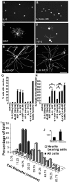
Figure 1.
Figure 1.
IL-6 does not initiate sprouting of adult rat sensory neurons but induces neurite elongation in the presence of a neurotrophin. A–F, Dark-field photomicrographs of cultured adult rat sensory neurons immunocytochemically stained with pan neuronal marker β (III) tubulin after 18 hr in vitro, grown in serum-free media supplemented with the factors indicated. Dissociated sensory neurons cultured in the presence of IL-6, either alone (10 ng/ml; A) or in the presence of its soluble receptor (10 pg/ml to 100 ng/ml; B), failed to exhibit neurite outgrowth after 18 hr in vitro (G). NGF (10 ng/ml) alone significantly increased the percentage of neurite-bearing cells (C, G). The percentage of neurite-bearing cells was not significantly different in the presence of NGF compared with NGF plus IL-6 (1 pg to 100 ng) or NGF plus IL-6 and its soluble receptor (sIL-6R; 10–100ng/ml; G). NT-3 (10ng/ml) alone increased the percentage of neurite-bearing cells (D, G); inclusion of IL-6 (1 pg to 100 ng/ml) alone or in the presence of its soluble receptor (10–100 ng/ml) failed to recruit an additional population of neurite-bearing cells (G). Preconditioning injury to the sciatic nerve results in neurite extension from 76.65% of dissociated cells in vitro (G). The addition of IL-6 (100 ng/ml) alone or in the presence of its soluble receptor (10 ng/ml) to NGF (10 ng/ml)-treated cultures significantly increased the average length of neurite extension compared with cells grown in the presence of NGF (10 ng/ml) alone (H; * and **p < 0.001, ANOVA). The addition of IL-6 (100 ng/ml) alone or in the presence of its soluble receptor (10 ng/ml) to NT-3 (10 ng/ml)-treated cultures also significantly increased the average length of neurite extension (F) compared with cells grown in the presence of NT-3 (10 ng/ml) alone (H; # and ##p<0.001, ANOVA). The level of neurite extension after the co-addition of IL-6 and NGF/NT-3 was not significantly different from that of cells that had received a preconditioning sciatic nerve lesion 7 d before plating (H). IL-6 induced neurite elongation in the presence of NT-3 in a subpopulation of large diameter sensory neurons in culture (I, J). The percentage of neurite-bearing cells is low in the presence of NT-3 (10 ng/ml) alone and is not significantly different when combined with IL-6 and its soluble receptor. This effect is preferentially confined to a population of large diameter sensory neurons. The histogram (I) shows the cell size distribution of all cells in vitro and the distribution of neurite-bearing cells in the presence of NT-3 plus IL-6 after 18 hr of culture. The percentage of neurite-bearing cells is small (because of few cells in the lumbar ganglia possessing trkC; McMahon et al., 1994) but is significantly distributed to the larger diameter profiles, with a diameter over 30 μm (J; *p < 0.001, ANOVA) %NBC, Percentage of neurite-bearing cells. Scale bar, 100 μm.
Figure 2.
Figure 2.
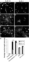
IL-6 –/– mice failed to display enhanced neurite extension after previous conditioning lesion in vitro. A–F illustrate photomicrographs of dissociated adult sensory neurons from IL-6 +/+ (A, C, E) and IL-6 –/– (B, D, F) mice after 18 hr in vitro. Cells from +/+ (A, G) and –/– (B, G) mice cultured in serum-free media failed to extend neurites after 18 hr in vitro. Cells cultured from IL-6 +/+ mice that had received a preconditioning lesion to their sciatic nerve 7 d before plating extended neurites with an average length of 869 ± 15.6 μm (C, G), significantly longer than from IL-6 –/– that had received the same lesion (D, G; *p < 0.001, ANOVA). Inclusion of 10 ng/ml IL-6 to preinjured cells from –/– mice increased the average length of neurite extension 800.2 ± 21.3 μm (F,G), while having no further influence on +/+ mice (E, G). Both +/+ and –/– mice retained the ability to respond to NT-3 [10 ng/ml; 455.8 ± 18.1 (+/+) vs 431.8 ± 14.5 μm (–/–); G] alone and in combination with IL-6 [10 ng/ml; 553.6 ± 7.2 (+/+) vs 510.3 ± 15.9 μm (–/–)] with no significant difference in neurite extension between genotypes. Scale bar, 100 μm.
Figure 3.
Figure 3.
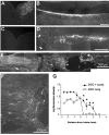
Immunofluorescent photomicrographs of CTB-labeled sciatic nerve axon collaterals within the spinal dorsal column and gracile nucleus of the adult wild-type mouse and the effect of spinal cord injury with or without preconditioning sciatic nerve injury. Horizontal 20 μm sections through the thoracic spinal cord (B) and a 15 μm transverse section through the gracile nucleus (A) of intact IL-6 +/+ mice. The sciatic projection within the ascending dorsal column pathway visualized by CTB-IR is seen as an uninterrupted tract that projects to the ipsilateral gracile nucleus (A). Four weeks after bilateral crush of the dorsal columns, the bundle of aligned fibers becomes irregular and retracts from the caudal edge of the lesion (D, arrowheads). Regeneration is abortive, as indicated by the large reactive end-bulbs and the absence of labeled terminals within the gracile nucleus (C). E, Low-power immunofluorescent photomicrograph of the lesioned IL-6 +/+ mouse spinal cord showing regeneration of injured dorsal column fibers evoked by a preconditioning injury to the sciatic nerve 7 d before dorsal column crush (scale bar, 500 μm). CTB-IR fibers can be seen growing up to the cavity and are also observed in a dense plexus rostral to the lesion site (asterisk). This region of regeneration is shown in higher power in F (scale bar, 200 μm). Pioneering CTB-IR axons were observed within the spinal cord white matter tracts considerable distances rostral to the lesion (arrow). In all spinal cord sections, caudal is to the right. G, Densitometric analysis of CTB labeling within the spinal cord 4 weeks after spinal cord injury with and without sciatic nerve preconditioning. Two-way ANOVA revealed a significant difference in CTB density between preconditioned and spinal cord-lesioned animals only. Post hoc analysis (Tukey) showed that all data points rostral to the lesion site in preinjured animals were significantly different from animals with spinal cord injury alone (p < 0.001).
Figure 4.
Figure 4.
Immunofluorescent photomicrographs of longitudinal sections of spinal cord from wild-type and knock-out mice showing 5-HT immunoreactivity under control conditions and 4 weeks after the dorsal column lesion. A, Under control conditions, 5-HT-containing terminals are entirely restricted to central gray matter regions (within arrowheads) in both IL-6 +/+ and –/– mice; +/+ is shown. Collateral branches extend from the medial gray border toward the spinal cord midline. B, In wild-type mice, 4 weeks after spinal cord injury, disruption of gray matter areas is apparent (crush marked by asterisk). Robust collateral sprouting of 5-HT-containing terminals is observed within novel regions of lateral white matter rostral to the region of spinal cord injury (B; arrows). D, High-power photomicrograph of the crush site region. In IL-6 –/– mice, extensive collateral sprouting of 5-HT-containing terminals is also apparent 4 weeks after spinal cord injury. Collateral sprouting is observed within white matter regions not normally occupied by descending raphe spinal axons (E, arrows).
Figure 5.
Figure 5.
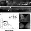
Immunofluorescent photomicrographs of CTB-IR fibers within the dorsal columns showing failure of spinal cord regeneration in IL-6 –/– mice after dorsal column injury in the presence or absence of a preconditioning injury to the sciatic nerve. In IL-6 –/– mice 4 weeks after spinal cord injury alone, CTB tracing shows that regenerating fibers caudal to the lesion site grow in a multidirectional tangled manner with most fibers not reaching the lesion perimeter (A, arrowheads). In contrast, preconditioning of the sciatic nerve 7 d before spinal cord injury resulted in a more robust attempt of damaged fibers to regenerate (see Fig. 2 B, C) compared with dorsal column crush alone. CTB-IR fibers grew up to the lesion in an ordered manner, however, growth stopped at the caudal end of the lesion and axons were never observed past the lesion site (indicated by arrowheads). In all spinal cord sections, caudal is to the right. Scale bar, 500 μm. Analysis of CTB fluorescent intensity measured at defined intervals caudal and rostral to the injury revealed a significant decrease in the degree of regeneration in IL-6 –/– mice compared with +/+ mice after a 7 d preconditioning injury (C). Inspection of the dorsal column nuclei revealed a regular termination of CTB-IR ascending A fibers in intact IL-6 –/– mice (D) and a complete absence of CTB-IR terminal labeling in IL-6 –/– mice (E) that had received a dorsal column lesion.
Figure 6.
Figure 6.
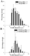
Sciatic axotomy does not result in differential sensory neuron cell loss in IL-6 +/+ or –/– mice. Applying a recursive translation protocol, we examined the cell size distribution of all cell profiles in ipsilateral L4 and L5 DRG 7 d after sciatic nerve transection in IL-6 +/+ and –/– mice (A). No significant difference was observed in cell size distributions of L4 and L5 DRG sensory neurons between genotypes after peripheral nerve lesions at the mid-sciatic level. B, CTB transport in sciatic afferents is not significantly different in IL-6 +/+ mice compared with –/– mice. The graph shows that the cell size distributions of CTB-IR profiles in +/+ and –/– mice are equivalent.
Figure 7.
Figure 7.
Sciatic axotomy fails to increase L5 DRGs GAP43 levels in IL-6 –/– mice. Photomicrographs of L5 DRGs sections from IL-6 +/+ (A–E) and IL-6 –/– (F–J) mice 5 weeks after sciatic nerve axotomy colabeled with antibodies against CTB and GAP43. A and F show CTB-IR and represent cell bodies of injured sciatic afferent fibers, the collaterals of which were transganglionically traced and subsequently revealed in the spinal cord. B and G illustrate GAP43-IR. C and H show the pseudo-colored overlay (yellow) of the two antibodies (CTB, green; GAP43, red). Significantly higher numbers of CTB-IR cell profiles display high levels of GAP43 in IL-6 +/+ mice compared with –/– mice (compare C and H with K) after sciatic axotomy, in comparison with their sham-operated controls (E, J, K). K illustrates that CTB-IR profiles in L5 DRGs from IL-6 +/+ mice express higher levels of GAP43-IR after a preconditioning injury in comparison with sham-operated controls, whereas preconditioning injury to IL-6 –/– mice failed to elevate GAP43 levels. Scale bars: A–C, F–H, 100 μm; D, E, I, J,50 μm.

Similar articles

Cited by

References

    1. Andersen LB, Schreyer DJ (1999) Constitutive expression of GAP-43 correlates with rapid, but not slow regrowth of injured dorsal root axons in the adult rat. Exp Neurol 155: 157–164. - PubMed
    1. Bjorklund A, Wiklund L, Descarries L (1981) Regeneration and plasticity of central serotonergic neurons: a review. J Physiol (Paris) 77: 247–255. - PubMed
    1. Bolin LM, Verity AN, Silver JE, Shooter EM, Abrams JS (1995) Interleukin-6 production by Schwann cells and induction in sciatic nerve injury. J Neurochem 64: 850–858. - PubMed
    1. Bomze HM, Bulsara KR, Iskandar BJ, Caroni P, Skene JH (2001) Spinal axon regeneration evoked by replacing two growth cone proteins in adult neurons. Nat Neurosci 4: 38–43. - PubMed
    1. Bourde O, Kiefer R, Toyka KV, Hartung HP (1996) Quantification of interleukin-6 mRNA in Wallerian degeneration by competitive reverse transcription polymerase chain reaction. J Neuroimmunol 69: 135–140. - PubMed

Publication types

MeSH terms