Prediction and identification of a permissive epitope insertion site in the vesicular stomatitis virus glycoprotein
- PMID: 15113889
- PMCID: PMC400361
- DOI: 10.1128/jvi.78.10.5079-5087.2004
Prediction and identification of a permissive epitope insertion site in the vesicular stomatitis virus glycoprotein
Abstract
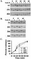
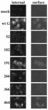
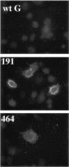
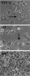
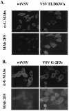
We developed a rational approach to identify a site in the vesicular stomatitis virus (VSV) glycoprotein (G) that is exposed on the protein surface and tolerant of foreign epitope insertion. The foreign epitope inserted was the six-amino-acid sequence ELDKWA, a sequence in a neutralizing epitope from human immunodeficiency virus type 1. This sequence was inserted into six sites within the VSV G protein (Indiana serotype). Four sites were selected based on hydrophilicity and high sequence variability identified by sequence comparison with other vesiculovirus G proteins. The site showing the highest variability was fully tolerant of the foreign peptide insertion. G protein containing the insertion at this site folded correctly, was transported normally to the cell surface, had normal membrane fusion activity, and could reconstitute fully infectious VSV. The virus was neutralized by the human 2F5 monoclonal antibody that binds the ELDKWA epitope. Additional studies showed that this site in G protein tolerated insertion of at least 16 amino acids while retaining full infectivity. The three other insertions in somewhat less variable sequences interfered with VSV G folding and transport to the cell surface. Two additional insertions were made in a conserved sequence adjacent to a glycosylation site and near the transmembrane domain. The former blocked G-protein transport, while the latter allowed transport to the cell surface but blocked membrane fusion activity of G protein. Identification of an insertion-tolerant site in VSV G could be important in future vaccine and targeting studies, and the general principle might also be useful in other systems.
Figures



Similar articles
-
Epitope mapping by deletion mutants and chimeras of two vesicular stomatitis virus glycoprotein genes expressed by a vaccinia virus vector.Virology. 1989 Jun;170(2):392-407. doi: 10.1016/0042-6822(89)90430-3. Virology. 1989. PMID: 2471352
-
Characterization of Antibody Interactions with the G Protein of Vesicular Stomatitis Virus Indiana Strain and Other Vesiculovirus G Proteins.J Virol. 2018 Nov 12;92(23):e00900-18. doi: 10.1128/JVI.00900-18. Print 2018 Dec 1. J Virol. 2018. PMID: 30232190 Free PMC article.
-
Monoclonal antibodies to the glycoprotein of vesicular stomatitis virus (New Jersey serotype): a method for preliminary mapping of epitopes.Virology. 1987 Dec;161(2):533-40. doi: 10.1016/0042-6822(87)90148-6. Virology. 1987. PMID: 2446424
-
Viral inactivation based on inhibition of membrane fusion: understanding the role of histidine protonation to develop new viral vaccines.Protein Pept Lett. 2009;16(7):779-85. doi: 10.2174/092986609788681823. Protein Pept Lett. 2009. PMID: 19601907 Review.
-
Structures of vesicular stomatitis virus glycoprotein: membrane fusion revisited.Cell Mol Life Sci. 2008 Jun;65(11):1716-28. doi: 10.1007/s00018-008-7534-3. Cell Mol Life Sci. 2008. PMID: 18345480 Free PMC article. Review.
Cited by
-
Selection of novel vesicular stomatitis virus glycoprotein variants from a peptide insertion library for enhanced purification of retroviral and lentiviral vectors.J Virol. 2006 Apr;80(7):3285-92. doi: 10.1128/JVI.80.7.3285-3292.2006. J Virol. 2006. PMID: 16537595 Free PMC article.
-
An unrelated monoclonal antibody neutralizes human immunodeficiency virus type 1 by binding to an artificial epitope engineered in a functionally neutral region of the viral envelope glycoproteins.J Virol. 2005 May;79(9):5616-24. doi: 10.1128/JVI.79.9.5616-5624.2005. J Virol. 2005. PMID: 15827176 Free PMC article.
-
Neutralization efficiency is greatly enhanced by bivalent binding of an antibody to epitopes in the V4 region and the membrane-proximal external region within one trimer of human immunodeficiency virus type 1 glycoproteins.J Virol. 2010 Jul;84(14):7114-23. doi: 10.1128/JVI.00545-10. Epub 2010 May 12. J Virol. 2010. PMID: 20463081 Free PMC article.
-
New generation of DNA-based immunotherapy induces a potent immune response and increases the survival in different tumor models.J Immunother Cancer. 2021 Apr;9(4):e001243. doi: 10.1136/jitc-2020-001243. J Immunother Cancer. 2021. PMID: 33795383 Free PMC article.
-
Protein expression/secretion boost by a novel unique 21-mer cis-regulatory motif (Exin21) via mRNA stabilization.Mol Ther. 2023 Apr 5;31(4):1136-1158. doi: 10.1016/j.ymthe.2023.02.012. Epub 2023 Feb 14. Mol Ther. 2023. PMID: 36793212 Free PMC article.
References
-
- Bachmann, M. F., H. Hengartner, and R. M. Zinkernagel. 1995. T helper cell-independent neutralizing B cell response against vesicular stomatitis virus: role of antigen patterns in B cell induction? Eur. J. Immunol. 25:3445-3451. - PubMed
-
- Bhella, R. S., S. T. Nichol, E. Wanas, and H. P. Ghosh. 1998. Structure, expression and phylogenetic analysis of the glycoprotein gene of Cocal virus. Virus Res. 54:197-205. - PubMed
-
- Brun, G., X. Bao, and L. Prevec. 1995. The relationship of Piry virus to other vesiculoviruses: a re-evaluation based on the glycoprotein gene sequence. Intervirology 38:274-282. - PubMed
Publication types
MeSH terms
Substances
Grants and funding
LinkOut - more resources
Full Text Sources
Other Literature Sources

