. 2004 Apr;113(8):1118-29.
doi: 10.1172/JCI19205.

Correction of metachromatic leukodystrophy in the mouse model by transplantation of genetically modified hematopoietic stem cells

Affiliations

Correction of metachromatic leukodystrophy in the mouse model by transplantation of genetically modified hematopoietic stem cells

Alessandra Biffi et al. J Clin Invest. 2004 Apr.

Abstract

Gene-based delivery can establish a sustained supply of therapeutic proteins within the nervous system. For diseases characterized by extensive CNS and peripheral nervous system (PNS) involvement, widespread distribution of the exogenous gene may be required, a challenge to in vivo gene transfer strategies. Here, using lentiviral vectors (LVs), we efficiently transduced hematopoietic stem cells (HSCs) ex vivo and evaluated the potential of their progeny to target therapeutic genes to the CNS and PNS of transplanted mice and correct a neurodegenerative disorder, metachromatic leukodystrophy (MLD). We proved extensive repopulation of CNS microglia and PNS endoneurial macrophages by transgene-expressing cells. Intriguingly, recruitment of these HSC-derived cells was faster and more robust in MLD mice. By transplanting HSCs transduced with the arylsulfatase A gene, we fully reconstituted enzyme activity in the hematopoietic system of MLD mice and prevented the development of motor conduction impairment, learning and coordination deficits, and neuropathological abnormalities typical of the disease. Remarkably, ex vivo gene therapy had a significantly higher therapeutic impact than WT HSC transplantation, indicating a critical role for enzyme overexpression in the HSC progeny. These results indicate that transplantation of LV-transduced autologous HSCs represents a potentially efficacious therapeutic strategy for MLD and possibly other neurodegenerative disorders.


Figures

Figure 1
Figure 1
Chimerism and hematologic reconstitution by transgene-expressing cells of transplanted mice. (A) FACS analysis of GFP expression in control (left panel) and transduced (right panel) purified progenitors. (B) GFP expression in PBMCs of primary and secondary transplanted mice, 3 months after BMT. Up to 90% of circulating donor CD45.1-, CD11b-, B220-, CD4-, and CD8-positive cells expressed GFP. The means and standard deviations of primary (n =16) and secondary (n = 4) mice are shown. (C) GFP expression by fluorescence microscopy (% GFP+) and transduction level by PCR (% vector sequence+), in CFCs from progenitors after transduction and from BM of primary and secondary recipients (50–100 CFCs scored for fluorescence and 20 CFCs analyzed by PCR per each mouse, n = 9). The great majority of CFCs contained LV sequences and expressed GFP along serial transplants. (D) Southern blot analysis of bone marrow (B) and spleen (S) DNA from transplanted mice. A standard plasmid curve, from 0.5 to 5 vector copies per genome, DNA markers (M) in kb, and control untransplanted mice (Co) are shown. AflII digestion (upper panel) showed 5 or more vector copies integrated per cell in all recipients. BamHI digestion (lower panel) showed a diffuse hybridization pattern in primary mice indicating oligo- to polyclonal engraftment and the absence of dominant clones. (E) and (F) GFP+ cells in liver (E) and kidney (F) sections of transplanted mice, 3 months after BMT, stained as indicated. GFP+ cells expressed F4/80 (3 panels on the right). Scale bar: 20 μm.
Figure 2
Figure 2
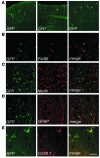
Identification of bone marrow–derived vector-expressing cells in the CNS of transplanted mice. Immunofluorescence analysis of cryostatic sections from the brains of transplanted mice. Fluorescent signals from single optical sections were sequentially acquired and are shown individually and after merging (merge). Immunostaining for GFP (green), F4/80, NeuN, GFAP, or CD45.1 (red) are indicated. (A) Representative sections from the cerebellums of transplanted mice, analyzed at 3 months (left panel, scale bar: 200 μm) and 6 months (middle and right panels, scale bar: 300 μm) after BMT. Three months after BMT, few ramified cells were identified in the upper cortical layers near the meninges. By 6 months after BMT, several GFP+ cells were present in the cortex, and small clusters of ramified GFP+ cells were detected throughout the parenchyma. (B–E) Representative brain sections from transplanted mice 9 months after BMT, immunostained as indicated. (B) GFP+ cells showed a ramified, microglial morphology and F4/80 immunoreactivity. Scale bar: 100 μm. (C and D) Overlay of GFP staining with the neuronal-specific marker NeuN (C) and the astrocytic marker GFAP (D) demonstrated separate localization of the two signals, with GFP+ ramified cells found between neurons and astrocytes. Scale bar: 300 μm. (E) CD45.1 immunostaining identified GFP+ cells as donor-derived. Scale bar: 70 μm.
Figure 3
Figure 3
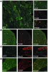
Identification of bone marrow–derived vector-expressing cells in the PNS of transplanted mice. Representative cryostatic sections of the dorsal root ganglion (A), sciatic nerve (B), and acoustic ganglion (C) of a transplanted mouse, 6 months after BMT, immunostained for GFP, F4/80, and NeuN, as indicated. (A) Left panel, GFP+ cells were found in the dorsal root ganglion, surrounding sensory neurons and showing a macrophage morphology. Scale bar: 60 μm. Right panel, all of the GFP+ cells coexpressed the macrophage marker F4/80. Scale bar: 200 μm. (B) GFP+ cells were detected in the endoneurial space of the sciatic nerve and expressed F4/80. Scale bar: 100 μm. (C) GFP+ cells were distributed between sensory neurons in the acoustic ganglion and did not express the neuronal marker NeuN. Scale bar: 200 μm. (D) Vector-expressing cells in the PNS of a representative secondary transplant recipient. Cryostatic section from the dorsal root ganglion 4 months after BMT, immunostained for GFP and F4/80. Scale bar: 150 μm.
Figure 4
Figure 4
Enhanced migration and activated morphology of vector-expressing cells in the CNS of As2–/– MLD mice. (A) Cryostatic sections of the corpus callosum of a representative transplanted MLD mouse 6 months after BMT, showing widespread GFP+ cells with a swollen ameboid morphology and coexpressing F4/80. Scale bar: 80 μm. (B) Cryostatic sections from the hippocampus of the same mouse were first immunostained as in A, examined under the fluorescent microscope (three panels on the left) and then stained with PAS (panel on the far right). The GFP+, F4/80+ microglia cells were PAS-reactive, indicating their content of lipid storage granules. Scale bar: 40 μm.
Figure 5
Figure 5
ARSA activity reconstitution and neurophysiological analysis of transplanted MLD mice. (A) Sulfatide assay on PBMCs from mice transplanted with ARSA-transduced (ARSA-LV) or GFP-transduced (As2–/–) HSCs, 7 months after BMT, and from WT mice. Sulfatide metabolism in WT and reconstituted As2–/– mice is shown by LRh-sulfatide reduction and appearance of galactosylceramide (LRh-GalCer) (average and range of picomoles normalized for total protein content). (B) PNC assay on PBMCs as in A. ARSA activity is expressed relative to the value obtained from WT mice. Full ARSA activity reconstitution and overexpression above the WT level was observed in ARSA-transplanted mice. (C–E) Neurophysiological assessment of central and peripheral motor conduction in 8-month-old MLD mice transplanted with ARSA- or GFP-transduced HSCs, and WT mice with the same genetic background (n = 15 mice per group). Significantly lower CCTs (C) and F wave latency values (D), and significantly higher sciatic MCV values (E) were recorded in ARSA- transplanted mice compared with GFP controls. Comparison with WT shows nearly complete prevention of motor conduction impairments (P > 0.05 for all parameters). (F–H) Neurophysiological assessment in the same groups of ARSA- and mock-transplanted mice as in C–E, at 12 months of age, and in age-matched, WT HSC–transplanted mice (n = 10 mice per group). The analysis shows maintenance of the therapeutic effect in ARSA-transplanted mice and significantly faster motor conduction as compared with WT HSC–transplanted ones (*P < 0.01 between ARSA-LV and As2–/–; ***P < 0.05 between ARSA-LV and WT HSC–transplanted mice). Data are expressed as single recordings and means.
Figure 6
Figure 6
Motor learning and coordination in treated and untreated MLD mice. Twelve-month-old mice transplanted with ARSA-transduced or WT HSCs and two cohorts of untreated As2–/– mice of 3 and 12 months of age were tested on an accelerating rotarod apparatus (n = 12–30 per group). Latencies to fall off the rotarod were recorded over 3 days, 3 consecutive trials per day. Means ± SEM of each day are indicated. Post hoc comparisons were made by one-way ANOVA using Scheffe’s test after significant main effect of the treatment was determined. Differences from 12-month-old untreated mice were considered significant at P < 0.05 (*P < 0.01; **P < 0.001). The decline in motor learning and coordination was completely prevented by ARSA-transduced HSC transplantation, whereas WT HSC transplantation was not significantly effective in preventing the development of the motor learning impairment.
Figure 7
Figure 7
Long-term protection from lipid storage in transplanted MLD mice. (A and B) Toluidine Blue–stained semithin sections of the cerebellum (A) and the hippocampal fimbria (B) of representative 12-month-old MLD mice transplanted with GFP-LV– and ARSA-LV–transduced As2–/– HSCs, or with WT HSCs. Abundant large metachromatic deposits are present in mock-treated and WT HSC–transplanted mice, whereas they are almost absent in mice transplanted with gene-corrected cells. Scale bar: 50 μm. For quantitative data and sample size, see Table 2.
Figure 8
Figure 8
Long-term protection from lipid storage and demyelination in transplanted MLD mice. Toluidine Blue–stained sections of the sciatic nerve of representative 12-month-old MLD mice transplanted with GFP-LV– and ARSA-LV–transduced As2–/– HSCs, or with WT HSCs. Several demyelinated fibers (arrows) and metachromatic granules in Schwann cells are present in mock-treated and WT HSC–transplanted mice, whereas they are almost absent in mice transplanted with gene-corrected cells. Scale bar: 40 μm. For quantitative data and sample size, see Table 2.

Comment in

  • Blood to brain to the rescue.
    Proia RL, Wu YP. Proia RL, et al. J Clin Invest. 2004 Apr;113(8):1108-10. doi: 10.1172/JCI21476. J Clin Invest. 2004. PMID: 15085187 Free PMC article.

Similar articles

Cited by

References

    1. Kolodney, E.H., and Fluharty, A.L. 1995. Metachromatic leukodystrophy and multiple sulfatase deficiency: sulfatide lipidosis. In The metabolic and molecular bases of inherited disease. 6th edition. C.R. Scriver, A.L. Beaudet, W.S. Sly, and D. Valle, editors. McGraw-Hill, New York, USA. 2693–7391.
    1. Neufeld EF. Lysosomal disease. Annu. Rev. Biochem. 1991;60:257–280. - PubMed
    1. Kay MA, Glorioso JC, Naldini L. Viral vectors for gene therapy: the art of turning infectious agents into vehicles of therapeutics. Nat. Med. 2001;7:33–40. - PubMed
    1. Glorioso JC, Mata M, Fink DJ. Therapeutic gene transfer to the nervous system using viral vectors. J. Neurovirol. 2003;9:165–172. - PubMed
    1. Consiglio A, et al. In vivo gene therapy of metachromatic leukodystrophy by lentiviral vectors: correction of neuropathology and protection against learning impairments in affected mice. Nat. Med. 2001;7:310–316. - PubMed

Publication types