CD28 delivers a unique signal leading to the selective recruitment of RelA and p52 NF-kappaB subunits on IL-8 and Bcl-xL gene promoters
- PMID: 15079071
- PMCID: PMC395929
- DOI: 10.1073/pnas.0308688101
CD28 delivers a unique signal leading to the selective recruitment of RelA and p52 NF-kappaB subunits on IL-8 and Bcl-xL gene promoters
Abstract
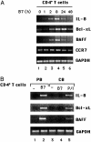
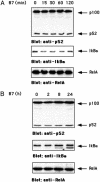
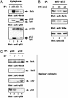
CD28 is one of the most important costimulatory receptors necessary for full T lymphocyte activation. The CD28 receptor can enhance T cell antigen receptor (TCR) signals, as well as deliver independent signals. Indeed, CD28 engagement by B7 can generate TCR-independent signals leading to IkappaB kinase and NF-kappaB activation. Here we demonstrate that the TCR-independent CD28 signal leads to the selective transcription of survival (Bcl-xL) and inflammatory (IL-8 and B cell activation factor, but not proliferative (IL-2), genes, in a NF-kappaB-dependent manner. CD28-stimulated T cells actively secrete IL-8, and Bcl-xL up-regulation protects T cells from radiation-induced apoptosis. The transcription of CD28-induced genes is mediated by the specific recruitment of RelA and p52 NF-kappaB subunits to target promoters. In contrast, p50 and c-Rel, which preferentially bind NF-kappaB sites on the IL-2 gene promoter after anti-CD3 stimulation, are not involved. Thus, we identify CD28 as a key regulator of genes important for both survival and inflammation.
Figures


Similar articles
-
Cyclosporin A inhibits the early phase of NF-kappaB/RelA activation induced by CD28 costimulatory signaling to reduce the IL-2 expression in human peripheral T cells.Int Immunopharmacol. 2005 Apr;5(4):699-710. doi: 10.1016/j.intimp.2004.11.018. Int Immunopharmacol. 2005. PMID: 15710339
-
The function of multiple IkappaB : NF-kappaB complexes in the resistance of cancer cells to Taxol-induced apoptosis.Oncogene. 2002 Sep 19;21(42):6510-9. doi: 10.1038/sj.onc.1205848. Oncogene. 2002. PMID: 12226754
-
CD28 costimulation regulates FOXP3 in a RelA/NF-κB-dependent mechanism.Eur J Immunol. 2011 Feb;41(2):503-13. doi: 10.1002/eji.201040712. Epub 2011 Jan 11. Eur J Immunol. 2011. PMID: 21268019
-
NF-κB family of transcription factors: biochemical players of CD28 co-stimulation.Immunol Lett. 2011 Mar 30;135(1-2):1-9. doi: 10.1016/j.imlet.2010.09.005. Epub 2010 Sep 21. Immunol Lett. 2011. PMID: 20863851 Review.
-
Regulation and function of IKK and IKK-related kinases.Sci STKE. 2006 Oct 17;2006(357):re13. doi: 10.1126/stke.3572006re13. Sci STKE. 2006. PMID: 17047224 Review.
Cited by
-
Regulation of p53 tumour suppressor target gene expression by the p52 NF-kappaB subunit.EMBO J. 2006 Oct 18;25(20):4820-32. doi: 10.1038/sj.emboj.7601343. Epub 2006 Sep 21. EMBO J. 2006. PMID: 16990795 Free PMC article.
-
The timing of differentiation and potency of CD8 effector function is set by RNA binding proteins.Nat Commun. 2022 Apr 27;13(1):2274. doi: 10.1038/s41467-022-29979-x. Nat Commun. 2022. PMID: 35477960 Free PMC article.
-
Intrinsic and extrinsic control of peripheral T-cell tolerance by costimulatory molecules of the CD28/ B7 family.Immunol Rev. 2011 May;241(1):180-205. doi: 10.1111/j.1600-065X.2011.01011.x. Immunol Rev. 2011. PMID: 21488898 Free PMC article. Review.
-
4-1BB costimulation promotes CAR T cell survival through noncanonical NF-κB signaling.Sci Signal. 2020 Mar 31;13(625):eaay8248. doi: 10.1126/scisignal.aay8248. Sci Signal. 2020. PMID: 32234960 Free PMC article.
-
TCR ligand potency differentially impacts PD-1 inhibitory effects on diverse signaling pathways.J Exp Med. 2023 Dec 4;220(12):e20231242. doi: 10.1084/jem.20231242. Epub 2023 Oct 5. J Exp Med. 2023. PMID: 37796477 Free PMC article.
References
-
- Lanzavecchia, A., Iezzi, G. & Viola, A. (1999) Cell 96, 1-4. - PubMed
-
- Wulfing, C. & Davis, M. M. (1998) Science 282, 2266-2269. - PubMed
-
- Michel, F., Attal-Bonnefoy, G., Mangino, G., Mise-Omata, S. & Acuto, O. (2001) Immunity 15, 935-945. - PubMed
-
- Tuosto, L. & Acuto, O. (1998) Eur. J. Immunol. 28, 2131-2142. - PubMed
-
- Kaga, S., Ragg, S., Rogers, K. A. & Ochi, A. (1998) J. Immunol. 160, 24-27. - PubMed
Publication types
MeSH terms
Substances
LinkOut - more resources
Full Text Sources
Research Materials

