. 2004 Apr;24(8):3373-86.
doi: 10.1128/MCB.24.8.3373-3386.2004.

The human beta-globin replication initiation region consists of two modular independent replicators

Affiliations

The human beta-globin replication initiation region consists of two modular independent replicators

Lixin Wang et al. Mol Cell Biol. 2004 Apr.

Abstract

Previous studies have shown that mammalian cells contain replicator sequences, which can determine where DNA replication initiates. However, the specific sequences that confer replicator activity were not identified. Here we report a detailed analysis of replicator sequences that dictate initiation of DNA replication from the human beta-globin locus. This analysis suggests that the beta-globin replication initiation region contains two adjacent, redundant replicators. Each replicator was capable of initiating DNA replication independently at ectopic sites. Within each of these two replicators, we identified short, discrete, nonredundant sequences, which cooperatively determine replicator activity. Experiments with somatic cell hybrids further demonstrated that the requirements for initiation at ectopic sites were similar to the requirements for initiation within native human chromosomes. The replicator clustering and redundancy exemplified in the human beta-globin locus may account for the extreme difficulty in identifying replicator sequences in mammalian cells and suggest that mammalian replication initiation sites may be determined by cooperative sequence modules.


Figures

FIG. 1.
FIG. 1.
Replication initiation assays. (A) Schematic illustration of the methodology used for nascent-strand abundance assay. DNA strands are depicted as solid gray lines, and 5′ primer RNAs are shown as solid black boxes. Newly replicated origin-proximal DNA was selected by size (600 to 2,500 bp) and by resistance to lambda exonuclease (an enzyme that digests DNA with a 5′ DNA tail but not DNA with a 5′ RNA tail). Nascent strands isolated in this way were then subjected to real-time PCR with primers encompassing the locus of interest. (B) An example of real-time PCR output. Standards of genomic DNA at fixed concentrations were used in a PCR along with an unknown sample. The abundance of the PCR products in nascent DNA was calculated based on the cycle in which fluorescence from the real-time PCR crossed the manually set threshold. (C) A calibration curve based on the data shown in panel B. (D to F) An example of data processing. (D) PCR amplification of three independent preparations of nascent strands from CV-1 E25B4 cells containing the β-globin IR at the B4 site. Histogram bars show the average amplification efficiency of PCR products from a series of probes (for probe sequences and locations, see Table 1). Error bars indicate standard deviations. (E) The same data shown in panel D, represented as the ratio of the abundance of specific sequences to the abundance of sequences amplified by lacZ primers. (F) A histogram depicting the average abundance of specific sequences in nascent strands, represented as the average of the measurements shown in panel E from three independent nascent-strand preparations. Error bars indicate the upper and lower ranges of the measured ratios.
FIG. 2.
FIG. 2.
Identification of two nonoverlapping, independent replicators within the human β-globin IR. (A) A schematic representation of the IR from the human globin locus. Replication initiates from the region between the two adult β-globin genes (top line). The IR (IR) encompasses the promoter and the majority of the β-like-globin gene (second line); preliminary analysis identified a core central region within the IR, which was essential but not sufficient for initiation (third line) (1). The present analysis divided the IR into two fragments designated bGRep-P and bGRep-I (bottom line). The gray boxes on the top line represent the globin genes; white boxes in the second line represent exons, while the dashed line represents the direction of transcription. (B to D) DNA fragments originating from the human β-globin IR were inserted into the FRT site in CV-1 E25B4 cells as described. The abundance of IR-derived DNA sequences in short, exonuclease-resistant, newly replicated DNA was determined by real-time quantitative PCR and quantified as shown in panel F. A dissection of the locus at the NcoI site (coordinate 62187) preserved the ability to initiate DNA replication in both fragments when each fragment was inserted at the ectopic site, suggesting that both fragments can function as independent nonoverlapping replicators. Real-time PCR analysis of the entire IR (B), analysis of bGRep-P, (C), and analysis of bGRep-I (D) are shown. Data are represented as the number of molecules amplified from RNA-primed nascent strands divided by the number of molecules amplified from the same preparation by the lacZ primers. Each histogram bar depicts the average of three independent measurements. Error bars represent the range of measured ratios.
FIG. 3.
FIG. 3.
An evolutionarily conserved alternate AT-rich stretch is not essential for replicator activity within the β-globin Rep-P replicator. (A) Insertion of the IR Rep-P fragment into an FRT-containing acceptor site in the simian genome using site-specific recombination. The insertion vector, used to clone putative replicator candidates, contains an FRT and a hygromycin resistance marker (hyg). The acceptor site has an identical FRT sequence inserted into the simian genome. Transfection of the vector into cells containing the target in the presence of excess FLP recombinase leads to frequent integrations of the entire insertion vector into the target. Integration disrupts the expression of the lacZ marker. Recombinant clones are selected based on hygromycin resistance and lack of lacZ expression, and Southern hybridization and PCR analyses are then used to verify that recombinant colonies contained single copies of the insertion vector in the acceptor sites. The filled grey arrows represent FRT sites; the double-headed arrow represents the location of the probe used in panel B; and the arrows designated P1 to P6 represent the locations of the primers used in panel C. (B) Analysis of the structure of recombinant clones using Southern hybridization. Lanes 1, 4, and 7 contain DNA from a colony with a single-copy integration of Rep-P at the target site; lanes 2, 5, and 8 contain DNA from a second colony containing the same insert exhibiting a similar structure; lanes 3, 6, and 9 contain DNA from a colony that exhibits a rearrangement of the inserted Rep-P fragment. DNAs were digested with HindIII (lanes 1 to 3), NotI (lanes 4 to 6), and AspI (lanes 7 to 9) and probed with a NotI fragment from the β-globin locus (double-headed arrow in panel A). Lane M shows molecular size markers ranging from 0.5 to 12.2 kb (Invitrogen, Ready-load 1 kb ladder). (C) PCR analysis of recombinant clones containing Rep-P. Lanes 1, 4, and 7 show the analysis of DNA from a colony with a single copy of Rep-P integrated at the target site, lanes 2, 5, and 8 show the analysis of DNA from a similar colony, and lanes 3, 6, and 9 show the analysis of DNA from a colony exhibiting integration at another genomic site. Lanes 1 to 3 show amplification products using primer pair P1 and P2; lanes 4 to 6 show amplification products using primer pair P3 and P4, and lanes 7 to 9 show amplification products using primer pair P5 and P6. Integration into the acceptor site will prevent amplification using primer pairs P1 and P2 but allow amplification using primer pairs P3 and P4 and P5 and P6. (D) DNA fragments containing Rep-P were mutated in vitro as indicated, inserted into the FRT site in CV-1 E25B4 cells, and tested for initiation activity as described in the legends to Fig. 1 and 2. The wild-type (WT) histogram bars show nascent-strand abundance data from the unaltered Rep-P. The B4 bar represents the nascent-strand abundance of a hygromycin marker at the FRT site in the absence of sequences from the β-globin replicator. Deletion of the AT-rich region, or replacement of this region with a non-AT-rich linker, did not affect initiation capacity.
FIG. 4.
FIG. 4.
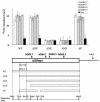
Genomic regions required for replicator activity within Rep-P. DNA fragments originating from bGRep-P (see Fig. 2) were inserted into the FRT site in CV-1 E25B4 cells and tested for initiation activity using the nascent-strand abundance assay described in the legends to Fig. 1 and 2. Deletion of a 300-bp fragment extending from the SnaBI site to the NcoI site (ΔS-N, coordinates 61870 to 62187) and deletion of an 800-bp fragment between the two BamHI sites (ΔB, coordinates 59882 to 60677) greatly diminished the representation of sequences from the inserted human β-globin fragment in nascent strands. Other deletions did not affect replicator activity, suggesting that the two 300- and 800-bp sequences (Rep-P-1 and Rep-P-2, outlined by double arrows) cooperate to confer the ability to initiate DNA replication at the ectopic site. WT, wild type.
FIG. 5.
FIG. 5.
Genomic regions required for replicator activity within Rep-I. DNA fragments originating from Rep-I (see Fig. 2) were inserted into the FRT site in CV-1 E25B4 cells and tested for initiation activity using the nascent-strand abundance assay as described in the legends to Fig. 1 and 2. Deletion of DNA fragments, including a region extending from the PmlI site to the EcoRI site designated Rep-I-1 and outlined by a double arrow, did not allow replication. These data suggested that this region was essential for the initiation of DNA replication. WT, wild type.
FIG. 6.
FIG. 6.
An asymmetric purine:pyrimidine stretch is required for replicator activity within Rep-P. DNA fragments containing Rep-P were mutated in vitro as indicated, inserted into the FRT site in CV-1 E25B4 cells, and tested for initiation activity as described in the legends to Fig. 1 and 2. Deletion of the CAAT box did not affect initiation capacity, whereas a deletion of the asymmetric purine:pyrimidine stretch (AG) blocked initiation from Rep-P sequences at the ectopic site. WT, wild type.
FIG. 7.
FIG. 7.
Requirements for replicator activity within the human β-globin locus in the context of an intact human chromosome 11. The entire human chromosome 11 was transferred to the chicken DT40 cell line, as described previously (16). The indicated regions were deleted from the human β-globin locus: ΔIRC, deletion of a 1.7-kb fragment between the PmeI and MfeI sites; ΔIRM, deletion of a 500-bp fragment between the SnaBI and BspMI sites; and ΔL, deletion of a fragment between two PmlI sites 5′ of the IR. (A) Illustration of the homologous recombination followed by CRE-mediated excision of the IR fragments (for details, see reference 16). The top line shows a schematic illustration of the entire β-globin locus; the β-like globin genes are shown as shaded boxes. The second line shows a magnification of the IR region between the δ and the β genes. The third line shows the structure of a recombinant in which a neomycin resistance gene (neo) flanked by LoxP sites (grey arrows) replaced a part of the IR by homologous recombination. The fourth line shows the structure of a chromosome created by Cre-mediated excision, which deleted a part of the IR, leaving a LoxP site. Double-headed arrows designated P or I represent the location of probes from the human β-globin locus that straddle the inserted LoxP sites. These probes were used in the hybridization experiments shown in panel B. Sites marked by the letter H represent HindIII restriction sites. (B) Southern blot analysis of the structure of ΔIRC. Genomic DNA (10 μg) was digested with HindIII, fractionated on a 0.6% agarose gel, immobilized on a nylon membrane, and hybridized with probes designated I and P as indicated. Lanes 1 contain DNA from a homologous recombinant (third line from the top in panel A); lanes 2 to 5 contain DNA from three different clones resulting from CRE-mediated deletion. Lane 3 contains DNA from the chosen clone ΔIRC. M, molecular size markers derived from 32P-labeled HindIII digestion of bacteriophage λ DNA ranging from 2.3 to 23 kb. The insertion of the neo marker into the globin locus introduced a HindIII site. The two probes, P and I, identified two separate HindIII fragments in the homologous recombinant (4.3 kb and 5.1 kb). After excision, one of the HindIII sites was eliminated and both probes identified a 7.4-kb fragment. Excision was also verified by lack of hybridization to a probe containing the neomycin gene (data not shown). (C) Initiation of DNA replication was measured by the nascent-strand abundance assay as described in the legends to Fig. 1 and 2, using primers from the human β-globin locus except for the CKNLYSC and the CKNLYSNG primers, which were derived from the chicken lysozyme locus (see Table 1 for sequence information). The primer pair CKNLYSNG, which is not abundant in nascent strands from chicken cells (45), served as a standard. Primer locations are illustrated below the histograms. The ΔIRC and ΔIRM chromosomes did not initiate DNA replication, whereas the unaltered chromosomes and the ΔL chromosomes initiated DNA replication from the IR. WT, wild type.
FIG. 8.
FIG. 8.
Summary of sequence features implicated in replication IRs and their locations within the two β-globin replicators. Diamond shapes indicate the locations of sequence features within the β-globin IR relative to the locations of the two replicators, bGRep-P and bGRep-I, and the essential regions within the replicators. Sequences whose roles in initiating DNA replication were tested directly by in vitro mutagenesis are depicted as shaded diamonds and designated essential or not essential.

Similar articles

Cited by

References

    1. Aladjem, M., L.-W. Rodewald, J. L. Kolman, and G. M. Wahl. 1998. Genetic dissection of a mammalian replicator in the human beta-globin locus. Science 281:1005-1009. - PubMed
    1. Aladjem, M., and G. M. Wahl. 1997. Mapping replication origins by leading strand analysis in the absence of protein synthesis. Methods Companion Methods Enzymol. 13:281-292. - PubMed
    1. Aladjem, M. I., M. Groudine, L. L. Brody, E. S. Dieken, R. E. K. Fournier, G. M. Wahl, and E. M. Epner. 1995. Participation of the human beta globin locus control region in initiation of DNA replication. Science 270:815-819. - PubMed
    1. Aladjem, M. I., L. W. Rodewald, C. M. Lin, S. Bowman, D. M. Cimbora, L. L. Brody, E. M. Epner, M. Groudine, and G. M. Wahl. 2002. Replication initiation patterns in the beta-globin loci of totipotent and differentiated murine cells: evidence for multiple initiation regions. Mol. Cell. Biol. 22:442-452. - PMC - PubMed
    1. Altman, A. L., and E. Fanning. 2001. The Chinese hamster dihydrofolate reductase replication origin beta is active at multiple ectopic chromosomal locations and requires specific DNA sequence elements for activity. Mol. Cell. Biol. 21:1098-1110. - PMC - PubMed

Publication types