

Covalently attached oligodeoxyribonucleotides induce RNase activity of a short peptide and modulate its base specificity
- PMID: 15047859
- PMCID: PMC390365
- DOI: 10.1093/nar/gkh514
Covalently attached oligodeoxyribonucleotides induce RNase activity of a short peptide and modulate its base specificity
Abstract
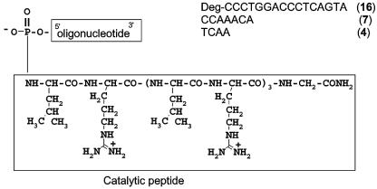
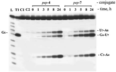
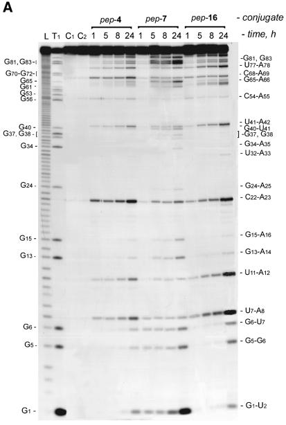
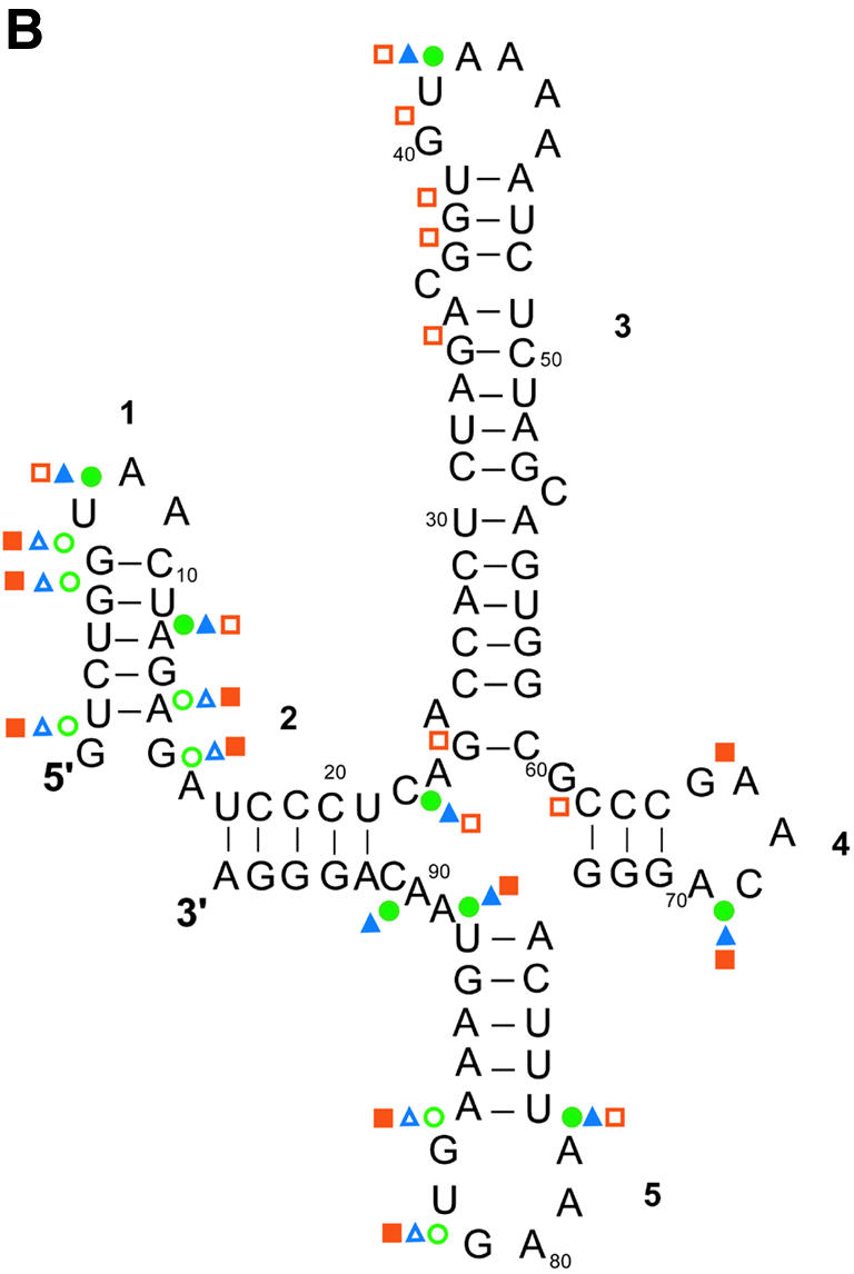
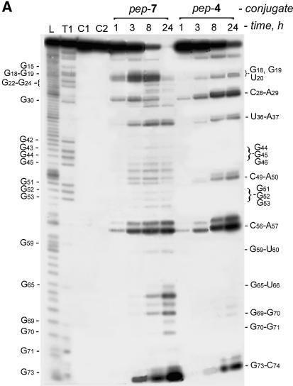
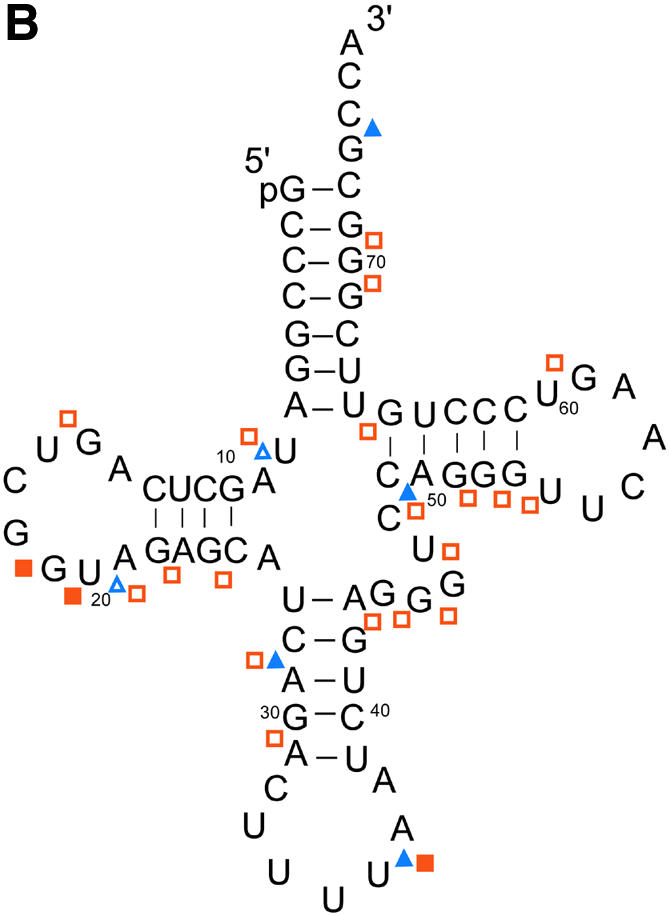
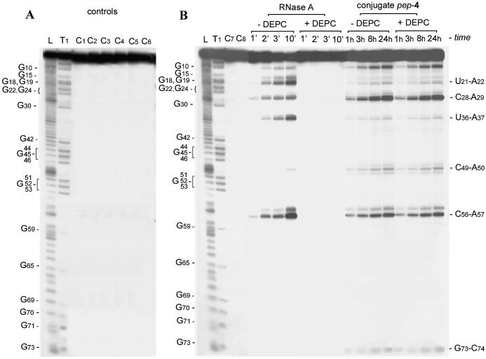
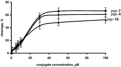
New artificial ribonucleases, conjugates of short oligodeoxyribonucleotides with peptides containing alternating arginine and leucine, were synthesized and characterized in terms of their catalytic activity and specificity of RNA cleavage. The conjugates efficiently cleave different RNAs within single-stranded regions. Depending on the sequence and length of the oligonucleotide, the conjugates display either G-X>>Pyr-A or Pyr-A>>G-X cleavage specificity. Preferential RNA cleavage at G-X phosphodiester bonds was observed for conjugate NH2-Gly-[ArgLeu]4-CCAAACA. The conjugates function as true catalysts, exhibiting reaction turnover up to 175 for 24 h. Our data show that in the conjugate the oligonucleotide plays the role of a factor which provides an 'active' conformation of the peptide via intramolecular interactions, and that it is the peptide residue itself which is responsible for substrate affinity and catalysis.
Figures
Similar articles
-
RNase T1 mimicking artificial ribonuclease.Nucleic Acids Res. 2007;35(7):2356-67. doi: 10.1093/nar/gkm143. Epub 2007 Mar 27. Nucleic Acids Res. 2007. PMID: 17389642 Free PMC article.
-
Site-Selective Artificial Ribonucleases: Renaissance of Oligonucleotide Conjugates for Irreversible Cleavage of RNA Sequences.Molecules. 2021 Mar 19;26(6):1732. doi: 10.3390/molecules26061732. Molecules. 2021. PMID: 33808835 Free PMC article. Review.
-
G-specific RNA-cleaving conjugates of short peptides and oligodeoxyribonucleotides.J Biomol Struct Dyn. 2006 Jun;23(6):591-602. doi: 10.1080/07391102.2006.10507084. J Biomol Struct Dyn. 2006. PMID: 16615805
-
[Artificial ribonucleases I. Targeted RNA cleavage by 5'-peptidyloligodeoxyribonucleotides containing arginine and leucine residues].Bioorg Khim. 1997 Jun;23(6):497-504. Bioorg Khim. 1997. PMID: 9265472 Russian.
-
Artificial ribonucleases.Adv Inorg Biochem. 1994;9:41-74. Adv Inorg Biochem. 1994. PMID: 7511321 Review.
Cited by
-
Non-enzymatic template-directed recombination of RNAs.Int J Mol Sci. 2009 Apr 21;10(4):1788-1807. doi: 10.3390/ijms10041788. Int J Mol Sci. 2009. PMID: 19468339 Free PMC article.
-
Peptide-oligonucleotide conjugates exhibiting pyrimidine-X cleavage specificity efficiently silence miRNA target acting synergistically with RNase H.Sci Rep. 2018 Oct 9;8(1):14990. doi: 10.1038/s41598-018-33331-z. Sci Rep. 2018. PMID: 30302012 Free PMC article.
-
"Bind, cleave and leave": multiple turnover catalysis of RNA cleavage by bulge-loop inducing supramolecular conjugates.Nucleic Acids Res. 2022 Jan 25;50(2):651-673. doi: 10.1093/nar/gkab1273. Nucleic Acids Res. 2022. PMID: 34967410 Free PMC article.
-
RNase T1 mimicking artificial ribonuclease.Nucleic Acids Res. 2007;35(7):2356-67. doi: 10.1093/nar/gkm143. Epub 2007 Mar 27. Nucleic Acids Res. 2007. PMID: 17389642 Free PMC article.
-
Site-Selective Artificial Ribonucleases: Renaissance of Oligonucleotide Conjugates for Irreversible Cleavage of RNA Sequences.Molecules. 2021 Mar 19;26(6):1732. doi: 10.3390/molecules26061732. Molecules. 2021. PMID: 33808835 Free PMC article. Review.
References
-
- Trawick B.N., Daniher,A.T. and Bashkin,J.K. (1998) Inorganic mimics of ribonucleases and ribozymes: from random cleavage to sequence-specific chemistry to catalytic antisense drugs. Chem. Rev., 98, 939–960. - PubMed
-
- Cowan J.A. (2001) Chemical nucleases. Curr. Opin. Chem. Biol., 5, 634–642. - PubMed
-
- Bashkin J.K, Frolova,E.I. and Sampath,U.S. (1994) Sequence-specific cleavage of HIV mRNA by a ribozyme mimic. J. Am. Chem. Soc., 116, 5981–5982.
-
- Ushijima K. and Takaku,H. (1998) Site-specific cleavage of tRNA by imidazole and/or primary amine groups bound at the 5′-end of oligodeoxyribonucleotides. Biochim. Biophys. Acta, 1379, 217–223. - PubMed
-
- Hovinen J., Guzaev,A., Azhayev,A. and Lonnberg,H. (1995) Imidazole tethered oligodeoxyribonucletides: synthesis and RNA cleaving activity. J. Org. Chem., 60, 2205–2209.
Publication types
MeSH terms
Substances
Grants and funding
LinkOut - more resources
Full Text Sources
Other Literature Sources
