

Identification of genes that synergize with Cbfb-MYH11 in the pathogenesis of acute myeloid leukemia
- PMID: 15044690
- PMCID: PMC387350
- DOI: 10.1073/pnas.0400930101
Identification of genes that synergize with Cbfb-MYH11 in the pathogenesis of acute myeloid leukemia
Abstract
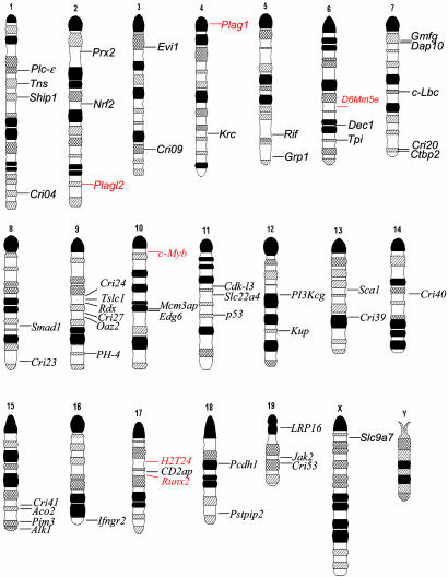
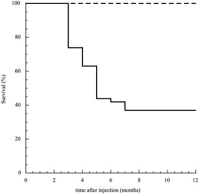
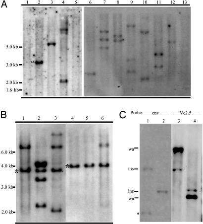
Acute myeloid leukemia subtype M4 with eosinophilia is associated with a chromosome 16 inversion that creates a fusion gene CBFB-MYH11. We have previously shown that CBFB-MYH11 is necessary but not sufficient for leukemogenesis. Here, we report the identification of genes that specifically cooperate with CBFB-MYH11 in leukemogenesis. Neonatal injection of Cbfb-MYH11 knock-in chimeric mice with retrovirus 4070A led to the development of acute myeloid leukemia in 2-5 months. Each leukemia sample contained one or a few viral insertions, suggesting that alteration of one gene could be sufficient to synergize with Cbfb-MYH11. The chromosomal position of 67 independent retroviral insertion sites (RISs) was determined, and 90% of the RISs mapped within 10 kb of a flanking gene. In total, 54 candidate genes were identified; six of them were common insertion sites (CISs). CIS genes included members of a zinc finger transcription factors family, Plag1 and Plagl2, with eight and two independent insertions, respectively. CIS genes also included Runx2, Myb, H2T24, and D6Mm5e. Comparison of the remaining 48 genes with single insertion sites with known leukemia-associated RISs indicated that 18 coincide with known RISs. To our knowledge, this retroviral genetic screen is the first to identify genes that cooperate with a fusion gene important for human myeloid leukemia.
Figures

Similar articles
-
Molecular characterization of 16p deletions associated with inversion 16 defines the critical fusion for leukemogenesis.Blood. 1995 Feb 1;85(3):772-9. Blood. 1995. PMID: 7833479
-
Plag1 and Plagl2 are oncogenes that induce acute myeloid leukemia in cooperation with Cbfb-MYH11.Blood. 2005 Apr 1;105(7):2900-7. doi: 10.1182/blood-2004-09-3630. Epub 2004 Dec 7. Blood. 2005. PMID: 15585652
-
Genomic acute myeloid leukemia-associated inv(16)(p13q22) breakpoints are tightly clustered.Oncogene. 1999 Jan 14;18(2):543-50. doi: 10.1038/sj.onc.1202321. Oncogene. 1999. PMID: 9927211
-
Function of the inv(16) fusion gene CBFB-MYH11.Curr Opin Hematol. 2001 Jul;8(4):201-5. doi: 10.1097/00062752-200107000-00004. Curr Opin Hematol. 2001. PMID: 11561156 Review.
-
Mechanism of leukemogenesis by the inv(16) chimeric gene CBFB/PEBP2B-MHY11.Oncogene. 2004 May 24;23(24):4297-307. doi: 10.1038/sj.onc.1207748. Oncogene. 2004. PMID: 15156186 Review.
Cited by
-
Runx1 is required for hematopoietic defects and leukemogenesis in Cbfb-MYH11 knock-in mice.Leukemia. 2015 Aug;29(8):1771-8. doi: 10.1038/leu.2015.58. Epub 2015 Mar 6. Leukemia. 2015. PMID: 25742748 Free PMC article.
-
Chemical biology. A small-molecule inhibitor of the aberrant transcription factor CBFβ-SMMHC delays leukemia in mice.Science. 2015 Feb 13;347(6223):779-84. doi: 10.1126/science.aaa0314. Science. 2015. PMID: 25678665 Free PMC article.
-
Contrasting requirements during disease evolution identify EZH2 as a therapeutic target in AML.J Exp Med. 2019 Apr 1;216(4):966-981. doi: 10.1084/jem.20181276. Epub 2019 Mar 19. J Exp Med. 2019. PMID: 30890554 Free PMC article.
-
The human pseudoxanthoma elasticum gene ABCC6 is transcriptionally regulated by PLAG family transcription factors.Hum Genet. 2008 Dec;124(5):451-63. doi: 10.1007/s00439-008-0570-0. Epub 2008 Oct 12. Hum Genet. 2008. PMID: 18850323
-
RUNX2 correlates with subtype-specific breast cancer in a human tissue microarray, and ectopic expression of Runx2 perturbs differentiation in the mouse mammary gland.Dis Model Mech. 2014 May;7(5):525-34. doi: 10.1242/dmm.015040. Epub 2014 Mar 13. Dis Model Mech. 2014. PMID: 24626992 Free PMC article.
References
-
- Look, A. T. (1997) Science 278, 1059-1064. - PubMed
-
- Liu, P., Tarle, S. A., Hajra, A., Claxton, D. F., Marlton, P., Freedman, M., Siciliano, M. J. & Collins, F. S. (1993) Science 261, 1041-1044. - PubMed
-
- Castilla, L. H., Wijmenga, C., Wang, Q., Stacy, T., Speck, N. A., Eckhaus, M., Marin-Padilla, M., Collins, F. S., Wynshaw-Boris, A. & Liu, P. P. (1996) Cell 87, 687-696. - PubMed
-
- Okuda T., van Duersen, J., Hiebert, S. W., Grosveld, G. & Downing, J. R. (1996) Cell 84, 321-330. - PubMed
-
- Wang, Q., Stacy, T., Miller, J. D., Lewis, A. F., Gu, T. L., Huang, X., Bushweller, J. H., Bories, J. C., Alt, F. W., Ryan, G., et al. (1996) Cell 87, 697-708. - PubMed
Publication types
MeSH terms
Substances
Associated data
- Actions
- Actions
- Actions
- Actions
- Actions
- Actions
- Actions
- Actions
- Actions
- Actions
- Actions
- Actions
- Actions
- Actions
- Actions
- Actions
- Actions
- Actions
- Actions
- Actions
- Actions
- Actions
- Actions
- Actions
- Actions
- Actions
- Actions
- Actions
- Actions
- Actions
- Actions
- Actions
- Actions
- Actions
- Actions
- Actions
- Actions
- Actions
- Actions
- Actions
- Actions
- Actions
- Actions
- Actions
- Actions
- Actions
- Actions
- Actions
- Actions
- Actions
- Actions
- Actions
- Actions
- Actions
- Actions
- Actions
- Actions
- Actions
- Actions
- Actions
- Actions
- Actions
- Actions
- Actions
- Actions
- Actions
- Actions
Grants and funding
LinkOut - more resources
Full Text Sources
Other Literature Sources
Molecular Biology Databases
