

Activation of human medial prefrontal cortex during autonomic responses to hypoglycemia
- PMID: 15026569
- PMCID: PMC395949
- DOI: 10.1073/pnas.0307048101
Activation of human medial prefrontal cortex during autonomic responses to hypoglycemia
Abstract
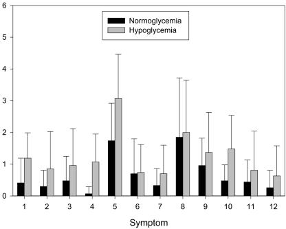
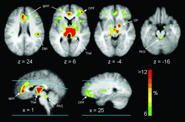
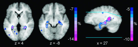
Studies in humans implicate the medial prefrontal cortex (MPFC) in complex cognitive and emotional states. We measured regional cerebral blood flow (CBF) four times each during euglycemia (5.2 +/- 0.2 mmol/liter) and hypoglycemia (3.0 +/- 0.3 mmol/liter) in nine normal human volunteers. Autonomic responses during hypoglycemia were manifested by increases in neurogenic symptoms, heart rate, and plasma levels of epinephrine, norepinephrine, and pancreatic polypeptide. Typical symptoms of hypoglycemia were mild, and none reflected evidence of cognitive or emotional stress. Quantitative CBF fell 6-8% in the cerebrum, brainstem, and cerebellum. Analysis of regional CBF differences identified neuronal activation during hypoglycemia in bilateral MPFC (areas 24 and 32) and bilateral thalamus. These results provide evidence that the MPFC participates in the autonomic responses to simple physiological stimuli in humans.
Figures
Comment in
-
The human cortex responds to an interoceptive challenge.Proc Natl Acad Sci U S A. 2004 Apr 27;101(17):6333-4. doi: 10.1073/pnas.0401510101. Epub 2004 Apr 19. Proc Natl Acad Sci U S A. 2004. PMID: 15096592 Free PMC article. No abstract available.
Similar articles
-
Effects of oral carbohydrate on autonomic nervous system counterregulatory responses during hyperinsulinemic hypoglycemia and euglycemia.Am J Physiol Endocrinol Metab. 2008 Sep;295(3):E618-25. doi: 10.1152/ajpendo.90470.2008. Epub 2008 Jul 8. Am J Physiol Endocrinol Metab. 2008. PMID: 18612042 Free PMC article. Clinical Trial.
-
Blood-to-brain glucose transport, cerebral glucose metabolism, and cerebral blood flow are not increased after hypoglycemia.Diabetes. 2001 Aug;50(8):1911-7. doi: 10.2337/diabetes.50.8.1911. Diabetes. 2001. PMID: 11473055
-
Hypoglycemic thalamic activation in type 1 diabetes is associated with preserved symptoms despite reduced epinephrine.J Cereb Blood Flow Metab. 2020 Apr;40(4):787-798. doi: 10.1177/0271678X19842680. Epub 2019 Apr 20. J Cereb Blood Flow Metab. 2020. PMID: 31006309 Free PMC article.
-
Functional imaging of brain responses to pain. A review and meta-analysis (2000).Neurophysiol Clin. 2000 Oct;30(5):263-88. doi: 10.1016/s0987-7053(00)00227-6. Neurophysiol Clin. 2000. PMID: 11126640 Review.
-
Functional anatomical abnormalities in limbic and prefrontal cortical structures in major depression.Prog Brain Res. 2000;126:413-31. doi: 10.1016/S0079-6123(00)26027-5. Prog Brain Res. 2000. PMID: 11105660 Review.
Cited by
-
Recurrent hypoglycemia is associated with loss of activation in rat brain cingulate cortex.Endocrinology. 2012 Apr;153(4):1908-14. doi: 10.1210/en.2011-1827. Epub 2012 Mar 6. Endocrinology. 2012. PMID: 22396449 Free PMC article.
-
Hypoglycemia-induced increases in thalamic cerebral blood flow are blunted in subjects with type 1 diabetes and hypoglycemia unawareness.J Cereb Blood Flow Metab. 2012 Nov;32(11):2084-90. doi: 10.1038/jcbfm.2012.117. Epub 2012 Aug 15. J Cereb Blood Flow Metab. 2012. PMID: 22892724 Free PMC article.
-
Autoregulation after ischaemic stroke.J Hypertens. 2009 Nov;27(11):2218-22. doi: 10.1097/HJH.0b013e328330a9a7. J Hypertens. 2009. PMID: 19644387 Free PMC article.
-
Differential functional brain network connectivity during visceral interoception as revealed by independent component analysis of fMRI TIME-series.Hum Brain Mapp. 2015 Nov;36(11):4438-68. doi: 10.1002/hbm.22929. Epub 2015 Aug 7. Hum Brain Mapp. 2015. PMID: 26249369 Free PMC article.
-
Neuroimaging of the periaqueductal gray: state of the field.Neuroimage. 2012 Mar;60(1):505-22. doi: 10.1016/j.neuroimage.2011.11.095. Epub 2011 Dec 14. Neuroimage. 2012. PMID: 22197740 Free PMC article. Review.
References
-
- Bush, G., Luu, P. & Posner, M. I. (2000) Trends Cogn. Sci. 4, 215-222. - PubMed
-
- Phan, K. L., Wager, T., Taylor, S. F. & Liberzon, I. (2002) Neuroimage 16, 331-348. - PubMed
-
- Bechara, A., Damasio, H., Tranel, D. & Damasio, A. R. (1997) Science 275, 1293-1295. - PubMed
-
- Critchley, H. D., Mathias, C. J. & Dolan, R. J. (2001) Neuron 29, 537-545. - PubMed
Publication types
MeSH terms
Grants and funding
LinkOut - more resources
Full Text Sources
Medical
