. 2004 Mar 10;23(5):1112-22.
doi: 10.1038/sj.emboj.7600125. Epub 2004 Feb 26.

SPAK kinase is a substrate and target of PKCtheta in T-cell receptor-induced AP-1 activation pathway

Affiliations

SPAK kinase is a substrate and target of PKCtheta in T-cell receptor-induced AP-1 activation pathway

Yingqiu Li et al. EMBO J. .

Abstract

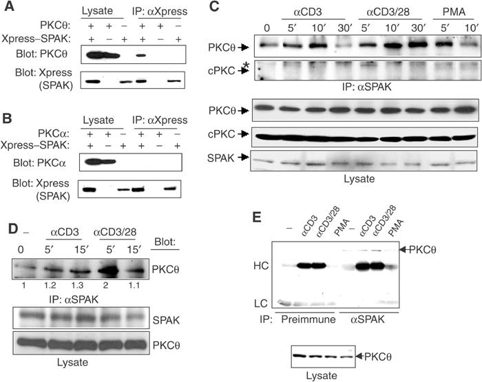
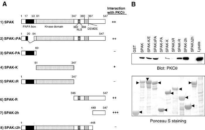
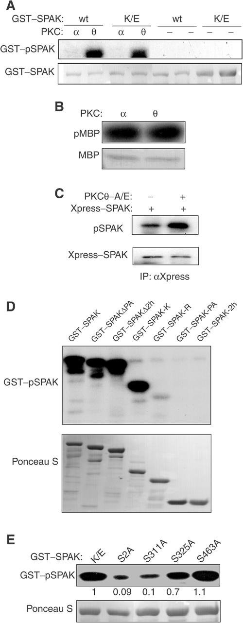
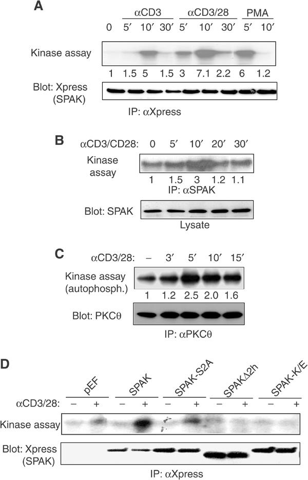
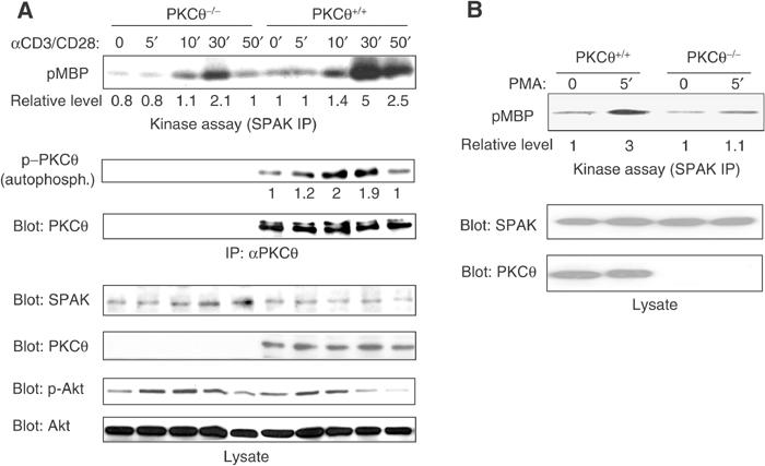
Protein kinase C-theta (PKCtheta) plays an important role in T-cell activation via stimulation of AP-1 and NF-kappaB. Here we report the isolation of SPAK, a Ste20-related upstream mitogen-activated protein kinase (MAPK), as a PKCtheta-interacting kinase. SPAK interacted with PKCtheta (but not with PKCalpha) via its 99 COOH-terminal residues. TCR/CD28 costimulation enhanced this association and stimulated the catalytic activity of SPAK. Recombinant SPAK was phosphorylated on Ser-311 in its kinase domain by PKCtheta, but not by PKCalpha. The magnitude and duration of TCR/CD28-induced endogenous SPAK activation were markedly impaired in PKCtheta-deficient T cells. Transfected SPAK synergized with constitutively active PKCtheta to activate AP-1, but not NF-kappaB. This synergistic activity, as well as the receptor-induced SPAK activation, required the PKCtheta-interacting region of SPAK, and Ser-311 mutation greatly reduced these activities of SPAK. Conversely, a SPAK-specific RNAi or a dominant-negative SPAK mutant inhibited PKCtheta- and TCR/CD28-induced AP-1, but not NF-kappaB, activation. These results define SPAK as a substrate and target of PKCtheta in a TCR/CD28-induced signaling pathway leading selectively to AP-1 (but not NF-kappaB) activation.


Figures

Figure 1
Figure 1
Interaction of PKCθ with SPAK. (A, B) EGY48(p8op–lacZ) yeast cells were cotransformed and assayed for growth and β-Gal activity following X-gal induction as described in Materials and methods. (C) SPAK was immunoprecipitated with an anti-Xpress mAb from 293T cells cotransfected with pEF–PKCθ plus pEF–Xpress–SPAK or empty vector. Immunoprecipitates (upper panel) or whole-cell lysates (two lower panels) were subjected to immunoblotting with anti-PKCθ or -Xpress antibodies. (D) Anti-PKCθ antibody or normal IgG was used to immunoprecipitate proteins from Jurkat-TAg cells transfected with Xpress–SPAK. The immunoprecipitates or lysates were probed with the indicated antibodies. (E) Jurkat T-cell lysates were incubated with empty glutathione–sepharose beads, GST-conjugated beads or GST–SPAK-conjugated beads, followed by SDS–PAGE analysis of bead-bound proteins and anti-PKCθ immunoblotting (upper panel). The lower panel shows the input of GST and GST–SPAK proteins determined by Coomassie blue staining.
Figure 2
Figure 2
Specific interaction of SPAK with PKCθ and its enhancement by CD3/CD28 costimulation. (A, B) Proteins immunoprecipitated with anti-Xpress antibody or cell lysates prepared from 293T cells transfected with the indicated combinations of wild-type PKCθ (A) or PKCα (B) with or without Xpress-tagged SPAK, were subjected to SDS–PAGE and immunoblotted with anti-PKC antibodies (upper panels), followed by reprobing with an anti-Xpress antibody (lower panels). (C) Endogenous SPAK was immunoprecipitated from unstimulated or stimulated Jurkat E6.1 cells. Immunoprecipitates (two upper panels) or lysates (three lower panels) were immunoblotted with anti-PKC or -SPAK antibodies. Arrows indicate the position of PKC or SPAK, and the asterisk corresponds to a nonspecific band recognized by the anti-cPKC antibody. (D) SPAK immunoprecipitates (8 × 10⁵ cell equivalents; upper panel) or cell lysates (1 × 10⁶ cell equivalents; two lower panels) from unstimulated or stimulated primary C57BL/6 mice were immunoblotted with the indicated antibodies. The numbers under the upper panel represent the fold increase in PKCθ–SPAK association compared to unstimulated cells (=1) as determined by densitometry. (E) Jurkat E6.1 cell stimulated as indicated for 10 (anti-CD3/CD28) or 5 (PMA) min were immunoprecipitated with preimmune serum or anti-SPAK serum, and immunoprecipitates (upper panel) or lysates (lower panel) were subjected to anti-PKCθ immunoblotting. The heavy chain (HC) and light chain (LC) of the immunoprecipitating antibody are indicated.
Figure 3
Figure 3
Mapping of the PKCθ-interacting domain of SPAK. (A) Schematic representation of the SPAK constructs expressed as GST fusion proteins. The proline/alanine-rich region (PAPA box), kinase domain, potential nuclear localization signal (NLS) and caspase cleavage site (DEMDE) are indicated. SPAK-2h corresponds to the cDNA insert from C51 identified in the yeast two-hybrid screen (Figure 1A). The numbers above the bars correspond to the amino-acid positions at the boundaries of each region. (B) Jurkat E6.1 cell lysates were incubated with the indicated GST or GST–SPAK proteins immobilized on glutathione–sepharose beads. Bead-bound proteins were analyzed as described in Materials and methods (upper panel). The lower panel shows the input of GST or GST–SPAK proteins (indicated by arrowheads) determined by Ponceau S staining.
Figure 4
Figure 4
Selective SPAK phosphorylation by PKCθ. (A) Recombinant wild-type (wt) or kinase-inactive (K/E) SPAK fusion proteins were used as substrates in in vitro kinase assays in the absence (−) or presence of recombinant PKCθ or PKCα enzymes. Phospho-SPAK (pSPAK) was detected by autoradiography (upper panel), and Ponceau S staining of the membrane shows the SPAK protein band (lower panel). (B) To normalize the activity of recombinant PKCθ or PKCα, a parallel kinase reaction was performed using MBP as a substrate. Phospho-MBP (pMBP) determined by autoradiography (upper panel) or the input MBP protein revealed by Ponceau S staining (lower panel) is shown. (C) 293T cells transfected with Xpress–SPAK in the absence or presence of constitutively active PKCθ were labeled with 32Pi and SPAK was immunoprecipitated with an anti-Xpress antibody. Immunoprecipitates were analyzed by SDS–PAGE and autoradiography (pSPAK; upper panel) or by anti-Xpress immunoblotting (lower panel). (D) PKCθ in vitro kinase assays were conducted using the indicated recombinant SPAK proteins as substrates, and analyzed as in (A). (E) GST fusion proteins of kinase-inactive SPAK (K/E) or SPAK-K/E with the indicated serine-to-alanine point mutations were used as substrates in PKCθ kinase reactions, and analyzed as in (D). The numbers refer to the phosphorylation level of each substrate relative to the phosphorylation of SPAK-K/E (=1).
Figure 5
Figure 5
Activation of SPAK by TCR/CD28 costimulation. (A) Transfected SPAK was immunoprecipitated from unstimulated (0′) or stimulated Jurkat-TAg cells cotransfected with a CD28 plasmid, and subjected to an in vitro kinase assay (upper panel). The membrane was reprobed with anti-Xpress antibody (lower panel). (B) Endogenous SPAK was immunoprecipitated from unstimulated or stimulated Jurkat E6.1 cells, and subjected to an in vitro kinase reaction (upper panel) as in (A). Lysates from the same cells were immunoblotted with an anti-SPAK antibody (lower panel). (C) Endogenous PKCθ was immunoprecipitated from unstimulated or stimulated Jurkat E6.1 cells, and subjected to an in vitro autophosphorylation reaction (upper panel) or anti-PKCθ immunoblotting (lower panel). (D) Jurkat-TAg cells were cotransfected with a CD28 plasmid plus empty vector (pEF) or the indicated pEF–SPAK constructs. After 20 h, the cells were left unstimulated (−) or stimulated (+) with crosslinked anti-CD3 plus anti-CD28 antibodies for 10 min. SPAK kinase activity (upper panel) or transfected SPAK expression (lower panel) was determined as in (A).
Figure 6
Figure 6
Deficient SPAK activation in PKCθ−/− T cells. Primary T cells purified from wild-type or PKCθ−/− mice were stimulated for the times as indicated. Endogenous SPAK was immunoprecipitated and assayed for kinase activity as in Figure 5. Lysates from the same cells were immunoblotted with anti-SPAK or -PKCθ antibodies. The numbers under the panels represent the fold increase in SPAK activity compared to unstimulated cells (=1) as determined by densitometry. In (A), the time course of PKCθ in vitro autophosphorylation (second panel from top) or its expression (third panel from top), as well as the activation and expression of Akt in the same cells (two lower panels), were also determined. (B) This experiment is representative of another, similar experiment.
Figure 7
Figure 7
SPAK is required for PKCθ-A/E-induced AP-1, but not NF-κB, activation. (A, B) Jurkat E6.1 cells were transfected with empty vector (left lane) or c-Myc–PKCθ-A/E (four right lanes) plus the indicated pSuper, pSuper–RNAi-1 or pSuper–RNAi-2 vectors, β-Gal reporter plasmid and AP-1 (B; upper panel) or NF-κB (B; lower panel) reporter genes. (A) Expression levels of SPAK mRNA (measured by real-time RT–PCR and expressed as arbitrary units), and SPAK or PKCθ protein (measured by immunoblotting with anti-SPAK or anti-c-Myc antibodies, respectively). (B) Normalized AP-1 (upper panel) or NF-κB (lower panel) activities in the same cells. (C) Jurkat-TAg cells were cotransfected with the indicated combinations of empty vector, c-Myc–PKCθ-A/E and/or different SPAK constructs together with AP-1–Luc and β-Gal reporter genes. Cell extracts were prepared 24 h later, and normalized AP-1 activity (upper panel) or protein expression levels (two lower panels) were determined. Mean values of duplicate determination±s.e. are shown. Similar results were obtained in two additional experiments.

Similar articles

Cited by

References

    1. Altman A, Isakov N, Baier G (2000) PKCθ: a new essential superstar on the T cell stage. Immunol Today 21: 567–573 - PubMed
    1. Arendt CW, Albrecht B, Soos TJ, Littman DR (2002) Protein kinase C-θ: signaling from the center of the T-cell synapse. Curr Opin Immunol 14: 323–330 - PubMed
    1. Avraham A, Jung S, Samuels Y, Seger R, Nen-Neriah Y (1998) Co-stimulation-dependent activation of a JNK-kinase in T lymphocytes. Eur J Immunol 28: 2320–2330 - PubMed
    1. Baier-Bitterlich G, Überall F, Bauer B, Fresser F, Wachter H, Grünicke H, Utermann G, Altman A, Baier G (1996) PKCθ isoenzyme selective stimulation of the transcription factor complex AP-1 in T lymphocytes. Mol Cell Biol 16: 1842–1850 - PMC - PubMed
    1. Bi K, Tanaka Y, Coudronniere N, Hong S, Sugie K, van Stipdonk MJB, Altman A (2001) Antigen-induced translocation of PKC-θ to membrane rafts is required for T cell activation. Nat Immunol 2: 556–563 - PubMed

Publication types

MeSH terms