. 2004 Mar;164(3):1099-114.
doi: 10.1016/S0002-9440(10)63197-5.

Morpho-regulation of ectodermal organs: integument pathology and phenotypic variations in K14-Noggin engineered mice through modulation of bone morphogenic protein pathway

Affiliations

Morpho-regulation of ectodermal organs: integument pathology and phenotypic variations in K14-Noggin engineered mice through modulation of bone morphogenic protein pathway

Maksim Plikus et al. Am J Pathol. 2004 Mar.

Abstract

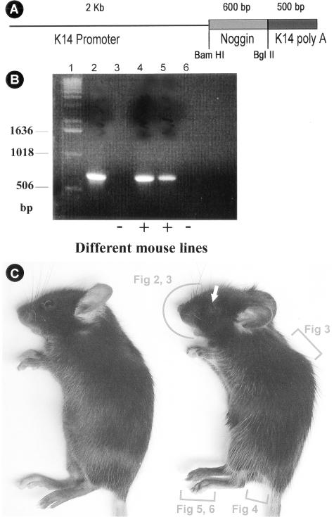
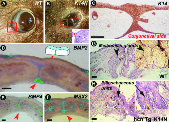
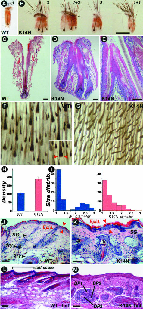
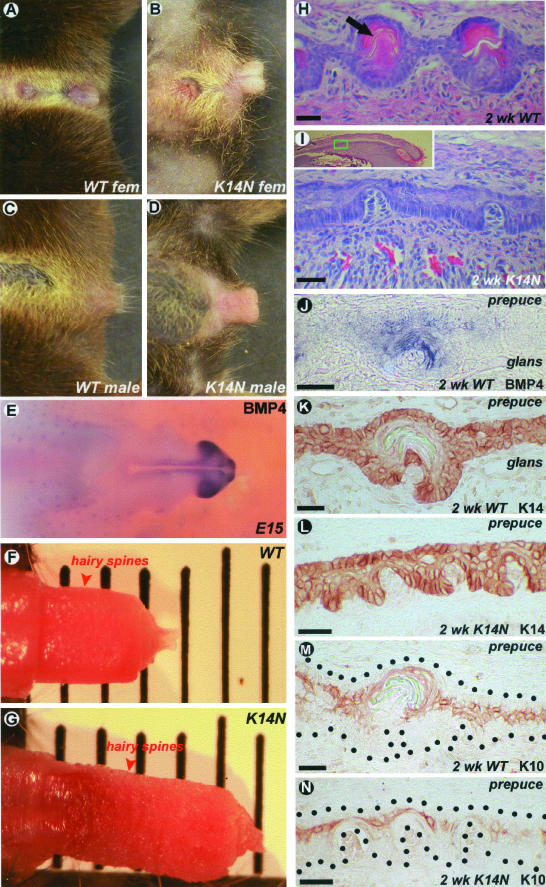
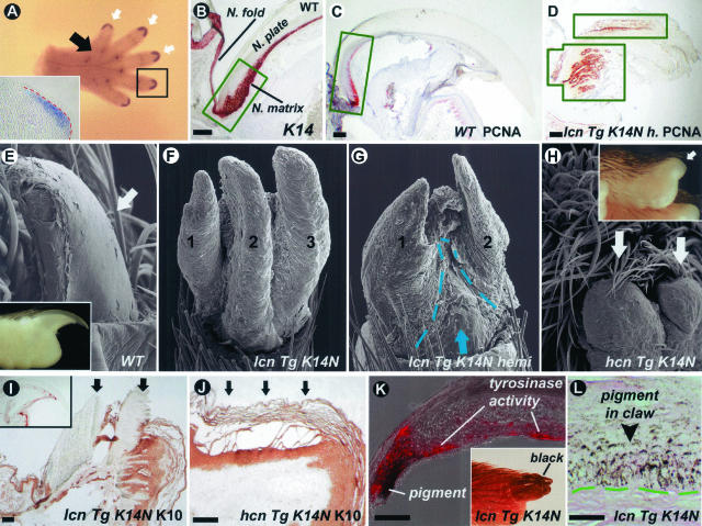
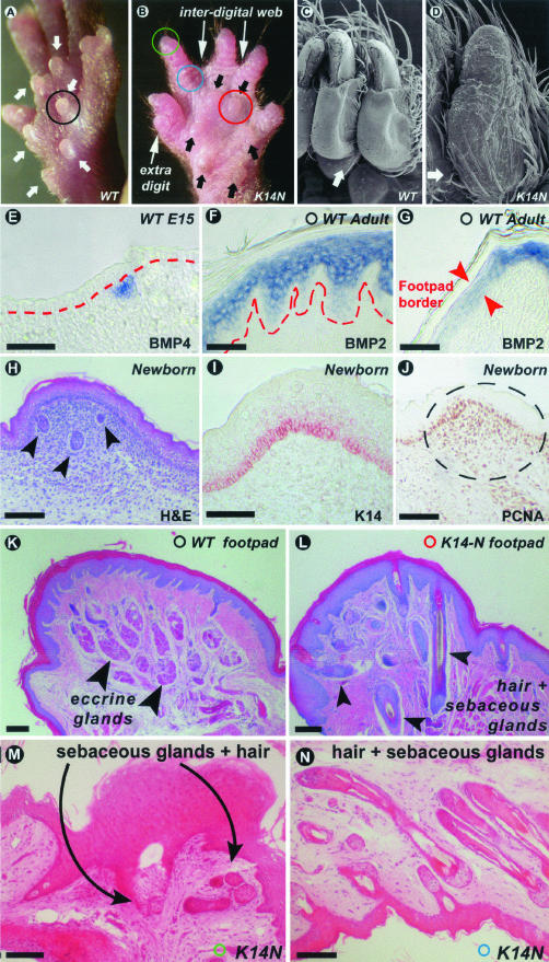
Ectodermal organs are composed of keratinocytes organized in different ways during induction, morphogenesis, differentiation, and regenerative stages. We hypothesize that an imbalance of fundamental signaling pathways should affect multiple ectodermal organs in a spatio-temporal-dependent manner. We produced a K14-Noggin transgenic mouse to modulate bone morphogenic protein (BMP) activity and test the extent of this hypothesis. We observed thickened skin epidermis, increased hair density, altered hair types, faster anagen re-entry, and formation of compound vibrissa follicles. The eyelid opening was smaller and ectopic cilia formed at the expense of Meibomian glands. In the distal limb, there were agenesis and hyperpigmentation of claws, interdigital webbing, reduced footpads, and trans-differentiation of sweat glands into hairs. The size of external genitalia increased in both sexes, but they remained fertile. We conclude that modulation of BMP activity can affect the number of ectodermal organs by acting during induction stages, influence the size and shape by acting during morphogenesis stages, change phenotypes by acting during differentiation stages, and facilitate new growth by acting during regeneration stages. Therefore during organogenesis, BMP antagonists can produce a spectrum of phenotypes in a stage-dependent manner by adjusting the level of BMP activity. The distinction between phenotypic variations and pathological changes is discussed.


Figures

Figure 1
Figure 1
Production of K14-Noggin mouse. A: K14 Noggin construct used to generate transgenic mouse. The size of insert used and restriction enzyme are indicated. B: Genotyping of K14-Noggin mutant mouse. Products of PCR reaction using specific primers (see Materials and Methods for primers used). Lane 1, DNA standard; lane 2, positive control—K14-Noggin founder mouse; lanes 3 and 6, wild-type mice; lanes 4 and 5, K14-Noggin-positive mice. C: Appearance of control C57BL/6J (left) and mutant K14-Noggin 2.5-month-old mice. Note the obvious hypertrichosis of the K14-Noggin mouse. The eye opening is small (white arrow). Digits are not distinctly separated from each other. Regions to be studied further in each figure are marked by red brackets.
Figure 2
Figure 2
Pathology in eyelid, an epithelial appendage to protect the eye. A: Normal size eye slits of 4-month-old control C57BL/6J mouse. B: Narrow eye slits in 4-month-old K14-Noggin mouse. Eyelids remain partially fused. Eyeballs are of the same size (not shown). Arrowheads point at inward-growing cilia. C–F: Eyelid suture of newborn control mouse. C: Strong K14 immunostaining is seen in the epidermal side, suture, and conjunctival side of the eyelid. D–F: Whole mount in situ hybridization of 1-mm-thick eyelid stripe. Green color is computer pseudo-coloring. BMP2 transcripts are expressed predominantly on the conjunctivae side whereas BMP4 is distributed more evenly throughout the eyelid suture. Msx2 transcript distribution is similar to BMP4. G and H: Corner of eyelids (commissure, corresponding to the areas outlined with red on A and B) of adult mice. Formation of ectopic pillosebaceous units at the expense of Meibomian glands in high copy number TG K14-Noggin mice (hcn TG K14-Noggin mice). Scale bars, 100 μm.
Figure 3
Figure 3
Pathology in vibrissae and pelage hairs. A and B: Control and K14-Noggin vibrissae HFs. K14-Noggin mice have compound follicles that share one orifice and one capsule. The number in B indicates the number of vibrissa filaments that share the same orifice. For example, 1 + 2 means one normal follicle with one filament from one orifice plus one compound follicle with two filaments growing from one orifice. C–E: H&E stain of control (C) and K14-Noggin (D, E) vibrissae HFs. K14-Noggin follicles share part of the same outer root sheath, open into the same canal but have a distinct dermal papillae and matrix, and produce a separate inner root sheath and fiber. F and G: View of inverted skin from the dorsal trunk region of the control and K14-Noggin mice. In the control mouse, all HFs are in anagen. Distinct primary (big) and secondary (small) HFs can be identified. In the K14-Noggin mouse, the density of HFs is increased and the difference between primary and secondary follicles is not obvious. H: Density of HFs per 3 mm2 in control (blue) and K14-Noggin (red) mice. Density is increased by ∼80% in the K14-Noggin mouse in comparison with the control mouse. I: Relative size distribution of HFs in control (blue) and K14-Noggin (red) mice (size corresponds to the diameter of the hair bulb; see inset in Figure 4F). In the control mouse, HFs clearly fractionate into two distinct groups: those with smaller size (primarily secondary HFs) and those with larger size (primary HFs). In the K14-Noggin mouse, there is no clear fractionation of HFs. The majority of HFs are of intermediate size. This could be because of increased proportion of secondary awl and auchene hairs (see Results). J and K: H&E staining of skin sections from the back of the control and K14-Noggin 2-week-old mice. Normal secondary and primary (bottom) HFs are seen in the control mouse. In some regions of the K14-Noggin mouse, there are enlarged HFs and hypertrophic sebaceous glands. Some hair fibers point to wrong directions. Spacing between follicles is reduced. Skin epidermis is thickened. L and M: H&E staining of longitudinal sections of the tail from the control and K14-Noggin mice. Different sizes of hypertrophic HFs pointing in different directions are seen in the K14-Noggin mouse. The total number of follicles has increased. Some follicles are dystrophic and appear to have multiple dermal papillae. The epidermal and dermal layers appear thicker. Scale bars: 1 mm (B); 100 μm (C–E and J–M).
Figure 4
Figure 4
Pathology in external genitalia. A and B: External genitalia of the control and K14-Noggin female mice. C and D: External genitalia of the control and K14-Noggin male mice. E: BMP4 expression pattern in the external genitalia of E15 mouse embryo. Very strong expression in the glans penis. F and G: Side-to-side comparison of the control and K14-Noggin male external genital. H and I: Hairy spines of the 2-week-old control and K14-Noggin mice. Hairy spines in the K14-Noggin mouse are not completely developed. J: BMP4 expression in the developing hairy spine of the 2-week-old control mouse. K–N: K14 and K10 keratin expression patterns in the preputial lamella and developing hairy spines on the border of the glans and prepuce from a 2-week-old wild-type and K14-Noggin mouse. Note weak K10 expression in the immature hairy spines of the K14-Noggin mouse (N). Scale bars 25 μm.
Figure 5
Figure 5
Pathology in claws. A: E15 wild-type mouse paw in situ with BMP4 probe. White arrows point to expression sites at the tips of the digits. Black arrow points to the group of expression sites corresponding to footpads. BMP4 expression in the mesenchyme of the developing claw is shown on the inset. B: Strong K14 expression in epithelial cells of proliferating claw matrix (green box) and beyond it. C and D: Expansion of proliferating area in the claw of low-TG copy number K14-Noggin mice (lcn TG K14-Noggin mice). PCNA-positive cells are limited to the claw matrix (green box) in wild-type mice, but extend all of the way to the tip of the digit in K14-Noggin mice (green boxes). There are multiple proliferating centers within one claw of the K14-Noggin mice. E to H: Progressive abnormalities in the claw of the K14-Noggin mice. Compared to the control claw from C57BL/6J mouse (E), claws in the low-TG copy number K14-Noggin mice split into separate plates (F) and are partially substituted by epidermis (G, demarcated with a green dotted line). No claw is identifiable in the high-TG copy number K14-Noggin mice (H, white arrows). I and J: K10 keratin expression in low (I) and high (J) TG copy number K14-Noggin mice. In low-TG copy number mice claws are K10-negative (arrows) despite the apparent macroscopic and microscopic abnormalities (see above). In the high-TG copy number mouse the epidermis that substitutes the claw expresses K10 (arrows). In the control mouse (K, inset) the claw is K10-negative. K and L: Pigmentation of the claw in low-TG copy number K14-Noggin mice. Abundant pigment deposits in the claw epithelium (L). Tyrosinase activities (red color, K) associated with pigment production are seen. Scale bars: 100 μm (B–D and I–K); 25 μm (L).
Figure 6
Figure 6
Pathology of the ventral side of the paw. A and B: Normal and hypoplastic foot pads from the wild-type and K14-Noggin mouse, respectively (white and black arrows). Presence of additional digit and interdigital webbing are marked in K14-Noggin mouse paw. C and D: Scanning EM normal glabrous skin and skin with ectopic HFs on the ventral side of the digits from wild-type and K14-Noggin mice, respectively. E: BMP4 expression in the mesenchyme of the developing footpad in E15 control mouse embryo. F and G: Suprabasal expression of BMP2 in the epidermis of the adult footpad (F) and on the border of the footpad (G). Note the sharp decline of BMP2 expression on the border of the footpad. H to J: Growing footpad of the newborn mouse. Active proliferation is seen in the mesenchymal condensation of the footpad as judged by PCNA staining (J). Eccrine glands are forming at this time (marked by arrowheads at H). K14 is expressed in the basal layer of the footpad epidermis (I). K and L: H&E of the footpad from control and K14-Noggin mice. Typical afollicular epidermis and eccrine glands are seen in the control mouse (K). In the K14-Noggin mouse, multiple HFs replace the eccrine glands. M and N: H&E of the skin on the digit from K14-Noggin mouse. M: Tip of the digit. Claw is absent. It is substituted by a hyperplastic patch of epidermis with sebaceous glands and HFs. N: Ventral side of the distal digit. Many HFs with sebaceous glands have formed, instead of sweat glands and just a few HFs in the normal. Scale bars, 100 μm.
Figure 7
Figure 7
Summary of multiple epidermal organ defects caused by disruption of BMP pathway in the skin. A: Defective regions are shown in gray shades. B: Phenotypes are summarized. They are grouped based on the developmental stages when defects occur.

Similar articles

Cited by

References

    1. Chuong C-M, editor. Austin: Landes Bioscience,; Molecular Basis of Epithelial Appendage Morphogenesis. 1998
    1. Wisniewski SA, Kobielak A, Trzeciak WH, Kobielak K. Recent advances in understanding of the molecular basis of anhidrotic ectodermal dysplasia: discovery of a ligand, ectodysplasin A and its two receptors. J Appl Genet. 2002;43:97–107. - PubMed
    1. Headon DJ, Overbeek PA. Involvement of a novel Tnf receptor homologue in HF induction. Nat Genet. 1999;22:370–374. - PubMed
    1. Brunner HG, Hamel BC, Bokhoven HVH. P63 gene mutations and human developmental syndromes. Am J Med Genet. 2002;112:284–290. - PubMed
    1. Chuong C-M, Chodankar R, Widelitz RB, Jiang TX. Evo-devo of feathers and scales: building complex epithelial appendages. Curr Opin Dev Genet. 2000;10:449–456. - PMC - PubMed

Publication types

LinkOut - more resources