. 2004 Mar 1;164(5):677-88.
doi: 10.1083/jcb.200308012. Epub 2004 Feb 23.

Mmm2p, a mitochondrial outer membrane protein required for yeast mitochondrial shape and maintenance of mtDNA nucleoids

Affiliations

Mmm2p, a mitochondrial outer membrane protein required for yeast mitochondrial shape and maintenance of mtDNA nucleoids

Matthew J Youngman et al. J Cell Biol. .

Abstract

The mitochondrial outer membrane protein, Mmm1p, is required for normal mitochondrial shape in yeast. To identify new morphology proteins, we isolated mutations incompatible with the mmm1-1 mutant. One of these mutants, mmm2-1, is defective in a novel outer membrane protein. Lack of Mmm2p causes a defect in mitochondrial shape and loss of mitochondrial DNA (mtDNA) nucleoids. Like the Mmm1 protein (Aiken Hobbs, A.E., M. Srinivasan, J.M. McCaffery, and R.E. Jensen. 2001. J. Cell Biol. 152:401-410.), Mmm2p is located in dot-like particles on the mitochondrial surface, many of which are adjacent to mtDNA nucleoids. While some of the Mmm2p-containing spots colocalize with those containing Mmm1p, at least some of Mmm2p is separate from Mmm1p. Moreover, while Mmm2p and Mmm1p both appear to be part of large complexes, we find that Mmm2p and Mmm1p do not stably interact and appear to be members of two different structures. We speculate that Mmm2p and Mmm1p are components of independent machinery, whose dynamic interactions are required to maintain mitochondrial shape and mtDNA structure.


Figures
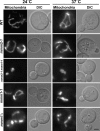
Figure 1.
Figure 1.
The combination of mmm2-1 and mmm1-1 disrupts mitochondrial shape. Wild-type strain YPH250, mmm1-1 strain YSB105, mmm1-1 mmm2-1 strain SB9, mmm1-1 mmm2-1 strain SB9 carrying the MMM1 plasmid, pSB201, and mmm1Δstrain YSB120 were grown at 24°C or 37°C, stained with Mitofluor 589 (Mitochondria) and then examined by fluorescence and DIC microscopy. Representative images are shown. Bar, 3 μm.
Figure 2.
Figure 2.
Mmm2p is required for mitochondrial shape and maintenance of mtDNA nucleoids. (A) Mitochondria and mtDNA in mmm2Δ, mmm1Δ, and mmm1Δ mmm2Δ cells. Wild-type strain MY4, mmm1Δ strain MY1, mmm2Δ strain MY2, and mmm1Δ mmm2Δ strain MY3 were grown in glycerol/ethanol medium, stained with Mitofluor 589 (Mitochondria) or DAPI (mtDNA), and then examined by DIC and fluorescence microscopy. Representative images of cells are shown. N, staining of the nucleus by DAPI. Bar, 3 μm. (B) Classification of mmm2Δ phenotypes. mmm2Δ strain MY2 was stained with DiOC6 and then examined by DIC and fluorescence microscopy. Cells were sorted into four groups based on their mitochondrial appearance. Class I, distorted and sausage-shaped mitochondria; Class II, spherical-shaped mitochondria; Class III, sheet-like mitochondria; Class IV, a mixture of distorted and spherical-shaped mitochondria. A representative from each group is shown, and the percentage of cells (out of 107 total cells examined) in each category is indicated to the right of each image. Bar, 3 μm.
Figure 3.
Figure 3.
Mmm2p is a mitochondrial outer membrane protein. (A) Mmm2p-HA is a mitochondrial protein. Strain RJ892, expressing Mmm2p-HA, was grown and homogenized (H) and then separated into a mitochondrial pellet (M) and a post-mitochondrial supernatant (PMS) by centrifugation. Aliquots of the fractions were analyzed by SDS-PAGE and Western blotting, using antibodies to the HA epitope (Mmm2p-HA), or antiserum to the mitochondrial protein, Tim23p, or to the cytosolic hexokinase protein. (B) Mmm2p is an integral membrane protein. Mitochondria from cells expressing Mmm2p-HA were incubated with buffer, 1.5 M sodium chloride, or 0.1 M sodium carbonate. After centrifugation, membrane pellets were analyzed by SDS-PAGE and Western blotting with HA antibodies (Mmm2p-HA), or antiserum to Tim23p or the β subunit of the F1-ATPase (F1β). (C) The COOH terminus of Mmm2p faces the cytosol. Mmm2p-HA mitochondria were incubated in the presence or absence of 100 μg/ml trypsin. In one aliquot of mitochondria, the outer membrane was disrupted by osmotic shock (OS). After centrifugation, mitochondrial proteins were analyzed by Western blotting using HA antibodies (Mmm2p-HA), or antiserum to Tim23p or cytochrome b2 (Cyt. b2). (D) Mmm2p is located in the outer membrane. Mmm2p-HA–containing mitochondria were sonicated, and membrane vesicles were loaded onto a sucrose density gradient. Fractions were collected and analyzed by Western blotting, using HA antibodies (Mmm2p-HA) or antiserum to the OM45 and F1β proteins. Fraction 1 corresponds to the top of the gradient.
Figure 4.
Figure 4.
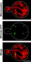
Mmm2p-GFP localizes to small spots along mitochondrial tubules. mmm2Δ strain RJ713, expressing the Mmm2p-GFP fusion protein from pAAH13, was stained with Mitotracker Red and examined by deconvolution microscopy. A total of 15 images were taken at 0.2-μm intervals in the z-axis, deconvolved, and then compressed into a single image. Mitotracker Red (Mitochondria), Mmm2p-GFP, and merged images with both Mmm2p-GFP and Mitotracker Red are shown. Bar, 3 μm.
Figure 5.
Figure 5.
Mmm2p is adjacent to a subset of mtDNA nucleoids. mmm2Δ strain RJ713, expressing the Mmm2p-GFP fusion protein from pAAH13, was stained with DAPI and examined by deconvolution microscopy. Each image represents a flattened, deconvolved composite of 15 images. (A) DIC, (B) Mmm2p-GFP, (C) DAPI, (D) merged images showing both Mmm2p-GFP and DAPI. DAPI staining of the yeast nucleus (N) is indicated. Bar, 3 μm.
Figure 6.
Figure 6.
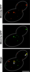
Mmm2p colocalizes with a fraction of Mmm1p. Strain AAH4, which expresses Mmm1p-GFP from the chromosomal MMM1::GFP gene and Mmm2p-RFP from pMY3, was examined by deconvolution microscopy. A single image containing 15 deconvolved and compressed images is shown. Mmm2p-RFP, Mmm1p-GFP, and merged images with both Mmm1p-GFP and Mmm2p-RFP are shown. Arrows indicate an example of colocalization of Mmm2p-RFP and Mmm1p-GFP. The cell outline, as seen by phase contrast, is shown by white lines. Bar, 3 μm.
Figure 7.
Figure 7.
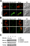
Mmm1p is necessary for the punctate distribution of Mmm2p, while Mmm2p is required for normal Mmm1p levels. (A) Wild-type strain MY4 and mmm1Δ strain MY5, each expressing Mmm2p-GFP from chromosomal MMM2::GFP, were stained with Mitofluor 589 and examined by DIC and fluorescence microscopy. Mitofluor 589 (Mitochondria), Mmm2p-GFP, merged images with both Mmm2p-GFP and Mitofluor 589, and DIC images are shown. Bar, 3 μm. (B) Wild-type strain SS12 and mmm2Δ strain AAH4, each carrying a chromosomal version of MMM1::GFP, were stained with Mitofluor 589 and examined by DIC and fluorescence microscopy. Mitofluor 589 (Mitochondria), Mmm1p-GFP, merged images with both Mmm1p-GFP and Mitofluor 589, and DIC images are shown. Bar, 3 μm. (C) Mitochondria (100 μg) were isolated from isogenic wild-type, mmm1Δ, mmm2Δ, and mmm1Δ mmm2Δ cells and Western blotted using antibodies to Mmm2p, Mmm1p, Tom70p, an outer membrane protein, and Tim23p, an inner membrane protein.
Figure 8.
Figure 8.
Mmm2p and Mmm1p are in large, but separate, membrane complexes. (A) Gel filtration analysis of Mmm2p and Mmm1p. Mitochondria were solubilized in digitonin-containing buffer and loaded onto a gel filtration column. Aliquots from each fraction were analyzed by Western blotting using antiserum to Mmm2p (open squares) or affinity-purified antibodies to Mmm1p (filled circles). After quantitation by densitometry, protein amounts are shown as a percent of total Mmm1p or Mmm2p proteins in the extract. (B) Analysis of the same column fractions as in A, but using antiserum to Tom40p (filled circles) or Tim23p (open squares). Molecular masses (shown above the graphs) were estimated by running protein calibration standards under identical conditions. (C) Mmm2p and Mmm1p do not coimmunoprecipitate. Mitochondria isolated from strain RJ1719, expressing Mmm1p-HA and Mmm2p-myc fusion proteins, or from strain RJ1302, expressing HA-Fzo1p and myc-Ugo1p fusion proteins, were solubilized in Triton X-100 and subjected to immunoprecipitation using antibodies to the myc epitope. Equal aliquots from the pellet (P) and supernatant (S) fraction were Western blotted using antibodies to either the HA or myc epitopes.

Similar articles

Cited by

References

    1. Adams, A., D. Gottschling, C. Kaiser, and T. Stearns. 1997. Methods in yeast genetics. Cold Spring Harbor Laboratory, Cold Spring Harbor, NY. 177 pp.
    1. Aiken Hobbs, A.E., M. Srinivasan, J.M. McCaffery, and R.E. Jensen. 2001. Mmm1p, a mitochondrial outer membrane protein, is connected to mitochondrial DNA (mtDNA) nucleoids and required for mtDNA stability. J. Cell Biol. 152:401–410. - PMC - PubMed
    1. Attardi, G., and G. Schatz. 1988. Biogenesis of mitochondria. Annu. Rev. Cell Biol. 4:289–333. - PubMed
    1. Azpiroz, R., and R.A. Butow. 1993. Patterns of mitochondrial sorting in yeast zygotes. Mol. Biol. Cell. 4:21–36. - PMC - PubMed
    1. Basson, M.E., R.L. Moore, J. O'Rear, and J. Rine. 1987. Identifying mutations in duplicated functions in Saccharomyces cerevisiae: recessive mutations in HMG-CoA reductase genes. Genetics. 117:645–655. - PMC - PubMed

Publication types

MeSH terms