Comparative Study
. 2004 Mar;78(5):2327-35.
doi: 10.1128/jvi.78.5.2327-2335.2004.

Cell cycle requirements for transduction by foamy virus vectors compared to those of oncovirus and lentivirus vectors

Affiliations
Comparative Study

Cell cycle requirements for transduction by foamy virus vectors compared to those of oncovirus and lentivirus vectors

Grant Trobridge et al. J Virol. 2004 Mar.

Abstract

Retroviral vectors based on foamy viruses (FV) are efficient gene delivery vehicles for therapeutic and research applications. While previous studies have shown that FV vectors transduce quiescent cell cultures more efficiently than oncoviral vectors, their specific cell cycle requirements have not been determined. Here we compare the transduction frequencies of FV vectors with those of onco- and lentiviral vectors in nondividing and dividing normal human fibroblasts by several methods. FV vectors transduced serum-deprived fibroblast cultures more efficiently than oncoretroviral vectors and at rates comparable to those of lentiviral vectors. However, in these cultures FV vectors only transduced a subpopulation of proliferating cells, as determined by bromodeoxyuridine staining for DNA synthesis. In contrast to lentiviral vectors, FV vectors were unable to transduce human fibroblasts arrested by aphidicolin (G(1)/S phase) or gamma-irradiation (G(2) phase), and a partial cell cycle that included mitosis but not DNA synthesis was required. We could not determine if mitosis facilitated nuclear entry of FV vectors, since cell-free vector preparations contained long terminal repeat circles, precluding their use as nuclear markers. In contrast to oncoviral vectors, both FV and lentiviral vectors efficiently transduced G(0) fibroblasts that were later stimulated to divide. In the case of FV vectors, this was due to the persistence of a stable transduction intermediate in quiescent cells. Our findings support the use of FV vectors as a safe and effective alternative to lentiviral vectors for ex vivo transduction of stem cells that are quiescent during culture but divide following transplantation.


Figures

FIG. 1.
FIG. 1.
Retroviral vectors used in this study. All vectors are shown in their integrated proviral form with the locations of LTRs, packaging signals (ψ), internal MLV LTR promoters (M), AP transgenes, cPPTs, and the Rev response element (RRE) indicated. Arrows indicate the start sites of AP transcription. The MLV vector LAPSN contains a downstream simian virus 40 promoter (S) and neomycin resistance gene (Neo) not relevant to the experiments described here.
FIG. 2.
FIG. 2.
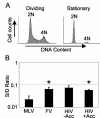
Transduction of serum-deprived cultures. Normal human fibroblasts were grown to confluence and serum starved for 4 days (Stationary) or were serum-starved for 3 days and then plated in medium with 10% serum 16 h prior to the addition of vector (Dividing). (A) Flow cytometry analysis of the DNA content by propidium iodide staining. (B) The number of AP+ foci in stationary cultures divided by the number of AP+ foci in dividing cultures (S/D ratio) is reported for three independent experiments with standard errors (error bars). HIV vector preparations were prepared with (HIV+Acc) and without (HIV−Acc) the accessory genes vif, vpr, vpu, and nef. An asterisk indicates significant differences (P < 0.05) compared to the MLV S/D ratio using Student's t test.
FIG. 3.
FIG. 3.
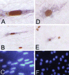
Transduction by FV vectors occurs only in cells that have undergone DNA synthesis. (A and D) High-power views (magnification, ×400) of a transduced human fibroblast and a rat 208F cell, respectively, after staining for BrdU incorporation as a marker of S phase (orange-brown nucleus) and for AP expression as a marker of transduction (purple cytoplasm). (B and E) Low-power views (magnification, ×100) of panels A and B, respectively. (C and F) Same fields as those in panels B and E, respectively, under UV illumination to detect all DAPI-stained nuclei.
FIG. 4.
FIG. 4.
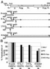
Transduction by FV vectors requires passage through mitosis. (A) Aphidicolin-arrested human fibroblasts were exposed to retroviral vectors and either allowed to progress through the cell cycle following removal of aphidicolin (S-cycle), released from aphidicolin arrest and irradiated 3 h later (S-G2 γ), irradiated 1 h prior to infection and then released from aphidicolin arrest (γ S-G2), or left in the presence of aphidicolin (Arrested). Solid lines delineate phases of the cell cycle traversed by the cells during each treatment, and dotted lines delineate blockage of these phases during the transduction period. Aphidicolin treatment is depicted by a white bar. The durations of cell cycle phases during the experiment are indicated at the top. (B) The percent transduction relative to that obtained in the S-cycle treatment is shown for each treatment in panel A. DNA synthesis was monitored by BrdU staining, and the percentage of cells that underwent DNA synthesis for each treatment is given in brackets. Progression (+M) or lack of progression (−M) through mitosis is indicated.
FIG. 5.
FIG. 5.
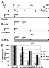
A partial cell cycle that includes mitosis but lacks S phase is sufficient for transduction by FV vectors. (A) Human fibroblasts were synchronized at the G1/S border and exposed to retroviral vectors at the beginning of S phase (S-cycle and S-G2-M-G1) or 9 h later (M-cycle and G2-M-G1). Aphidicolin was added 9 h after the initial withdrawal to prevent additional DNA synthesis in the S-G2-M-G1 and G2-M-G1 treatments. Solid lines delineate phases of the cell cycle traversed by the cells, and dotted lines delineate blockage of these phases during the transduction period. Aphidicolin treatment is depicted by a white bar. The durations of cell cycle phases during the experiment are indicated at the top. (B) The percent transduction relative to that obtained in the S-cycle treatment is shown for each treatment in panel A.
FIG. 6.
FIG. 6.
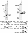
Southern blot analysis of LTR circles. Purified DNA from FV vector and wild-type FV stocks was digested with StuI, SwaI, EagI, PacI, and/or NheI as indicated and analyzed by Southern blotting. The vector preparation DNA was also digested with DpnI to remove transfected plasmid DNA. Diagrams of the predicted LTR-containing fragments (not to scale) are shown below the corresponding Southern blots with the predicted sizes of linear, one-LTR circle (1-LTRc), and two-LTR circle (2-LTRc) DNA fragments. The position of the FV probe used is shown with a black bar.
FIG. 7.
FIG. 7.
Transduction after release from serum deprivation. Retroviral vectors were added to confluent, serum-deprived human fibroblasts, and 2, 4, 6, or 10 days later (days before release) the cells were stimulated to divide by replating in larger wells and increasing the concentration in serum from 0.5 to 10%. Forty-eight hours after replating, AP staining was performed; the number of AP+ foci in released cultures divided by the number of AP foci in dividing cultures (R/D ratio) is reported for three independent experiments with standard errors (error bars).

Similar articles

Cited by

References

    1. Alexander, I. E., D. W. Russell, and A. D. Miller. 1997. Transfer of contaminants in adeno-associated virus vector stocks can mimic transduction and lead to artifactual results. Hum. Gene Ther. 8:1911-1920. - PubMed
    1. Bieniasz, P. D., O. Erlwein, A. Aguzzi, A. Rethwilm, and M. O. McClure. 1997. Gene transfer using replication-defective human foamy virus vectors. Virology 235:65-72. - PubMed
    1. Bieniasz, P. D., R. A. Weiss, and M. O. McClure. 1995. Cell cycle dependence of foamy retrovirus infection. J. Virol. 69:7295-7299. - PMC - PubMed
    1. Brown, P., G. Nemo, and D. C. Gajdusek. 1978. Human foamy virus: further characterization, seroepidemiology, and relationship to chimpanzee foamy viruses. J. Infect. Dis. 137:421-427. - PubMed
    1. Bukrinsky, M., S. Haggerty, M. Dempsey, N. Sharova, A. Adzhubel, L. Spitz, P. Lewis, D. Goldfarb, M. Emerman, and M. Stevenson. 1993. A nuclear localization signal within HIV-1 matrix protein that governs infection of non-dividing cells. Nature 365:666-669. - PMC - PubMed

Publication types

MeSH terms

Substances

LinkOut - more resources