

doi: 10.1073/pnas.0305895101.
Epub 2004 Feb 6.
Chemical-genetic inhibition of a sensitized mutant myosin Vb demonstrates a role in peripheral-pericentriolar membrane traffic
Affiliations
- PMID: 14766983
- PMCID: PMC357019
- DOI: 10.1073/pnas.0305895101
Item in Clipboard
Chemical-genetic inhibition of a sensitized mutant myosin Vb demonstrates a role in peripheral-pericentriolar membrane traffic
Proc Natl Acad Sci U S A.
.
Abstract
Selective, in situ inhibition of individual unconventional myosins is a powerful approach to determine their specific physiological functions. Here, we report the engineering of a myosin Vb mutant that still hydrolyzes ATP, yet is selectively sensitized to an N(6)-substituted ADP analog that inhibits its activity, causing it to remain tightly bound to actin. Inhibition of the sensitized mutant causes inhibition of accumulation of transferrin in the cytoplasm and increases levels of plasma-membrane transferrin receptor, suggesting that myosin Vb functions in traffic between peripheral and pericentrosomal compartments.
Figures
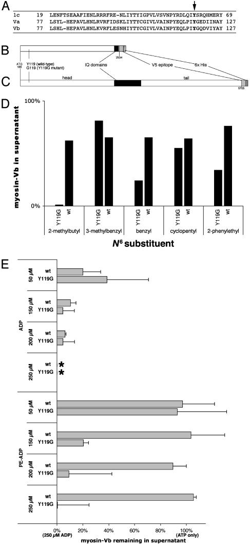
Characterization of mutant and wild-type myosin Vb. (A) Sequence alignment of myosin 1c, myosin Va, and myosin Vb showing conserved tyrosine (arrow). (B) Diagram of truncated myosin Vb insert. (C) Diagram of full-length myosin Vb insert. Numbers below the diagrams refer to nucleotides from GenBank accession no. U60416 (10). (D) Effects of 100 μM concentrations of five ADP analogs. (E) Effects of ADP and PE-ADP on myosin/ATP/actin-binding equilibria for truncated wild-type (wt) and mutant (Y119G) myosin Vb. Cosedimentation data are plotted as the proportion of myosin remaining in the supernatant relative to the data for 100 μM ATP only (100%) and the proportion remaining for 100 μM ATP plus 250 μM ADP (0%; *) on the same blot. Error bars represent standard deviations. A representative Western blot is shown in Fig. 6, which is published as supporting information on the PNAS web site.
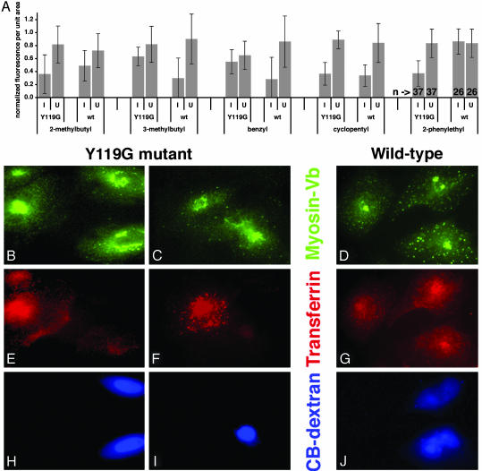
Screen of ADP analogs for the ability to alter transferrin uptake in HeLa cells expressing sensitized myosin Vb. Cells expressing mutant or wild-type myosin Vb were microinjected with Cascade Blue dextran and a 2.5 mM concentration of each ADP analog. Cells were then incubated with fluorescent transferrin. (A) Plots of cytoplasmic fluorescence per unit area. Because fluorescence levels varied between coverslips and experiments, fluorescence measurements were normalized to the level of the brightest cell in the same field. Error bars represent standard deviation. I, injected; U, uninjected; Y119G, cells expressing sensitized mutant myosin Vb; wt, cells expressing control wild-type myosin Vb; bottom line, N6 substituent. Numbers of cells are shown for the PE-ADP groups at the far right. (B–J) Representative cells from the PE-ADP experiment. HeLa cells expressing mutant (B, C, E, F, H, and I) and control wild-type myosin Vb (D, G, and J) were cultured on coverslips. Cells were imaged for anti-V5 to detect myosin Vb (B–D), Alexa 546-transferrin (E–G), and Cascade Blue, which accumulates in nuclei, to identify injected cells (H–J). The tubulovesicular distribution of mutant myosin Vb in injected cells (B, two cells on right) was observed in most, but not all, cases. (Bar = 10 μm.) Additional cells are shown in Fig. 7, which is published as supporting information on the PNAS web site.
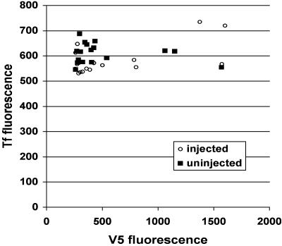
Inhibition of a truncated, tailless Y119G myosin Vb fragment has no effect on transferrin uptake. HeLa cells expressing truncated mutant myosin Vb (Fig. 1B) were microinjected for 2 min with PE-ADP and Cascade Blue dextran, allowed to recover for 5 min, and incubated with Alexa 546-transferrin for 10 min. V5 fluorescence, a measure of expression levels of the myosin Vb construct, is plotted on the x axis, and transferrin fluorescence is plotted on the y axis. ○, values from injected cells; ▪, values from uninjected cells.
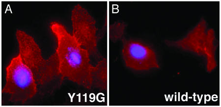
Inhibition of mutant myosin Vb increases levels of plasma-membrane transferrin receptor. HeLa cells expressing sensitized mutant (A) and wild-type (B) Myosin Vb were serum-starved for 30 min and some were injected (blue nuclei) with PE-ADP and Cascade Blue dextran. Cells were allowed to recover for 10 min at 37°C before fixation. Surface TfR was detected by indirect immunofluorescence.
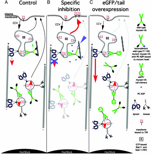
Model reconciling results from chemical-genetic and tail-fragment approaches to inactivating myosin Vb during transferrin uptake in HeLa cells. (A) Normal state in which myosin Vb antagonizes retrograde budding of vesicles from early endosomes (EE) along microtubules, after which it is distributed within the cytoplasm by equilibrium between anterograde and retrograde microtubule-based motility. (B) Inhibition of mutant myosin Vb by PE-ADP prevents retrograde exit from the early endosome, shunting transferrin and TfR into peripheral recycling pathways (red arrow). (C) Overexpression of the enhanced GFP/myosin Vb tail (15) prevents this antagonism, causing pericentriolar accumulation of endocytosed transferrin. Late endosomes, pericentriolar recycling vesicles, lysosomes, Golgi complex, and intermediate membrane compartments are omitted.
Similar articles
-
Myosin-Vb functions as a dynamic tether for peripheral endocytic compartments during transferrin trafficking.BMC Cell Biol. 2008 Aug 7;9:44. doi: 10.1186/1471-2121-9-44. BMC Cell Biol. 2008. PMID: 18687135 Free PMC article.
-
Myosin vb is associated with plasma membrane recycling systems.Mol Biol Cell. 2001 Jun;12(6):1843-57. doi: 10.1091/mbc.12.6.1843. Mol Biol Cell. 2001. PMID: 11408590 Free PMC article.
-
Human myosin-Vc is a novel class V myosin expressed in epithelial cells.J Cell Sci. 2002 Mar 1;115(Pt 5):991-1004. doi: 10.1242/jcs.115.5.991. J Cell Sci. 2002. PMID: 11870218
-
Chemical-genetic inhibition of sensitized mutant unconventional myosins.Methods Mol Biol. 2007;392:231-40. doi: 10.1007/978-1-59745-490-2_16. Methods Mol Biol. 2007. PMID: 17951722
-
Holding the reins on myosin V.Proc Natl Acad Sci U S A. 2005 Sep 27;102(39):13719-20. doi: 10.1073/pnas.0507068102. Epub 2005 Sep 19. Proc Natl Acad Sci U S A. 2005. PMID: 16172373 Free PMC article. Review. No abstract available.
Cited by
-
The Bump-and-Hole Tactic: Expanding the Scope of Chemical Genetics.Cell Chem Biol. 2018 Oct 18;25(10):1171-1184. doi: 10.1016/j.chembiol.2018.07.001. Epub 2018 Aug 2. Cell Chem Biol. 2018. PMID: 30078633 Free PMC article. Review.
-
Myosin Vb interacts with Rab8a on a tubular network containing EHD1 and EHD3.Mol Biol Cell. 2007 Aug;18(8):2828-37. doi: 10.1091/mbc.e07-02-0169. Epub 2007 May 16. Mol Biol Cell. 2007. PMID: 17507647 Free PMC article.
-
Myosin-Vb functions as a dynamic tether for peripheral endocytic compartments during transferrin trafficking.BMC Cell Biol. 2008 Aug 7;9:44. doi: 10.1186/1471-2121-9-44. BMC Cell Biol. 2008. PMID: 18687135 Free PMC article.
-
CART: an Hrs/actinin-4/BERP/myosin V protein complex required for efficient receptor recycling.Mol Biol Cell. 2005 May;16(5):2470-82. doi: 10.1091/mbc.e04-11-1014. Epub 2005 Mar 16. Mol Biol Cell. 2005. PMID: 15772161 Free PMC article.
-
Mitochondrial iron trafficking and the integration of iron metabolism between the mitochondrion and cytosol.Proc Natl Acad Sci U S A. 2010 Jun 15;107(24):10775-82. doi: 10.1073/pnas.0912925107. Epub 2010 May 21. Proc Natl Acad Sci U S A. 2010. PMID: 20495089 Free PMC article. Review.
References
-
- Bishop, A. C., Buzko, O. & Shokat, K. M. (2001) Trends Cell Biol. 11, 167–172. - PubMed
-
- Gulick, A. M., Bauer, C. B., Thoden, J. B. & Rayment, I. (1997) Biochemistry 36, 11619–11628. - PubMed
-
- Gillespie, P. G., Gillespie, S. K. H., Mercer, J. A., Shah, K. & Shokat, K. M. (1999) J. Biol. Chem. 274, 31373–31381. - PubMed
-
- Holt, J. R., Gillespie, S. K. H., Provance, D. W., Shah, K., Shokat, K. M., Corey, D. P., Mercer, J. A. & Gillespie, P. G. (2002) Cell 108, 371–381. - PubMed
Publication types
MeSH terms
Substances
Grants and funding
LinkOut - more resources
Full Text Sources
Other Literature Sources
Molecular Biology Databases
Miscellaneous
