. 2004 Jan 29;32(2):598-610.
doi: 10.1093/nar/gkh195. Print 2004.

Modification of de novo DNA methyltransferase 3a (Dnmt3a) by SUMO-1 modulates its interaction with histone deacetylases (HDACs) and its capacity to repress transcription

Affiliations

Modification of de novo DNA methyltransferase 3a (Dnmt3a) by SUMO-1 modulates its interaction with histone deacetylases (HDACs) and its capacity to repress transcription

Yan Ling et al. Nucleic Acids Res. .

Abstract

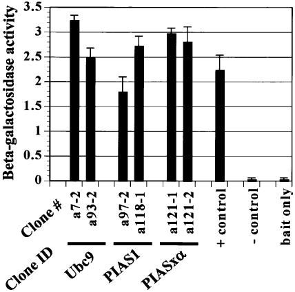
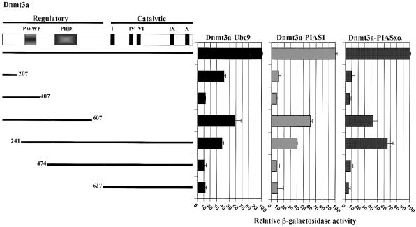
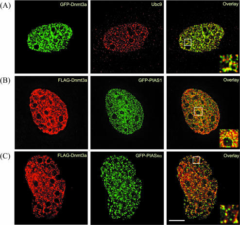
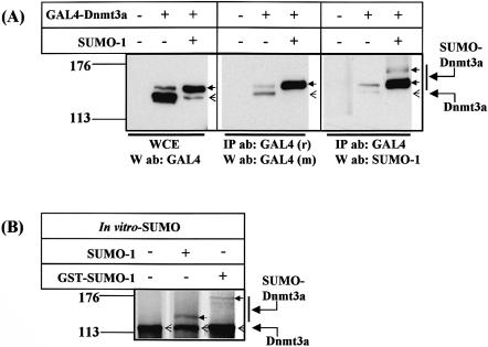
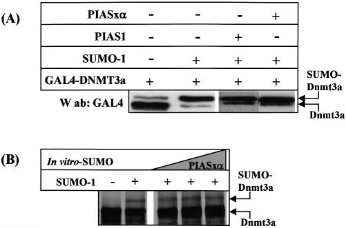
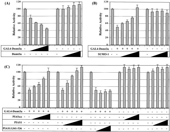
The de novo DNA methyltransferase Dnmt3a is one of three mammalian DNA methyltransferases that has been shown to play crucial roles in embryonic development, genomic imprinting and transcriptional silencing. Despite its importance, very little is known about how the enzymatic activity and transcriptional repression functions of Dnmt3a are regulated. Here we show that Dnmt3a interacts with multiple components of the sumoylation machinery, namely the E2 sumo conjugating enzyme Ubc9 and the E3 sumo ligases PIAS1 and PIASxalpha, all of which are involved in conjugating the small ubiquitin-like modifier polypeptide, SUMO-1, to its target proteins. Dnmt3a is modified by SUMO-1 in vivo and in vitro and the region of Dnmt3a responsible for interaction maps to the N-terminal regulatory domain. Functionally, sumoylation of Dnmt3a disrupts its ability to interact with histone deacetylases (HDAC1/2), but not with another interaction partner, Dnmt3b. Conditions that enhance the sumoylation of Dnmt3a in vivo abolish its capacity to repress transcription. These studies reveal a new level of regulation governing Dnmt3a whereby a post-translational modification can dramatically regulate its interaction with specific protein partners and alter its ability to repress transcription.


Figures

Figure 1
Figure 1
Dnmt3a binds to Ubc9, PIAS1 and PIASxα in a yeast two-hybrid screen. Full-length Dnmt3a was fused to the GAL4-DBD and used as bait in a yeast two-hybrid screen against a human fetal liver cDNA library in AH109 yeast cells under high stringency selection conditions. Clones scoring positive for growth on selective media were confirmed using a beta-galactosidase assay in Y187 yeast cells, then sequenced. The identity of two representative, independently isolated clones is shown below the graph of beta-galactosidase activity (as relative units). The positive control (+) is the interaction between p53 and the SV40 T antigen, the negative control (–) is the interaction between p53 and lamin B, and ‘bait only’ is the GAL4-Dnmt3a construct alone under the appropriate selection conditions. Expression of full-length Dnmt3a in AH109 cells was confirmed by western blotting (data not shown). Values are the mean of three independent experiments and the error bar is the standard deviation from the mean.
Figure 2
Figure 2
Dnmt3a interacts with Ubc9, PIAS1 and PIASxα in vivo and in vitro. (A) Dnmt3a co-immunoprecipitates with Ubc9 in whole cell extracts from transfected COS-7 cells using either an antibody directed against a tagged Dnmt3a or against Ubc9. Endogenous levels of Ubc9 were high (not shown), and we could detect the Dnmt3a–Ubc9 interaction with or without co-transfection of a Ubc9 expression vector. (B) Dnmt3a co-immunoprecipitates with PIAS1 and PIASxα using antibodies directed against either Dnmt3a (or a tagged Dnmt3a) or the PIAS proteins. In each case, COS-7 cells were transfected with 2.0 µg of each of the indicated expression constructs for 48 h. Whole cell extracts were then prepared and immunoprecipitated with the antibodies indicated at the left of each panel (IP ab), washed, the remaining bound proteins resolved on an SDS–PAGE gel, then subjected to western analysis with the antibody indicated at the left of each panel (W ab). (C) Dnmt3a binds to Ubc9, PIAS1 and PIASxα in vitro in a GST pull-down assay. GST alone, or the GST fusion proteins indicated across the top of each panel (bound to glutathione-Sepharose beads), were incubated with in vitro transcribed and translated 35S-labeled proteins (IVT) and washed extensively. GST alone served as a control for non-specific binding to IVT-Dnmt3a in the top panel while IVT-luciferase served as a control for non-specific binding to GST-Dnmt3a in the lower panel. The presence of specifically bound protein is indicated with an arrow at the right of each panel. In some cases, the IVT reaction yielded two closely spaced products, most likely due to initiation at an internal methionine residue.
Figure 3
Figure 3
Ubc9, PIAS1 and PIASxα interact with Dnmt3a via a region within the N-terminal regulatory domain. A schematic diagram of the Dnmt3a protein is shown at the top with the PWWP, PHD and catalytic domains indicated. Each of the Dnmt3a deletion mutants, fused to the GAL4-DBD, was co-transformed with GAL4-AD fusions of Ubc9 (left), PIAS1 (middle), or PIASxα (right) into AH109 yeast cells. Thick lines denote the regions of Dnmt3a that were fused to the GAL4-DBD. Three colonies growing under high stringency selection for each transformation were selected, grown in liquid culture, then used for a beta-galactosidase assay (relative units). Graphs show the mean of three independent experiments (colonies) relative to the full-length Dnmt3a set at 100%. Error bars are the standard deviation from the mean.
Figure 4
Figure 4
Dnmt3a co-localizes with Ubc9, PIAS1 and PIASxα in transfected cells. (A) NIH3T3 cells grown on glass cover slips were co-transfected for 24 h with GFP-Dnmt3a and Ubc9, (B) FLAG-Dnmt3a and GFP-PIAS1 or (C) FLAG-Dnmt3a and GFP-PIASxα. Immunofluorescence labeling was performed with an anti-Ubc9 antibody in (A) or the anti-FLAG-M2 antibody in (B–C) followed by incubation with an appropriate secondary antibody as described in Materials and Methods. Images were collected with an Olympus IX70 inverted microscope equipped with a 100× 1.35 NA oil immersion objective. The two images are merged in the right-most panel where yellow color represents co-localization. Boxed areas are enlarged and shown in the lower-right corner of each overlay panel. The scale bar in the lower right panel corresponds to 5 µm.
Figure 5
Figure 5
Dnmt3a is sumoylated in vivo and in vitro. (A) GAL4-DBD-tagged Dnmt3a (2 µg) was co-transfected with or without a SUMO-1 expression vector (1.5 µg) into COS-7 cells. Whole cell extracts (WCE) were prepared in the presence of NEM and the migration position of the transfected Dnmt3a determined by western blotting with an anti-GAL4-DBD antibody (left panel). Dnmt3a was also immunoprecipitated from the transfected WCEs with a rabbit polyclonal GAL4-DBD antibody (‘r’), and the molecular weight of the total pool of GAL4-Dnmt3a determined by western blotting with a mouse monoclonal GAL4-DBD antibody (‘m’, middle panel). The presence of slower migrating forms of Dnmt3a in the ‘+SUMO-1’ lane indicates that Dnmt3a is sumoylated. When the blot is stripped and re-probed with a SUMO-1 antibody, only the slower migrating species are detectable and these higher molecular weight forms are specifically enriched in the ‘+SUMO-1’ reaction (right panel). (B) Dnmt3a is sumoylated in vitro. In vitro sumoylation reactions were carried out in the presence of 35S-labeled in vitro transcribed/translated (IVT) Dnmt3a, 200 ng recombinant SAE1/SAE2, 1 µg Ubc9, 2 mM ATP, ATP regeneration system, and 10 µg of SUMO-1 or 5 µg GST-SUMO-1 as indicated at the top of each panel. Molecular weight markers (kDa), are indicated at the left. The open arrow denotes the position of the unmodified GAL4-Dnmt3a and the filled arrow shows the migration position of the sumo-modified Dnmt3a.
Figure 6
Figure 6
PIAS1 and PIASxα do not enhance sumoylation of Dnmt3a in vivo or in vitro. (A) Two micrograms of full-length GAL4-Dnmt3a was transfected without (lane 1) or with 1.5 µg of SUMO-1 expression vector (lane 2) into COS-7 cells and whole cell extracts were prepared for western analysis with anti-GAL4-DBD antibody. In lanes 3 and 4, four micrograms of PIAS1 or PIASxα expression vector, respectively, are co-transfected with the SUMO-1 and GAL4-Dnmt3a expression vectors. (B) In vitro sumoylation reactions containing 35S-labeled-IVT Dnmt3a, performed as described in Figure 5B, were supplemented with recombinant GST-PIASxα (1, 5, 10 µg) and the reaction products analyzed on a 6% SDS–PAGE gel.
Figure 7
Figure 7
Sumoylation differentially affects several known protein–protein interactions involving Dnmt3a. (A) HCT116 cells were transfected for 24 h with 2.0 µg of each of the expression constructs listed along the top. Whole cell extracts were prepared and subjected to immunoprecipitation with a mouse anti-GAL4-DBD tag antibody to pull-down Dnmt3a. Subsequent western blotting with antibodies against HDAC1 (left panel), or HDAC2 (right panel), revealed that Dnmt3a interacts with both HDAC1 and HDAC2 and that this interaction is markedly reduced under conditions favoring sumoylation of Dnmt3a (co-transfection of 1.5 µg of SUMO-1 expression vector). Levels of HDAC1/2 and GAL4-Dnmt3a were not affected by SUMO-1 over-expression (lower two panels, ‘Inputs’). (B) HCT116 cells were transfected with tagged Dnmt3a and Dnmt3b1 and whole cell extracts prepared as described above. Immunoprecipitations were carried out with anti-GAL4-DBD antibody and western blotting of the bound material was performed with an anti-FLAG antibody to detect the presence of Dnmt3b1. SUMO-1 expression did not alter the ability of Dnmt3a to interact with Dnmt3b1 (upper panel), nor did it affect the levels of the transfected proteins (lower two panels, ‘Inputs’). Similar results were obtained using COS-7 cells (not shown).
Figure 8
Figure 8
SUMO-1 modification of Dnmt3a eliminates its capacity to repress transcription. (A) Full-length Dnmt3a represses transcription when fused to the GAL4-DBD. Increasing amounts (indicated by the black wedge) of GAL4-Dnmt3a (0.5, 1.0, 2.5, 5.0 µg) or untethered Dnmt3a (0.5, 1.0, 2.5, 5.0 µg) were co-transfected into HCT116 cells along with 10 ng of an SV40-Renilla luciferase construct (to control for transfection efficiency), and 1.0 µg of a firefly luciferase reporter gene driven by five GAL4 DNA binding sites. Both firefly and Renilla luciferase activities were measured from the same whole cell extract preparation after 24 h using the Promega Dual Luciferase Assay kit. (B) GAL4-Dnmt3a (4.0 µg) was co-transfected with increasing amounts of SUMO-1 expression vector (1.0, 2.5, 5.0, 8.0 µg) into HCT116 cells. Transfection of equivalent amounts of SUMO-1 expression vector in the absence of GAL4-Dnmt3a did not alter promoter activity. (C) Effects of PIAS proteins on Dnmt3a transcriptional repression. GAL4-Dnmt3a (4.0 µg) was co-transfected with increasing amounts of PIASxα or PIAS1 expression vectors (1.0, 2.5, 5.0, 8.0 µg) or a mutant form of PIAS1 (Δ341–536, 2.5, 5.0, 8.0 µg) deleted for the RING-finger-like domain, into HCT116 cells and reporter activity determined after 24 h. Transfection of PIAS1 or PIASxα (2.5, 5.0, 8.0 µg) expression plasmids in the absence of GAL4-Dnmt3a did not affect reporter gene activity. All values were normalized for transfection efficiency (firefly luciferase/Renilla luciferase) and then set relative to the reporter activity of the 5× GAL4-BS-luciferase construct alone set at 100% (first bar in each series). Values are the average of two independent experiments and error bars are the range. Total DNA content was kept constant in each transfection by addition of ‘empty’ parental expression vector.

Similar articles

Cited by

References

    1. Robertson K.D. (2002) DNA methylation and chromatin—unraveling the tangled web. Oncogene, 21, 5361–5379. - PubMed
    1. Baylin S.B., Esteller,M., Rountree,M.R., Bachman,K.E., Schuebel,K. and Herman,J.G. (2001) Aberrant patterns of DNA methylation, chromatin formation and gene expression in cancer. Hum. Mol. Genet., 10, 687–692. - PubMed
    1. Jones P.A. and Baylin,S.B. (2002) The fundamental role of epigenetic events in cancer. Nature Rev. Genet., 3, 415–428. - PubMed
    1. Bestor T.H. (2000) The DNA methyltransferases. Hum. Mol. Genet., 9, 2395–2402. - PubMed
    1. Li E. (2002) Chromatin modification and epigenetic reprogramming in mammalian development. Nature Rev. Genet., 3, 662–673. - PubMed

Publication types

MeSH terms