. 2004 Jan 28;24(4):916-27.
doi: 10.1523/JNEUROSCI.4733-03.2004.

Postsynaptic density 95 controls AMPA receptor incorporation during long-term potentiation and experience-driven synaptic plasticity

Affiliations

Postsynaptic density 95 controls AMPA receptor incorporation during long-term potentiation and experience-driven synaptic plasticity

Ingrid Ehrlich et al. J Neurosci. .

Abstract

The regulated delivery of AMPA-type glutamate receptors (AMPARs) to synapses is an important mechanism underlying synaptic plasticity. Here, we ask whether the synaptic scaffolding protein PSD-95 (postsynaptic density 95) participates in AMPAR incorporation during two forms of synaptic plasticity. In hippocampal slice cultures, the expression of PSD-95-green fluorescent protein (PSD-95-GFP) increases AMPAR currents by selectively delivering glutamate receptor 1 (GluR1)-containing receptors to synapses, thus mimicking long-term potentiation (LTP). Mutational analysis shows that the N terminal of PSD-95 including the first two PDZ [PSD-95/Discs large (Dlg)/zona occludens-1 (ZO-1)] domains is necessary and sufficient to mediate this effect. Further supporting a role in synaptic plasticity, wild-type PSD-95 occludes LTP and dominant negative forms block LTP. Moreover, we demonstrate that PSD-95 also participates in AMPAR delivery during experience-driven plasticity in vivo. In the barrel cortex from experience-deprived animals, the expression of PSD-95-GFP selectively increases AMPAR currents, mimicking experience-driven plasticity. In nondeprived animals, PSD-95-GFP produces no additional potentiation, indicating common mechanisms between PSD-95-mediated potentiation and experience-driven synaptic strengthening. A dominant negative form of PSD-95 blocks experience-driven potentiation of synapses. Pharmacological analysis in slice cultures reveals that PSD-95 acts downstream of other signaling pathways involved in LTP. We conclude that PSD-95 controls activity-dependent AMPAR incorporation at synapses via PDZ interactions not only during LTP in vitro but also during experience-driven synaptic strengthening by natural stimuli in vivo.


Figures
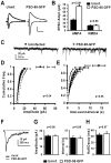
Figure 1.
Figure 1.
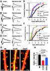
Expression of PSD-95–GFP specifically potentiates AMPA-mediated EPSCs. A, Synaptic currents recorded simultaneously from nearby pyramidal neurons held at –60 and +40 mV, one uninfected and one expressing PSD-95–GFP. B, The AMPA component of EPSCs was significantly increased from 28.5 ± 2.5 to 64.8 ± 4.9 pA (control and infected neurons, respectively; n = 51), where as the late NMDA component was not altered (13.1±1.3 and 13.5±1.8 pA, control and infected neurons, respectively; n = 33). Calibration: 20 pA, 50 msec. C, Miniature EPSCs recorded at –60 mV from a pair of CA1 neurons. D, E, Cumulative histograms of amplitude and interevent interval for AMPA minis (n = 6 cells/group). The mini amplitude increases, whereas the interevent-interval decreases significantly (t test for each binned data point). F, Overlaid average miniature NMDA currents recorded from a pair of CA1 neurons recorded at –60 mV in 0 mm Mg 2+ and 5 μm CNQX. G, Plots of amplitude (23.3 ± 1.1 and 23.4 ± 1.0 pA, control and infected, respectively) and interevent interval (10.4 ± 1.2 and 10.1 ± 1.2 sec, control and infected, respectively) for average NMDA minis indicate no significant change (n = 10 cells/group; t test). H, Time to half decay of average NMDA minis (32.3 ± 2.9 and 34.3 ± 3.6 msec, control and PSD-95–GFP-expressing neurons, respectively) also shows no difference (n = 10 cells/group; t test).
Figure 2.
Figure 2.
Membrane localization and PDZ interactions are necessary and sufficient for potentiation of AMPA currents. A–D, Synaptic currents measured in paired recordings from nearby control and infected neurons expressing mutant forms of PSD-95–GFP at –60 and +40 mV. Calibration: 20 pA, 50 msec. E, F, Cumulative distributions of AMPA-EPSC ratios (AMPAInf/AMPAUninf) in paired recordings are used to compare AMPA current potentiation by different mutants (p values from KS tests). E, The small potentiating effect of PSD-95-C3, 5S (n = 30) and PSD-95-AAAA (n = 33) was highly significantly different from wt PSD-95 (p = 0.002 and p = 0.004, respectively). F, Like GFP (n = 47), PSD-95-Double (n = 24) did not potentiate AMPA currents and was significantly different from wt PSD-95 (p < 0.001), whereas PSD-95–PDZ1–2 (n = 21) potentiated AMPA–EPSCs similar to wt-PSD-95 (p = 0.63). G, Subcellular distribution of mutant forms of PSD-95 compared with wt PSD-95–GFP. Dual-wavelength two-photon images of CA1 pyramidal neurons in slice cultures (9 d in vitro) coexpressing DsR-T1 and GFP-tagged PSD-95 constructs for ∼40–48 hr. Scale bars, 2μm. H, Spine/dendrite ratio shows that wt-PSD-95 is strongly accumulated in spines (ratio, 2.51 ± 0.07; n = 312 spines), C3, 5S is passively distributed (ratio, 1.11 ± 0.06; n = 285 spines), and the AAAA mutant is accumulated in spines (ratio, 1.78 ± 0.06; n = 323 spines), but less than wt (from three neurons each; KS tests).
Figure 3.
Figure 3.
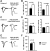
Expression of PSD-95 mimics LTP by driving GluR1 to synapses. A, C, E, AMPA-EPSCs recorded simultaneously from nearby CA1 neurons at –60 and +40 mV in 100μm APV. Calibration: 10 pA, 40 msec. A, Coexpression of PSD-95–GFP and GluR1–GFP resulted in potentiated and more rectified AMPA current. B, The relative AMPA-EPSC amplitude (100 ± 9.0 vs 159 ± 13.5%; n = 35) and rectification (2.3 ± 0.2; n = 43 vs 3.9 ± 0.3; n = 40; t test) were significantly increased in control versus transfected neurons. C, Coexpression of PSD-95–GFP and GluR1(T887A)–GFP resulted in no change in AMPA-current amplitude or rectification. D, Relative AMPA-EPSCs (100±10.1 and 95.6±8.8%; n = 29) and rectification (2.0±0.1, n = 29 vs 2.3 ± 0.2, n = 27; t test) were not significantly different in control versus transfected neurons. E, Co-expression of PSD-95–GFP and GluR1–C-tail–GFP did not change AMPA-EPSC amplitude. F, Relative AMPA-EPSCs were not significantly different in control versus transfected neurons (100 ± 13.4 vs 108.3 ± 11.7%; n = 31).
Figure 4.
Figure 4.
PSD-95 does not affect the constitutive pathway of AMPAR delivery. A, B, AMPA-EPSCs recorded simultaneously from nearby CA1 neurons at –60 and +40 mV in 100μm APV. Calibration: 30 pA, 40 msec. A, Expression of GluR2(R607Q)–GFP did not alter AMPA-EPSC amplitude, but rectification was increased. B, PSD-95–GFP was coexpressed with GluR2(RQ)–GFP. AMPA-EPSC amplitude was strongly increased, but rectification was only slightly increased. C, Relative AMPA-EPSCs were not significantly different in control versus GluR2(RQ)-transfected neurons (100 ± 17.2 and 84.9 ± 7.6%; n = 15), but significantly increased in control versus neurons coexpressing PSD-95–GFP and GluR2(RQ)–GFP (100±11.2 vs 164.8± 22.3%; n = 17). D, Rectification was strongly increased in control versus GluR2(RQ)–GFP-transfected neurons (2.2 ± 0.2, n = 16 vs 3.7 ± 0.4, n = 13; t test) and slightly increased in control versus PSD-95–GFP and GluR2(RQ)–GFP-expressing neurons (2.0 ± 0.2, n = 16 vs 2.7 ± 0.2, n = 16; t test). Importantly, rectification in neurons coexpressing PSD-95 and GluR2(RQ) was significantly smaller than in neurons expressing GluR2(RQ) alone (t test). E, Changes in synaptic AMPA currents recorded during infusion of peptide pep2m (2 mm) into two nearby neurons. EPSC amplitudes decreased in the uninfected neuron, but there was little change in a neuron expressing PSD-95–GFP. Calibration: 40 pA, 20 msec. F, The residual AMPA-EPSC 25 min after infusion of pep2m is significantly smaller in control than infected neurons (55.8 ± 10.7 vs 84 ± 9.5% of initial value; n = 11; t test).
Figure 5.
Figure 5.
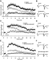
Expression of wt PSD-95 occludes and expression of putative dominant negative forms of PSD-95 blocks LTP. A, C, E, Time courses of relative changes in AMPA-mediated EPSCs by pairing-induced LTP for control and infected neurons. Induced (paired) pathways are shown in large and control (unpaired) pathways in small symbols for infected and control neurons. The time of delivery of the pairing protocol is indicated by the bar. A, Expression of PSD-95–GFP-occluded LTP. Transmission onto infected neurons returned almost to baseline 30–35 min after pairing (1.17 ± 0.20; n = 12) and was significantly different from the potentiation observed in control neurons (1.88 ± 0.22; n = 13; p = 0.028; t test). Ci Expression of PSD-95C3,5S–GFP blocked LTP. In PSD-95C3,5S–GFP-expressing neurons, transmission returned almost to baseline levels 30–35 min after pairing (1.17 ± 0.16; n = 10). This was significantly different from potentiation in the control neurons (1.89 ± 0.23; n = 7; p = 0.02; t test). E, Expression of PSD-95AAAA–GFP blocked LTP. In infected neurons, transmission returned close to baseline levels 30–35 min after pairing (1.21 ± 0.15; n = 11), which was significantly different from control neurons (2.10 ± 0.29; n = 11; p = 0.014; t test). B, D, F, Example traces of EPSCs in uninfected and infected neurons before and ∼32–35 min after pairing. Traces are the averages of 30 sweeps. Calibration: 40 pA, 20 msec.
Figure 6.
Figure 6.
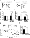
Experimental approach, and effects of sensory deprivation in barrel cortex. A, Sketch of the experimental protocol. Sindbis virus was injected at P11 or P12 and rats were allowed or deprived of sensory experience by trimming all contralateral whiskers for 2 d. Cortical slices were obtained and the barrel cortex was identified by trans-illumination. Paired recordings from neighboring layer II/III pyramidal neurons, infected and control, were obtained and EPSCs elicited by stimulation of layer IV. B, C, Sensory deprivation decreased the AMPA/NMDA ratio at synapses. B, EPSCs recorded in uninfected layer II/III pyramidal neurons at holding potentials of –60 and +40 mV from animals with intact or deprived whiskers. Calibration: 10 pA, 40 msec. C, The AMPA/NMDA ratio was significantly lower in deprived (1.5 ± 0.2; n = 16) than in nondeprived animals (2.6 ± 0.3; n = 20; t test).
Figure 7.
Figure 7.
Expression of wt PSD-95 mimics and occludes experience-driven AMPAR delivery in barrel cortex. A, B, Paired recordings of EPSCs from layer II/III pyramidal neurons held at –60 and +40 mV, one neuron uninfected and one expressing wt PSD-95–GFP. Calibration: 20 pA, 40 msec. A, In intact whisker animals, there was no difference in the AMPAR- or NMDAR-mediated EPSC in a control and a neuron expressing PSD-95–GFP. B, In deprived animals, the AMPA-EPSC was potentiated by the expression of PSD-95–GFP, whereas the NMDA current was unaffected. C, Summary of relative changes in AMPAR- and NMDAR-mediated EPSCs. The AMPA component was not significantly different between control and infected neurons in intact animals (100 ± 17.7 and 112.7 ± 12.8%; n = 19) but potentiated during contralateral deprivation (100 ± 18.9 and 168.5 ± 25.4%, control and infected, respectively; n = 20). In all cases, NMDA currents were not significantly changed (Intact, 100 ± 21.0 and 83.1 ± 13.0%, control and infected, n = 17; Deprived, 100 ± 19.7 and 93.8 ± 13.4%, control and infected, n = 15).
Figure 8.
Figure 8.
Expression of dominant negative PSD-95 blocks experience-driven AMPAR delivery to synapses in barrel cortex. A, B, EPSCs evoked in layer IV and recorded simultaneously from nearby layer II/III pyramidal neurons held at –60 and +40 mV. Calibration: 10 pA, 40 msec. A, In intact whisker animals, the AMPAR- but not the NMDAR-mediated EPSC was depressed in a neuron expressing PDS-95AAAA–GFP. B, In deprived animals, there was no change in AMPA or NMDA currents in the control versus the infected neuron. C, Summary of relative changes in AMPAR- and NMDAR-mediated EPSCs. The AMPA component was significantly depressed by PSD-95AAAA when whiskers were intact (100 ± 11.1 and 54.5 ± 7.0%, control and infected; n = 18), but no change was seen during contralateral deprivation (100 ± 14.3 and 100.3 ± 15.2%, control and infected; n = 20). In all cases, NMDA currents were not significantly changed (Intact, 100 ± 16.7 and 93.5 ± 15.1%, control and infected, n = 14; Deprived, 100 ± 16.4 and 90.9 ± 11.4%, control and infected, n = 16).
Figure 9.
Figure 9.
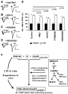
PSD-95 acts downstream of synaptic activity and other signaling pathways involved in LTP. A–D, AMPA-EPSCs measured in paired recordings from CA1 neurons held at –60 mV to compare amplitudes in uninfected and PSD-95–GFP-expressing neurons. Slices had been subjected to different pharmacological treatments (10 mm Mg 2+, 20 μm KN-93, 20 μm PD98059, 20 μm SB203580). Calibration: 30 pA, 40 msec. E, Summary and comparison of changes in AMPA-EPSCs. *Significant potentiation within one experimental group. In high Mg 2+, PSD-95–GFP increased AMPA currents from 100 ± 17.2 to 204.7 ± 28.3% (n = 22; p = 0.003), in KN-93 from 100 ± 12.0 to 184.2 ± 15.1% (n = 27; p = 0.001), in PD98059 from 100 ± 10.1 to 227.7 ± 16.1% (n = 39; p < 0.0005), and in SB203580 from 100 ± 15.5 to 196.1 ± 11.6% (n = 40; p < 0.0005). Comparison between different treatments was done for AMPAInf/AMPAUninf ratios from each group using KS tests (see Materials and Methods; p values above bars); no significant differences were detected. F, Model for the role of PSD-95 in activity-dependent AMPAR delivery to synapses. Strong synaptic activity leads to signaling events that trigger changes in several proteins, including PSD-95. Synaptic accumulation, multimerization and formation of PDZ interactions by PSD-95 result in the recruitment of extrasynaptic GluR1-containing receptors to synapses, possibly in a complex with PSD-95 and other proteins. Interestingly, an increased level of PSD-95 can drive this process, whereas an increased level of GluR1 does not.

Similar articles

Cited by

References

    1. Aoki C, Miko I, Oviedo H, Mikeladze-Dvali T, Alexandre L, Sweeney N, Bredt DS (2001) Electron microscopic immunocytochemical detection of PSD-95, PSD-93, SAP-102, and SAP-97 at postsynaptic, presynaptic, and nonsynaptic sites of adult and neonatal rat visual cortex. Synapse 40: 239–257. - PubMed
    1. Arnold DB, Clapham DE (1999) Molecular determinants for subcellular localization of PSD-95 with an interacting K+ channel. Neuron 23: 149–157. - PubMed
    1. Barria A, Malinow R (2002) Subunit-specific NMDA receptor trafficking to synapses. Neuron 35: 345–353. - PubMed
    1. Barria A, Muller D, Derkach V, Griffith LC, Soderling TR (1997) Regulatory phosphorylation of AMPA-type glutamate receptors by CaM-KII during long-term potentiation. Science 276: 2042–2045. - PubMed
    1. Beique J-CA, R (2003) PSD-95 regulates synaptic transmission and plasticity in rat cerebral cortex. J Physiol (Lond) 546: 859–867. - PMC - PubMed

Publication types

MeSH terms

Substances