. 2004 Feb 2;164(3):441-9.
doi: 10.1083/jcb.200304135. Epub 2004 Jan 26.

Extracellular HMGB1, a signal of tissue damage, induces mesoangioblast migration and proliferation

Affiliations

Extracellular HMGB1, a signal of tissue damage, induces mesoangioblast migration and proliferation

Roberta Palumbo et al. J Cell Biol. .

Abstract

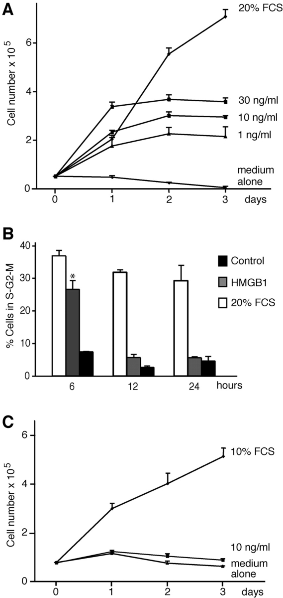
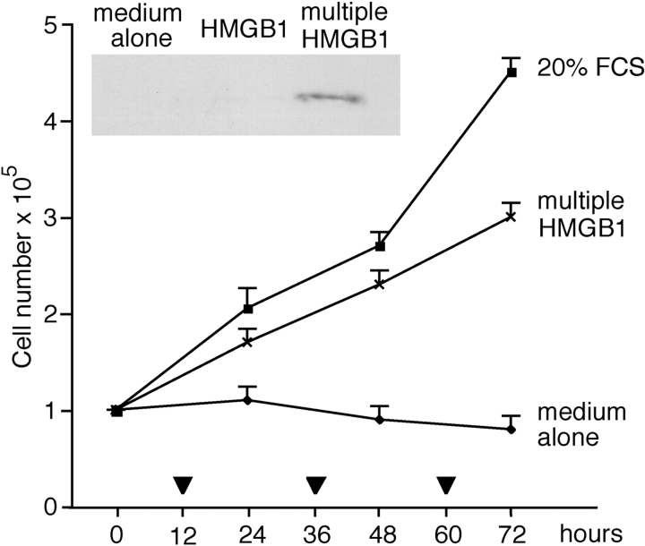
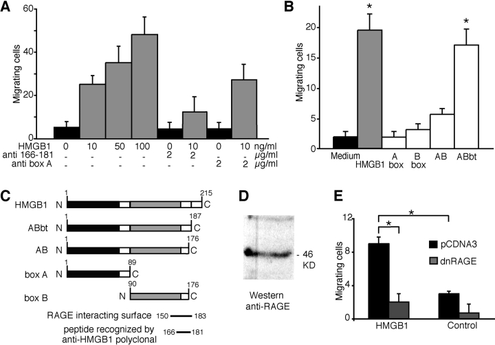
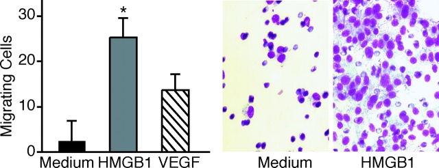
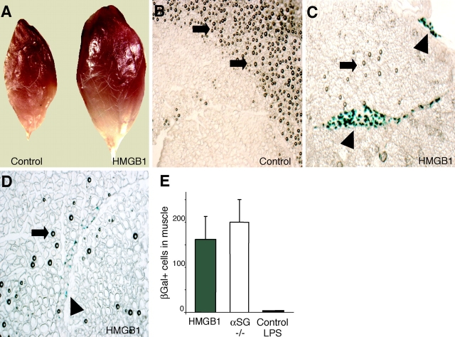
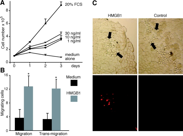
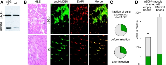
High mobility group box 1 (HMGB1) is an abundant chromatin protein that acts as a cytokine when released in the extracellular milieu by necrotic and inflammatory cells. Here, we show that extracellular HMGB1 and its receptor for advanced glycation end products (RAGE) induce both migration and proliferation of vessel-associated stem cells (mesoangioblasts), and thus may play a role in muscle tissue regeneration. In vitro, HMGB1 induces migration and proliferation of both adult and embryonic mesoangioblasts, and disrupts the barrier function of endothelial monolayers. In living mice, mesoangioblasts injected into the femoral artery migrate close to HMGB1-loaded heparin-Sepharose beads implanted in healthy muscle, but are unresponsive to control beads. Interestingly, alpha-sarcoglycan null dystrophic muscle contains elevated levels of HMGB1; however, mesoangioblasts migrate into dystrophic muscle even if their RAGE receptor is disabled. This implies that the HMGB1-RAGE interaction is sufficient, but not necessary, for mesoangioblast homing; a different pathway might coexist. Although the role of endogenous HMGB1 in the reconstruction of dystrophic muscle remains to be clarified, injected HMGB1 may be used to promote tissue regeneration.


Figures

Figure 1.
Figure 1.
Effect of HMGB1 on embryonic mesoangioblast proliferation. (A) D16 cells were grown in RPMI medium containing no addition, HMGB1 at the indicated concentrations, or 20% FCS. HMGB1 induced cell proliferation at all concentrations tested, but the cell number reached a plateau after 48 h. Each point represents the mean ± SD (n = 3). The experiment was repeated three times. (B) D16 cell division was analyzed by FACS®. After 6 h in the presence of 30 ng/ml HMGB1 the DNA content increases, but returns to the normal diploid content after 24 h. Asterisk indicates statistical significance (P < 0.001). (C) 3T3 fibroblasts (treated as the D16 cells in A) do not divide in the presence of HMGB1.
Figure 2.
Figure 2.
Continued HMGB1 stimulation sustains mesoangioblast proliferation. D16 cells were placed at time 0 in RPMI medium containing 30 ng/ml HMGB1; a similar amount of HMGB1 was also added at the times indicated with a triangle. Multiply stimulated D16 cells kept growing. Each point represents the average ± SD of two experiments performed in duplicate. Inset: Western blot of HMGB1 in the medium bathing D16 cells 48 h after the beginning of the experiment. HMGB1 was still present in the medium of restimulated cells, but not in the medium of cells stimulated once at time 0. This experiment was repeated two times with similar results.
Figure 3.
Figure 3.
HMGB1 has chemotactic activity on embryonic mesoangioblasts. (A) D16 cells were subjected to chemotaxis assays with 10, 50, or 100 ng/ml HMGB1. Data represent the average ± SD of four experiments performed in duplicate; the effect of increasing HMGB1 concentrations is highly significant (P < 0.001 in ANOVA analysis). Addition of anti-HMGB1 antibodies recognizing the peptide 166–181 significantly reduced the chemotactic response (P < 0.05 in comparison to the sample without antibody), whereas the addition of monoclonal anti-box A antibodies had no effect. (B) Chemotactic activity on D16 cells of various HMGB1 fragments (all at 10 ng/ml). ABbt has a chemotactic effect comparable with full-length HMGB1 (P < 0.05 of proteins vs. medium alone). In contrast, boxes A and B and the AB didomain have no significant chemotactic activity. Bars represent the average ± SD of three experiments performed in duplicate. Asterisks indicate statistical significance (P< 0.01). (C) Schematic representation of full-length HMGB1, boxes A and B, the didomain AB, and tailless HMGB1 (ABbt). (D) Western blot with anti-RAGE antibodies on total D16 cell extract. (E) D16 cells transfected with dnRAGE-expressing plasmid or pCDNA3 empty vector were assayed for chemotaxis in response to medium with or without HMGB1. Cells transfected with dnRAGE exhibited a significant decrease in migration in comparison to cells transfected with control plasmid. Asterisks indicate statistical significance (P < 0.05).
Figure 4.
Figure 4.
HMGB1 induces the transit of mesoangioblasts through an endothelial monolayer. D16 cells were placed in the upper compartment of Boyden apparatuses. The lower chambers contained RPMI alone (medium), RPMI plus 100 ng/ml HMGB1, or RPMI plus 10 ng/ml VEGF; chambers were separated by a confluent endothelial cell monolayer grown on polycarbonate filters. HMGB1 significantly stimulated D16 transmigration (P < 0.01). Bars represent the aver- age ± SD of three experiments performed in duplicate. Panels beside the bar graph show D16 cells stained with Giemsa after migration, toward medium alone or containing HMGB1. Asterisk indicates statistical significance (P < 0.05).
Figure 5.
Figure 5.
HMGB1 attracts mesoangioblasts in vivo. D16 cells were first transduced with a lentiviral vector encoding nuclear LacZ, and were then injected through the femoral artery of mice where heparin-Sepharose beads (either unloaded or loaded with HMGB1) had been injected in the tibialis anterior muscle. Mice were killed after 24 h. (A) Tibialis anterior muscles injected with HMGB1-loaded and control beads. (B–D) Cryosections of muscles treated with control heparin-Sepharose beads (control) or HMGB1-coated heparin-Sepharose beads (HMGB1). Arrows indicate the beads. Sections were stained with X-gal, and mesoangioblasts (arrowheads) appear blue. Mesoangioblasts were found in large clusters (C) or as isolated cells (D) only in muscles injected with HMGB1-coated beads. (E) Number of migrating D16 cells in tibialis anterior muscles of wild-type mice treated with HMGB1-loaded beads (gray bar; n = 2) or control beads coinjected with 1 μg LPS (black bar; n = 3). White bar represents the number of D16 cells found in tibialis anterior muscles of α-SG−/− dystrophic mice (n = 2).
Figure 6.
Figure 6.
Effect of HMGB1 on adult mesoangioblasts. (A) Mesoangioblasts of the G1 clone, obtained from mouse bone marrow, were grown in RPMI medium containing 1, 10, or 30 ng/ml HMGB1. For comparison, G1 cells were also grown in RPMI medium alone or in RPMI plus 20% FCS. (B) Migration of G1 cells toward the lower chamber of Boyden apparatuses containing RPMI (medium) or RPMI plus 10 ng/ml HMGB1 (HMGB1). In the migration experiment, the chambers were separated by a filter; in the transmigration experiment, the chambers were separated by a filter overgrown with a monolayer of endothelial cells. Each bar represents the average ± SD of three experiments, and the arrows indicate statistical significance (P < 0.05). (C) G1 cells were labeled with DiI and then injected through the femoral artery of mice where heparin-Sepharose beads (either loaded with HMGB1 or unloaded) had been implanted in the tibialis anterior muscle (arrows). Top, phase contrast; bottom, fluorescence. G1 cells (red fluorescence) migrate in the vicinity of HMGB1-loaded cells; no G1 cells are detected near control beads.
Figure 7.
Figure 7.
HMGB1 expression and mesoangioblast migration in α-SG / dystrophic muscles. (A) Western blot analysis of HMGB1 expression levels in tibialis anterior muscles from α-SG−/− dystrophic and wild-type mice. (B) Hematoxylin and eosin staining (H&E) of tibialis anterior muscle sections from α-SG−/− dystrophic and wild-type mice. Different sections from the same muscles were processed for immunofluorescence (right). A considerable number of foci containing inflammatory cells were evident in α-SG−/− dystrophic muscles, and these contained HMGB1 in the cytoplasm in addition to the nucleus (anti-HMGB1, green; DAPI, red pseudocolor). (C) Fraction of dnRAGE expressing D16 cells before and after injection and homing to tibialis anterior muscles of α-SG−/− dystrophic mice. About 500 cells were counted before and after injection in two mice. The difference before and after migration is not statistically different. (D) Number of migrating D16 cells in tibialis anterior muscles of α-SG−/− dystrophic mice injected with control beads (n = 2) or HMGB1-loaded beads (n = 2). Cells expressing dnRAGE and GFP are indicated in green; the nonexpressing cells are indicated in gray. The difference in total number of cells, and in cells not expressing dnRAGE, is significant (P < 0.05). The difference in cells expressing dnRAGE is not statistically significant.

Similar articles

Cited by

References

    1. Abraham, E., J. Arcaroli, A. Carmody, H. Wang, and K.J. Tracey. 2000. HMG-1 as a mediator of acute lung inflammation. J. Immunol. 165:2950–2954. - PubMed
    1. Agresti, A., and M.E. Bianchi. 2003. HMGB proteins and gene expression. Curr. Opin. Genet. Dev. 13:170–178. - PubMed
    1. Andersson, U., H. Wang, K. Palmblad, A.C. Aveberger, O. Bloom, H. Erlandsson-Harris, A. Janson, R. Kokkola, M. Zhang, H. Yang, and K.J. Tracey. 2000. High mobility group 1 protein (HMG-1) stimulates proinflammatory cytokine synthesis in human monocytes. J. Exp. Med. 192:565–570. - PMC - PubMed
    1. Bianchi, M.E. 1988. Interaction of a protein from rat liver nuclei with cruciform DNA. EMBO J. 7:843–849. - PMC - PubMed
    1. Bianchi, M.E., and A. Manfredi. 2004. Chromatin and cell death. Biochim. Biophys. Acta. In press. - PubMed

Publication types

MeSH terms