. 2004 Apr;15(4):1918-30.
doi: 10.1091/mbc.e03-09-0684. Epub 2004 Jan 23.

SNAP-23 functions in docking/fusion of granules at low Ca2+

Affiliations

SNAP-23 functions in docking/fusion of granules at low Ca2+

Evelina Chieregatti et al. Mol Biol Cell. 2004 Apr.

Abstract

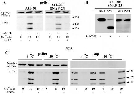
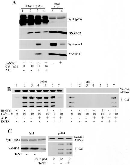
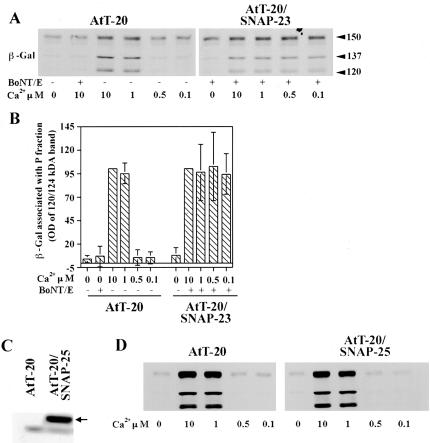
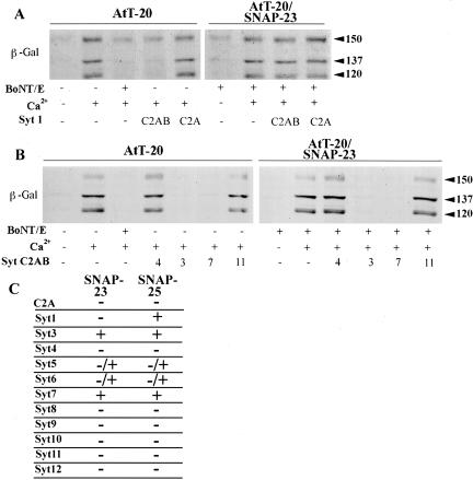
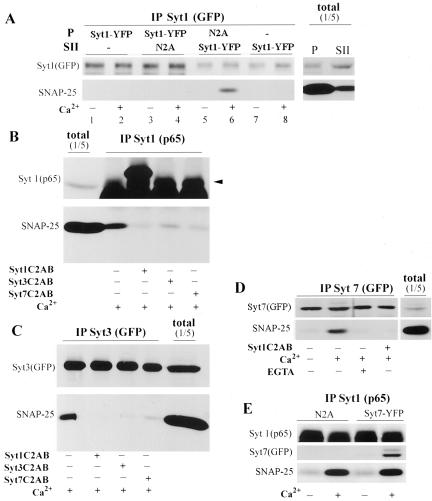
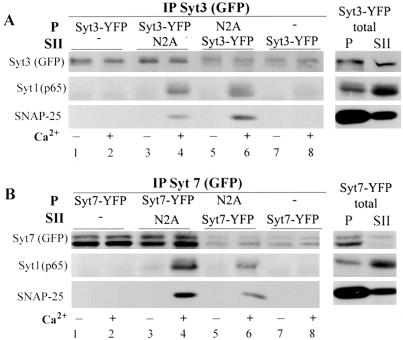
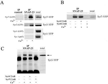
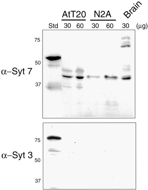
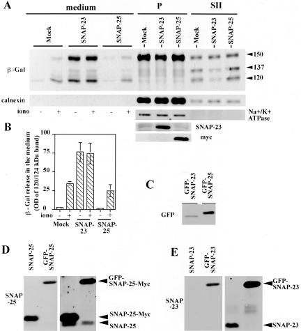
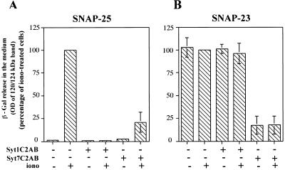
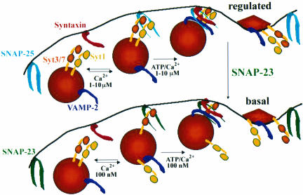
Ca(2+)-triggered exocytosis of secretory granules mediates the release of hormones from endocrine cells and neurons. The plasma membrane protein synaptosome-associated protein of 25 kDa (SNAP-25) is thought to be a key component of the membrane fusion apparatus that mediates exocytosis in neurons. Recently, homologues of SNAP-25 have been identified, including SNAP-23, which is expressed in many tissues, albeit at different levels. At present, little is known concerning functional differences among members of this family of proteins. Using an in vitro assay, we show here that SNAP-25 and SNAP-23 mediate the docking of secretory granules with the plasma membrane at high (1 microM) and low (100 nM) Ca(2+) levels, respectively, by interacting with different members of the synaptotagmin family. In intact endocrine cells, expression of exogenous SNAP-23 leads to high levels of hormone secretion under basal conditions. Thus, the relative expression levels of SNAP-25 and SNAP-23 might control the mode (regulated vs. basal) of granule release by forming docking complexes at different Ca(2+) thresholds.


Figures

Figure 1.
Figure 1.
SNAP-25- and SNAP-23-dependent docking. (A) Plasma membrane-containing fraction P from AtT-20 cells (AtT-20) and AtT-20 cells expressing SNAP-23 (AtT-20/SNAP-23, stable transfection; Koticha et al., 1999) were preincubated with or without 500 nM BoNT/E and mixed with granules (fraction SII) derived from N2A transiently transfected with POMC-β-Gal (see MATERIALS AND METHODS). Samples were incubated in the presence or absence of 10 μM free Ca2+ to induce docking and further incubated with or without 20 mM EGTA. Granules that cofractionated with the plasma membrane in the pellet after centrifugation at 7200 × g for 10 h (docked granules) were analyzed by Western blot with antibodies against the plasma membrane marker Na+/K+ATPase and β-Galactosidase. (B) Plasma-containing fraction P from AtT-20/SNAP-23 were incubated with 500 nM BoNT/E. Samples were analyzed by Western blot with antibodies against SNAP-25 and SNAP-23. (C) The docking assay was done with plasma membrane fraction P derived from N2A cells and granules derived from N2A transiently transfected with POMC-β-Gal. Samples were incubated at 4 and 30°C in the presence or absence of 10 μM free Ca2+ to induce docking and further incubated in the presence of EGTA as in A. After centrifugation, granules docked to the plasma membrane (pellet, 100% of total sample) and undocked granules in the supernatants (50% of total sample) were analyzed by Western blot as described in A.
Figure 2.
Figure 2.
The Ca2+-dependent docking complex includes Syt1 and SNAP-25, but not syntaxin 1 and VAMP-2. (A) Fractions containing plasma membrane (P) and granule (SII) were derived from wild-type N2A cells (N2A). The plasma membrane-containing P fraction was incubated for 30 h at 30°C with or without 100 nM BoNT/C in a buffer containing 100 mM NaCl, 100 mM KCl, 10 mM HEPES, and 1 mM dithiothreitol. P membranes were centrifuged at 7200 × g for 10 h, resuspended in Kglu buffer, mixed with granules in SII, and further incubated in the absence and in the presence of 10 μM free Ca2+ to induce docking. All samples were diluted in the IP buffer containing 50 μM free Ca2+ (see MATERIALS AND METHODS). Samples were immunoprecipitated with mouse monoclonal anti-Syt1 antibodies (p65) and analyzed by Western blot with antibodies against Syt1, SNAP-25, syntaxin, and VAMP2. Equal volumes of P and SII from cells were also analyzed (total). A representative experiment of a total of three independent ones is shown. (B) Plasma membranes in fraction P from N2A cells were incubated with or without BoNT/C as in A and mixed with granules derived from N2A transiently transfected with POMC-β-Gal. Samples were incubated at 30°C in the presence or absence of 10 μM free Ca2+, and where indicated, with 1 mM ATP and 1 mM MgCl2 (ATP). Samples were further incubated with or without 20 mM EGTA and analyzed as in Figure 1A. The entire pellet and one-half the supernatants were loaded into the SDS-PAGE acrylamide gel (C) Granules in fraction SII derived from N2A transiently transfected with POMC-β-Gal were incubated with and without 150 nM TeNT for 30 min at 30°C. The same fraction of each sample was analyzed by Western blot with antibodies against VAMP-2. The remaining TeNT-treated granules were mixed with the plasma membrane-containing fraction P from N2A cells and docking was induced and analyzed as in Figure 1C. Experiments in B and C were done twice, with similar results.
Figure 3.
Figure 3.
SNAP-23-dependent granule docking functions at lower [Ca2+] than that supported by SNAP-25. (A) Granule docking to plasma membranes from wild-type AtT-20 cells (SNAP-25-dependent docking) and to plasma membranes from AtT-20 cells expressing SNAP-23 treated with BoNT/E (SNAP-23-dependent docking) in the presence of the indicated free [Ca2+] was measured as in Figure 1A. (B) Three experiments, including that shown in A, are analyzed. Columns are mean OD values of the 120/124-kDa band of processed POMC-β-Gal ± SD. (C) Plasma membrane-containing fraction P from AtT-20 cells (AtT-20) and AtT-20 cells expressing SNAP-25A-Myc (AtT-20/SNAP-25, stable transfection) were analyzed by Western blot with antibodies against SNAP-25. The arrow indicates exogenous SNAP-25A-Myc protein. (D) Granule docking to plasma membranes from wild-type AtT-20 cells and to plasma membranes from AtT-20 cells expressing SNAP-25 in the presence of the indicated free [Ca2+] was measured as in A.
Figure 4.
Figure 4.
Syt1 C2AB inhibits specifically SNAP-25-dependent docking. (A and B) Plasma membranes from AtT-20 cells and from AtT-20 cells expressing SNAP-23 were pretreated with or without BoNT/E, mixed with granules, and incubated with 5 μM GST-Syt1C2AB or GST-Syt1C2A (A) or with 5 μM of the GST-C2AB domain of the indicated Synaptotagmins (B). Samples were further incubated in the absence or in the presence of 100 μM Ca2+ to induce docking. Granules docked to the plasma membrane were analyzed by Western blot as in Figure 1A. (C) The table shows the effect of 5 μM C2AB domains of the indicated Synaptotagmins and of 5 μM Syt1C2A (C2A) on SNAP-23 and SNAP-25-dependent docking. Data were derived from experiments (n = 2 for each peptide) as in A and B.
Figure 5.
Figure 5.
SNAP-25-dependent docking complex is formed in trans by SNAP-25 at the plasma membrane and Syt1 on the granule. (A) Fractions containing plasma membrane (P) and granule (SII) were derived either from wild-type N2A cells (N2A) or from N2A cells transiently transfected with Syt1-YFP, as indicated. P and SII were mixed and incubated in the absence and in the presence of 10 μM free Ca2+ to induce docking. All samples were diluted in the IP buffer containing 50 μM free Ca2+ (see MATERIALS AND METHODS). Samples were immunoprecipitated with mouse monoclonal anti-GFP antibodies and analyzed by Western blot with rabbit anti-GFP antibodies (to detect Syt1-YFP) and with antibodies against SNAP-25, as indicated. Equal volumes of P and SII from cells transfected with Syt1-YFP were analyzed (total, right). (B-D) Plasma membranes and granules from N2A cells (B) and N2A cells transiently transfected with Syt3-YFP (C) and Syt7-YFP (D) were mixed and preincubated with or without 5 μM GST-Syt1C2AB, 5 μM GST-Syt3C2AB, and 5 μM GST-Syt7C2AB. After incubation in the absence or in the presence of 100 μM free Ca2+ to induce docking, samples were diluted in the IP buffer (as in A) and immunoprecipitated with the indicated antibodies. The immunoprecipitated material was analyzed by Western blot with Syt1 antibodies (p65, to detect endogenous Syt1) and GFP antibodies (to detect Syt3-YFP and Syt7-YFP) and SNAP-25 antibodies. Arrowhead in B shows the immunoprecipitated Syt1. (E) Plasma membranes and granules from N2A cells or N2A cells transiently transfected with Syt7-YFP were used for the docking reaction. Samples were immunoprecipitated with Syt1 antibodies (p65) and analyzed by Western Blot with the indicated antibodies.
Figure 6.
Figure 6.
Syt3 and Syt7 in the plasma membrane and granule fraction function in SNAP-25-dependent docking. (A and B) Fractions containing plasma membrane (P) and granule (SII) were derived from wild-type N2A cells (N2A) and cells transiently transfected with Syt3-YFP and with Syt7-YFP cDNA as indicated. P and SII were mixed and incubated in the absence and in the presence of 10 μM free Ca2+ to induce docking. Samples were diluted in the IP buffer and immunoprecipitated as in Figure 5A. Samples were analyzed by Western blot with GFP antibodies (to detect Syt3-YFP and Syt7-YFP), with p65 antibodies (to detect endogenous Syt1), and with SNAP-25 antibodies, as indicated.
Figure 7.
Figure 7.
Interaction of SNAP-23 with Syt7 (or Syt3), but not with Syt1, supports SNAP-23-dependent granule docking. (A) Fractions containing plasma membrane (P) and granule (SII) were derived from G14 cells transiently cotransfected with SNAP-23 and Syt1-YFP or Syt3-YFP or Syt7-YFP and mixed in the docking assay. Incubations were carried out as in Figure 5B. Samples were immunoprecipitated with anti-SNAP-23 polyclonal antibody (Koticha et al., 1999) and analyzed by Western blot with antibodies against GFP (to detect Syt1-YFP, Syt3-YFP, andSyt7-YFP) and p65 (to detect endogenous Syt1). (B and C) Plasma membranes (P) and granules (SII) from N2A cells transiently transfected with Syt7-YFP (B) and Syt3-YFP (C) were mixed and incubated with or without 5 μM GST-Syt1C2AB, 5 μM GST-Syt3C2AB, 5 μM GST-Syt7C2AB, and 100 μM Ca2+ as indicated. Samples were immunoprecipitated and analyzed by Western blot as in A.
Figure 8.
Figure 8.
Syt7 is expressed in AtT-20 and N2A cells. The standards (Std) correspond to 1.5 ng of a fragment of Syt 7 (residues 1-260) or 15 ng of a fragment of Syt 3 (residues 1-423) fused to GST, and immunoblot analysis was carried out using isoform specific antibodies as described in Tucker et al. (2003). The indicated amounts of postnuclear supernatants derived from AtT-20 cells, N2A cells, and brain were mixed with sample buffer and loaded onto the SDS gel.
Figure 9.
Figure 9.
Expression of SNAP-23 in N2A cells leads to high levels of hormone release in the basal state. (A) N2A cells were transiently cotransfected with POMC-β-Gal-pcDNA3 and control pcB7 vector (mock), SNAP-23-pcB7 (SNAP-23), or SNAP-25A-Myc-pcB7 (SNAP-25). Cells were kept in basal and stimulated (+ 5 μM Ionomycin) conditions as described in Secretion Assay in MATERIALS AND METHODS. The medium and the P and SII fractions derived from the cells were analyzed by Western blot with the indicated antibodies. (B) Three independent experiments including that shown in B were analyzed. Columns are mean OD values of the 120/124-kDa band of processed POMC-β-Gal in the medium ± SD. (C-E) In parallel experiments N2A cells were transfected with SNAP-25AMyc-pEGFP.C1 (GFP-SNAP-25) (C and D), SNAP-23-pEGFP.C1 (GFP-SNAP-23) (C and E), SNAP-25A-Myc-pcB7 (SNAP-25) (D), and SNAP-23-pcB7 (SNAP-23) (E). Equal amounts (10 μg) of P fractions derived from these cells were analyzed with antibodies against GFP, SNAP-23, and SNAP-25, as indicated. (D and E) Same blot exposed for 20 s (left) and 4 min (right).
Figure 10.
Figure 10.
C2AB domain of Syt1 inhibits regulated exocytosis but does not impair SNAP-23-dependent granule release in the basal state. (A) N2A cells were transiently cotransfected with POMC-β-Gal-pcDNA3, SNAP-25A-Myc-pcB7 (SNAP-25), and pcDNA/GW/d-TOPO-Syt1C2AB-EYFP (Syt1C2AB) or pcDNA/GW/d-TOPO-Syt7C2AB-EYFP (Syt7C2AB). Cells were kept in basal and stimulated (+ 5 μM Ionomycin) conditions and secretion was measured as in Figure 8. (B) N2A cells were transiently cotransfected with POMC-β-Gal-pcDNA3, SNAP-23-pcB7 (SNAP-23), and pcDNA/GW/d-TOPO-Syt1C2AB-EYFP (Syt1C2AB) or pcDNA/GW/d-TOPO-Syt7C2AB-EYFP (Syt7C2AB), and secretion was analyzed as in A. Columns are mean OD values of the 120/124-kDa POMC-β-Gal band in the medium (derived from three independent experiments) shown as percentage of the ionomycin-treated sample ± SD.
Figure 11.
Figure 11.
SNAP-25- and SNAP-23-dependent docking/fusion of granules. In endocrine cells with endogenous levels of SNAP-25 and SNAP-23 granules positioned at the cell cortex dock to the plasma membrane in response to an increase in intracellular [Ca2+] by forming a complex with SNAP-25, Syt1, and Syt3 (or Syt7). This step is followed by other ATP- and Ca2+-dependent steps that lead to SNARE complex formation and fusion. Differently, in endocrine cells overexpressing SNAP-23, granules dock and fuse at resting Ca2+ levels by forming a complex with SNAP-23 and Syt7.

Similar articles

Cited by

References

    1. Baldini, G., Baldini, G., Wang, G., Weber, M., Zweyer, M., Bareggi, R., Witkin, J.W., and Martelli, A.M. (1998). Expression of Rab3D N135I inhibits regulated secretion of ACTH in AtT-20cells. J. Cell Biol. 140, 305-313. - PMC - PubMed
    1. Binz, T., Blasi, J., Yamasaki, S., Baumeister, A., Link, E., Sudhof, T.C., Jahn, R., and Niemann, H. (1994). Proteolysis of SNAP-25 by types E and A botulinal neurotoxins. J. Biol. Chem. 269, 1617-1620. - PubMed
    1. Bittner, M.A., and Holz, R.W. (1992). Kinetic analysis of secretion from permeabilized adrenal chromaffin cells reveals distinct components. J. Biol. Chem. 267, 16219-16225. - PubMed
    1. Blasi, J., Chapman, E.R., Yamasaki, S., Binz, T., Niemann, H., and Jahn, R. (1993). Botulinum neurotoxin C1 blocks neurotransmitter release by means of cleaving HPC-1/syntaxin. EMBO J. 12, 4821-4828. - PMC - PubMed
    1. Borgonovo, B., Cocucci, E., Racchetti, G., Podini, P., Bachi, A., and Meldolesi, J. (2002). Regulated exocytosis: a novel, widely expressed system. Nat. Cell Biol. 4, 955-962. - PubMed

Publication types

MeSH terms

LinkOut - more resources