. 2004 Feb;10(2):287-98.
doi: 10.1261/rna.5138804.

L-Myc protein synthesis is initiated by internal ribosome entry

Affiliations

L-Myc protein synthesis is initiated by internal ribosome entry

Catherine L Jopling et al. RNA. 2004 Feb.

Abstract

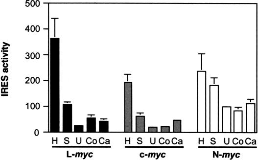
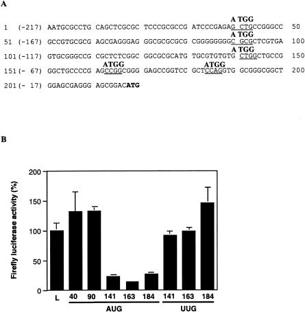
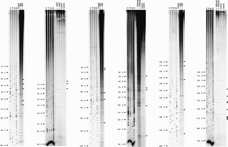
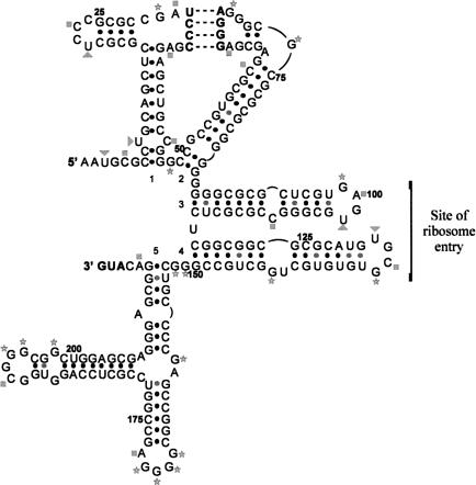
An internal ribosome entry segment (IRES) has been identified in the 5' untranslated region (5' UTR) of two members of the myc family of proto-oncogenes, c-myc and N-myc. Hence, the synthesis of c-Myc and N-Myc polypeptides can involve the alternative mechanism of internal initiation. Here, we show that the 5' UTR of L-myc, another myc family member, also contains an IRES. Previous studies have shown that the translation of mRNAs containing the c-myc and N-myc IRESs can involve both cap-dependent initiation and internal initiation. In contrast, the data presented here suggest that internal initiation can account for all of the translation initiation that occurs on an mRNA with the L-myc IRES in its 5' UTR. Like many other cellular IRESs, the L-myc IRES appears to be modular in nature and the entire 5' UTR is required for maximum IRES efficiency. The ribosome entry window within the L-myc IRES is located some distance upstream of the initiation codon, and thus, this IRES uses a "land and scan" mechanism to initiate translation. Finally, we have derived a secondary structural model for the IRES. The model confirms that the L-myc IRES is highly structured and predicts that a pseudoknot may form near the 5' end of the mRNA.


Figures

FIGURE 1.
FIGURE 1.
Sequence of the L-myc 5′ UTR. The sequence of the 5′ region of the L-myc gene from the transcription initiation site to the first initiation codon. Exon 1, intron A, and exon 2 are indicated with arrows, and the start codon is italicized. The short isoform of the L-myc 5′ UTR is composed of exon 1 and 10 nucleotides of exon 2, whereas the long isoform also retains intron A. A DNA fragment encoding the short isoform was amplified from HL60 cDNA using the primers LF2 and LR2 (see Materials and Methods). Shown in bold is the sequence used to design the primer LF2 (5′-AATGCGCCTG CAGCTCGCGCTCCC-3′). To ensure that only the short 5′ UTR was amplified, the oligonucleotide LR2 was designed to complement the sequence from the 3′ end of exon 1 and the 5′ end of exon 2 (5′-GGCGGCTGGAGCGAGGGGAGCCGACATG-3′). This sequence is underlined.
FIGURE 2.
FIGURE 2.
Effect of the L-myc 5′ UTR on reporter gene expression. (A) (i) Schematic representation of the firefly luciferase expression cassette of pGL3 and phpL. As detailed in Stoneley et al. (2000), phpL was created from pGL3 by introducing an inverted repeat sequence into the SpeI site. (ii) Inserting the L-myc 5′ UTR sequence between the EcoRI and NcoI sites of pGL3 and phpL created the constructs pGLsL and phpLsL, respectively. In these constructs, the 3′ end of the 5′ UTR sequence is followed immediately by the firefly luciferase initiation codon. Hence, the relative positions of the initiation codon and the 5′ UTR are the same in these constructs as in the L-myc mRNA. (B) HeLa cells were transfected with the constructs pGL3, phpL, pGLsL, and phpLsL in conjunction with the plasmid pcDNA3.1/HisB/lacZ. Lysates were prepared from the cells 48 h post-transfection, and the activity of firefly luciferase was determined as described in Materials and Methods. To account for variations in transfection efficiency firefly luciferase activity was normalized to that of β-galactosidase.
FIGURE 3.
FIGURE 3.
The L-myc 5′ UTR contains an IRES. (A) Schematic representation of the expression cassette of the dual-luciferase dicistronic constructs pRF, pRMF, pRNF, and pRLF. The construct pRLF was generated inserting the L-myc 5′ UTR sequence into pRF between the EcoRI and NcoI sites. The remaining constructs have been described elsewhere (Stoneley et al. 1998; Jopling and Willis 2001). As described in Figure 2A ▶ (ii), the 5′ UTR sequence is inserted into these constructs such that the relative positions of the initiation codon and the 5′ UTR are the same as in the L-myc mRNA. The sequence of the junction between the L-myc 5′ UTR and the firefly luciferase-coding region is identical to that shown in Figure 2A ▶ (ii). (B) The constructs detailed above were transfected into HeLa cells in combination with pcDNA3.1/HisB/lacZ. Renilla luciferase, firefly luciferase, and β-galactosidase activities were determined 48 h post-transfection. Renilla luciferase (gray) and firefly luciferase (black) activities were normalized to β-galactosidase activity. (C) The L-myc 5′ UTR sequence was inserted into the construct phpRF between the EcoRI and NcoI sites. HeLa cells were transfected with pRF, phpRF, pRLF, and phpRLF in conjunction with pcDN3.1/HisB/lacZ. Normalized Renilla luciferase (gray) and firefly luciferase (black) activities were then determined as described previously. (D) Total RNA was isolated from HeLa cells that had been transfected with pGLsL, pRF, and pRLF. Poly(A)+ RNA was purified from these RNA samples, and Northern analysis was performed essentially as described in West et al. (1995). Transcripts containing firefly luciferase-coding sequence were detected using a radiolabeled probe (Coldwell et al. 2000).
FIGURE 4.
FIGURE 4.
A comparison of the c-, N-, and L-myc IRES efficiency in a range of cell lines. The constructs pRF, pRLF, pRMF, and pRNF were transfected into HeLa (H; cervical carcinoma), SaOS2 (S; osteosarcoma), U2OS (U; osteosarcoma), Cos-1 (Co; SV40-transformed epithelial cells), and Cal51 (Ca; breast carcinoma) cells in conjunction with pcDNA3.1/HisB/lacZ. Cell lysates were prepared 48 h after transfection, and the activities of firefly luciferase, Renilla luciferase (data not shown), and β-galactosidase were determined. After normalizing firefly luciferase activity to that of β-galactosidase, the IRES activity was calculated using the ratio (firefly luciferase activity from pRLF, pRMF, or pRNF)/(firefly luciferase activity from pRF).
FIGURE 5.
FIGURE 5.
Deletion analysis of the L-myc IRES. (A) Diagrammatic representation of the truncated versions of the L-myc 5′ UTR used in the deletion analysis. DNA fragments were amplified using PCR (using oligonucleotides as described in Materials and Methods) and inserted into the dicistronic construct pRF at the EcoRI and NcoI sites. (B) The constructs pRF, pRLF, and the dicistronic constructs containing the truncated 5′ UTR sequences, as described above, were transfected into HeLa cells, and lysates were prepared 48 h post-transfection. All constructs were cotransfected with pcDNA3.1/HisB/lacZ. Renilla luciferase (gray) and firefly luciferase (black) activities were determined and normalized to β-galactosidase activities.
FIGURE 6.
FIGURE 6.
Locating the ribosome entry window in the L-myc IRES. (A) The sequence of the short L-myc 5′ UTR indicating the positions at which initiation codons were introduced. Site-directed mutagenesis was used to alter the underlined sequence to ATGG using oligonucleotides (see Materials and Methods). In addition, the sequences at 141 to 144, 163 to 166, and 184 to 187 were also altered to UUGG. Mutated L-myc 5′ UTR PCR fragments were then inserted into the dicistronic construct pRF. The position of each mutated sequence is numbered from the A of the ATGG or U of UUGG. (B) HeLa cells were transfected with pRLF (L) and the dicistronic constructs containing the mutated L-myc sequences (AUG 40, AUG 90, AUG 141, AUG 163, AUG 184, UUG 141, UUG 163, and UUG 184) in conjunction with pcDNA3.1/HisB/lacZ. Lysates were prepared 48 h post-transfection. The data are represented as the percentage firefly luciferase activity compared with the activity from pRLF.
FIGURE 7.
FIGURE 7.
Chemical and enzymatic probing of the L-myc IRES. Renatured in vitro-transcribed L-myc IRES RNA was treated with DMS, kethoxal, CMCT, or RNase V1 (data not shown) as described in Materials and Methods. Primer extension was then performed with these samples and a mock-treated L-myc IRES RNA sample (control) using [α-32P]dCTP and an oligonucleotide (see Materials and Methods). The products of these reactions, together with the corresponding DNA-sequencing reaction were subjected to electrophoresis on a denaturing 6% polyacrylamide/urea gel. The black arrows indicate sites of chemical modification.
FIGURE 8.
FIGURE 8.
Secondary structural model of the L-myc IRES. The secondary structural model of the L-myc IRES was derived using the chemical and enzymatic accessibility data to constrain the Mfold algorithm. Five regions of helical structure are indicated (1–5). Nucleotides involved in pairing interactions are indicated by a black dot. The sequences involved in forming the potential pseudoknot are indicated in bold, and the region of ribosome entry is also indicated. The position of modifications are indicated as follow; (squares) sites of DMS modification (A,C); (triangles) sites of CMCT modification (U); (stars) sites of kethoxal modification (G). Numbering is from 5′ to 3′.

Similar articles

Cited by

References

    1. Belsham, G.J. and Jackson, R.J. 2000. Translation initiation on Picornavirus RNA. In Translational control of gene expression (eds. N. Sonenberg et al.), pp. 869–900. Cold Spring Harbor Laboratory Press, Cold Spring Harbor, NY.
    1. Birrer, M.J., Dosaka, H., Segal, S., Nau, M., Degreve, J., Kaye, F., Sausville, E., and Minna, J. 1988a. Multiple L-myc messenger-RNA forms have transforming activity for primary rat embryo fibroblasts and an established rat fibroblast cell-line. Clinical Res. 36: A589–A589.
    1. Birrer, M.J., Segal, S., Degreve, J.S., Kaye, F., Sausville, E.A., and Minna, J.D. 1988b. L-Myc cooperates with Ras to transform primary rat embryo fibroblasts. Mol. Cell. Biol. 8: 2668–2673. - PMC - PubMed
    1. Blackwood, E.M., Luscher, B., and Eisenman, R.N. 1992. Myc and Max associate in vivo. Genes & Dev. 6: 71–80. - PubMed
    1. Bonnal, S., Boutonnet, C., Prado-Lourenco, L., and Vagner, S. 2003. IRESdb: The internal ribosome entry site database. Nucleic Acids Res. 31: 427–428. - PMC - PubMed

Publication types

LinkOut - more resources