Decreased expression of the CD3zeta chain in T cells infiltrating the synovial membrane of patients with osteoarthritis
- PMID: 14715568
- PMCID: PMC321327
- DOI: 10.1128/cdli.11.1.195-202.2004
Decreased expression of the CD3zeta chain in T cells infiltrating the synovial membrane of patients with osteoarthritis
Abstract
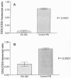
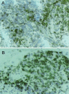
Osteoarthritis (OA) is a heterogeneous disease which rheumatologists consider to be noninflammatory. However, recent studies suggest that, at least in certain patients, OA is an inflammatory disease and that patients often exhibit inflammatory infiltrates in the synovial membranes (SMs) of macrophages and activated T cells expressing proinflammatory cytokines. We report here that the expression of CD3zeta is significantly decreased in T cells infiltrating the SMs of patients with OA. The CD3zeta chain is involved in the T-cell signal transduction cascade, which is initiated by the engagement of the T-cell antigen receptor and which culminates in T-cell activation. Double immunofluorescence of single-cell suspensions derived from the SMs from nine patients with OA revealed significantly increased proportions of CD3epsilon-positive (CD3epsilon+) cells compared with the proportions of CD3zeta-positive (CD3zeta+) T cells (means +/- standard errors of the means, 80.48% +/- 3.92% and 69.02% +/- 6.51%, respectively; P = 0.0096), whereas there were no differences in the proportions of these cells in peripheral blood mononuclear cells (PBMCs) from healthy donors (94.73% +/- 1.39% and 93.79% +/- 1.08%, respectively; not significant). The CD3zeta+ cell/CD3epsilon+ cell ratio was also significantly decreased for T cells from the SMs of patients with OA compared with that for T cells from the PBMCs of healthy donors (0.84 +/- 0.17 and 0.99 +/- 0.01, respectively; P = 0.0302). The proportions of CD3epsilon+ CD3zeta+ cells were lower in the SMs of patients with OA than in the PBMCs of healthy donors (65.04% +/- 6.7% and 90.81% +/- 1.99%, respectively; P = 0.0047). Substantial proportions (about 15%) of CD3epsilon+ CD3zeta-negative (CD3zeta-) and CD3epsilon-negative (CD3epsilon-) CD3zeta- cells were found in the SMs of patients with OA. Amplification of the CD3zeta and CD3delta transcripts from the SMs of patients with OA by reverse transcriptase PCR consistently exhibited stronger bands for CD3delta cDNA than for CD3zeta cDNA The CD3zeta/CD3delta transcript ratio in the SMs of patients with OA was significantly lower than that in PBMCs from healthy controls (P < 0.0001). These results were confirmed by competitive MIMIC PCR. Immunoreactivities for the CD3zeta protein were detected in the SMs of 10 of 19 patients with OA, and they were of various intensities, whereas SMs from all patients were CD3epsilon+ (P = 0.0023). The decreased expression of the CD3zeta transcript and protein in T cells from the SMs of patients with OA relative to that of the CD3epsilon transcript is suggestive of chronic T-cell stimulation and supports the concept of T-cell involvement in OA.
Figures


Similar articles
-
Immune Contributions to Osteoarthritis.Curr Osteoporos Rep. 2017 Dec;15(6):593-600. doi: 10.1007/s11914-017-0411-y. Curr Osteoporos Rep. 2017. PMID: 29098574 Review.
-
T cells and T-cell cytokine transcripts in the synovial membrane in patients with osteoarthritis.Clin Diagn Lab Immunol. 1998 Jul;5(4):430-7. doi: 10.1128/CDLI.5.4.430-437.1998. Clin Diagn Lab Immunol. 1998. PMID: 9665944 Free PMC article.
-
Lung carcinomas do not induce T-cell apoptosis via the Fas/Fas ligand pathway but down-regulate CD3 epsilon expression.Cancer Immunol Immunother. 2008 Mar;57(3):325-36. doi: 10.1007/s00262-007-0372-6. Epub 2007 Aug 1. Cancer Immunol Immunother. 2008. PMID: 17668204 Free PMC article.
-
Reduced T-cell receptor CD3zeta-chain protein and sustained CD3epsilon expression at the site of mycobacterial infection.Immunology. 2001 Nov;104(3):269-77. doi: 10.1046/j.1365-2567.2001.01323.x. Immunology. 2001. PMID: 11722641 Free PMC article.
-
Synovial inflammation, immune cells and their cytokines in osteoarthritis: a review.Osteoarthritis Cartilage. 2012 Dec;20(12):1484-99. doi: 10.1016/j.joca.2012.08.027. Epub 2012 Sep 7. Osteoarthritis Cartilage. 2012. PMID: 22960092 Review.
Cited by
-
Immune Mediators in Osteoarthritis: Infrapatellar Fat Pad-Infiltrating CD8+ T Cells Are Increased in Osteoarthritic Patients with Higher Clinical Radiographic Grading.Int J Rheumatol. 2016;2016:9525724. doi: 10.1155/2016/9525724. Epub 2016 Dec 14. Int J Rheumatol. 2016. PMID: 28070192 Free PMC article.
-
Investigation of candidate genes for osteoarthritis based on gene expression profiles.Acta Orthop Traumatol Turc. 2016 Dec;50(6):686-690. doi: 10.1016/j.aott.2016.04.002. Epub 2016 Nov 18. Acta Orthop Traumatol Turc. 2016. PMID: 27866912 Free PMC article.
-
Obesity, Inflammation, and Immune System in Osteoarthritis.Front Immunol. 2022 Jul 4;13:907750. doi: 10.3389/fimmu.2022.907750. eCollection 2022. Front Immunol. 2022. PMID: 35860250 Free PMC article. Review.
-
Human autoimmune diseases are specific antigen-driven T-cell diseases: identification of the antigens.Immunol Res. 2007;38(1-3):359-72. doi: 10.1007/s12026-007-0044-9. Immunol Res. 2007. PMID: 17917046 Review.
-
Immune Contributions to Osteoarthritis.Curr Osteoporos Rep. 2017 Dec;15(6):593-600. doi: 10.1007/s11914-017-0411-y. Curr Osteoporos Rep. 2017. PMID: 29098574 Review.
References
-
- Allen, M. E., S. P. Young, R. H. Michell, and P. A. Bacon. 1995. Altered T lymphocyte signaling in rheumatoid arthritis. Eur. J. Immunol. 25:1547-1554. - PubMed
-
- Alsalameh, S., J. Mollenhauer, N. Hain, K. P. Stock, J. R. Kalden, and G. R. Burmester. 1990. Cellular immune responses toward human articular chondrocytes. T cell reactivities against chondrocyte and fibroblast membranes in destructive joint diseases. Arthritis Rheum. 33:1477-1486. - PubMed
-
- Altman, R. D. 1991. Classification of disease: osteoarthritis. Semin. Arthritis Rheum. 20:40-47. - PubMed
-
- Carson, C. W., L. D. Beal, G. G. Hunder, C. M. Johnson, and W. Newman. 1994. Soluble E-selectin is increased in inflammatory synovial fluid. J. Rheumatol. 21:605-611. - PubMed
Publication types
MeSH terms
Substances
Grants and funding
LinkOut - more resources
Full Text Sources
Medical

