. 2004 Jan;24(2):823-36.
doi: 10.1128/MCB.24.2.823-836.2004.

Distinct domains in the SHP-2 phosphatase differentially regulate epidermal growth factor receptor/NF-kappaB activation through Gab1 in glioblastoma cells

Affiliations

Distinct domains in the SHP-2 phosphatase differentially regulate epidermal growth factor receptor/NF-kappaB activation through Gab1 in glioblastoma cells

Gurpreet S Kapoor et al. Mol Cell Biol. 2004 Jan.

Abstract

The transcription factor nuclear factor kappaB (NF-kappaB) plays an important role in inflammation and cancer, is activated by a variety of stimuli including tumor necrosis factor alpha, interleukin-1, UV irradiation, and viruses, as well as receptor tyrosine kinases, such as epidermal growth factor receptor (EGFR). Although previous studies suggest that EGFR can induce NF-kappaB, the mechanism of this activation remains unknown. In this study, we identify the components of the EGFR-induced signalosome in human glioblastoma cells required to regulate NF-kappaB activation. Immunoprecipitation analyses with ErbB-modulated cells indicate that association between SHP-2 and Grb2-associated binder 1 (Gab1) is the critical step in the formation of the signalosome linking EGFR to NF-kappaB activation. We also show that EGFR-induced NF-kappaB activation is mediated by the PI3-kinase/Akt activation loop. Overexpression of SHP-2, Gab1, and myristoylated Akt significantly upregulated NF-kappaB transcriptional activity and DNA binding activity in glioblastoma cells. Interestingly, overexpression of either one of the two SH2 domain mutants of SHP-2, R32E or R138E, slightly reduced NF-kappaB activity relative to that of wild-type SHP-2, indicating that the SH2 domains of SHP-2 are required for EGFR-induced NF-kappaB activation. On the other hand, ectopic overexpression of either a Gab1 mutant incapable of binding to SHP-2 (Y627F) or a phosphatase-inactive SHP-2 mutant (C459S) caused a significant increase in NF-kappaB activity. Moreover, SHP-2 C459S-expressing cells displayed higher Gab1 phosphotyrosine content, suggesting that SHP-2 regulates Gab1 phosphorylation through its phosphatase domain, which confers a negative regulatory effect on NF-kappaB activity. These results indicate that SHP-2/Gab1 association is critical for linking EGFR to NF-kappaB transcriptional activity via the PI3-kinase/Akt signaling axis in glioblastoma cells and that SHP-2 acts as a dual regulator of NF-kappaB activation.


Figures
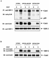
FIG. 1.
FIG. 1.
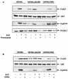
EGFR activation by ligand or oncogenic mutation induces Gab1 and SHP-2 association in glioblastoma cells. (A) U87MG, U87MG.ΔEGFR, and U87MG/T691 cells were serum starved for 24 h and subsequently either stimulated with EGF (50 ng/ml) or left unstimulated. Cell lysates were subjected to immunoprecipitation with anti-SHP-2 antibody followed by SDS-PAGE and immunoblotting with anti-phosphotyrosine (pY20) and anti-Gab1 antibodies. The membrane was stripped and reprobed with anti-SHP-2 antibody to determine the amount of SHP-2 protein precipitated from each sample. (B) Unstimulated and EGF-stimulated cell lysates were subjected to immunoprecipitation with anti-Gab1 antibody followed by SDS-PAGE and immunoblotting with anti-SHP-2 antibody. IP, immunoprecipitation; IB, immunoblotting.
FIG. 2.
FIG. 2.
The PI3-kinase/Akt pathway is required for EGFR-mediated oncogenic signaling in glioblastoma cells. (A and B) Serum-starved U87MG, U87MG.ΔEGFR, and U87MG/T691 cells were pretreated for 25 min with wortmannin (100 nmol/ml) or U0126 (10 μmol/ml) when indicated before a 5-min stimulation with 50 ng of EGF/ml. Total cellular proteins (30 μg) were resolved by SDS-PAGE and immunoblotted with anti-phospho-Akt, anti-phospho-p42/44MAPK, and anti-phospho-GSK-3 antibodies. Membranes were stripped and reprobed with anti-total-Akt antibody.
FIG. 3.
FIG. 3.
Overexpression of Gab1 and SHP-2 upregulates Akt kinase activity in glioblastoma cells. U87MG and U87MG/T691 cells were plated at a density of 1.5 × 10⁶ cells per 10-cm-diameter dish. The following day, cells were transiently transfected with approximately 5 μg of the empty vector, Gab1(WT), or SHP-2(WT) construct. Transfected cells were left untreated or incubated with EGF (100 ng/ml) for 5 min after 24 h of serum starvation. Cell lysates with equal protein content were immunoprecipitated with phospho-specific Akt monoclonal antibody. Immune complexes were subjected to an Akt kinase assay using GSK-3 fusion protein as a substrate according to the manufacturer's recommendations (nonradioactive Akt kinase assay kit from Cell Signaling Technology). Kinase assay reaction mixtures were resolved by SDS-PAGE and immunoblotted with anti-phospho-GSK-3α/β antibody. Immunoreactive bands were quantified by Scion Image Beta Release 3b software and then plotted using the Microsoft Excel program.
FIG. 4.
FIG. 4.
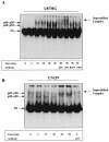
EGFR-induced upregulation of NF-κB DNA-binding activity in glioblastoma cells. (A and B) U87MG cells and LN229 cells were treated for the indicated time intervals with EGF (50 ng/ml) after 24 h of serum starvation. Nuclear protein was isolated, and the binding reaction was performed with consensus NF-κB oligonucleotide (Promega) labeled with [γ32-P]ATP in the presence of 2 μg of poly(dI-dC). The reaction mixture was electrophoresed on a 4% native polyacrylamide gel that was then vacuum dried and processed for autoradiography. The experiment shown here is representative of three independent experiments. For NF-κB supershift analysis, 2.5 μg of nuclear extract was preincubated with 2 μg of p65, p50, RelB, and c-Rel antibodies at room temperature for 10 min, and then EMSA was performed as described above.
FIG. 5.
FIG. 5.
Akt overexpression increases NF-κB activity in glioblastoma cells. A 5× NF-κB-Luciferase reporter gene was cotransfected into U87MG and U87MG/T691 cells with either active (myristoylated) or kinase-dead Akt and pSV-β-galactosidase expression constructs (0.5 μg each). Forty-eight hours after transfection, cells were serum starved for 24 h and cell lysates were made. Cells lysates were assayed for luciferase activity according to the manufacturer's instruction (Promega). All activities were normalized by β-galactosidase activity from three different experiments. The results are reported as the means ± standard deviation of fold induction, considering 1 as the relative luciferase activity of the cells transfected with corresponding empty vector.
FIG. 6.
FIG. 6.
Gab1 and SHP-2 overexpression increases NF-κB activity in glioblastoma cells via the PI3-kinase pathway. (A) U87MG cells and U87MG/T691 cells were cotransfected with a 5× NF-κB-luciferase reporter gene, the pSV-β-galactosidase vector, and one of the following constructs (0.5 μg each): an empty vector, a wild-type Gab1 construct, a wild-type SHP-2 construct, or a vector expressing a Gab1 mutant incapable of binding the p85 subunit of PI3-kinase (YF3). (B) LN229 cells were cotransfected with a 5× NF-κB-luciferase reporter gene, the pSV-β-galactosidase vector, and 0.5 μg of one of the following constructs: an active (myristoylated) Akt or a kinase-dead Akt construct, a wild-type Gab1 construct, a wild-type SHP-2 construct, a vector expressing a Gab1 mutant incapable of binding the p85 subunit of PI3-kinase (YF3), a vector expressing constitutive active EGFR (ΔEGFR), or a vector expressing the ErbB2/Neu receptor mutant (T691stop). In addition, U87MG, U87MG/T691, and LN229 cells were cotransfected with a 5× NF-κB-Luciferase reporter gene, the pSV-β-galactosidase vector, a wild-type Gab1 construct, and a vector expressing wild-type SHP-2 (A and B). Forty-eight hours after transfection, cells were serum starved for 24 h, followed by cell lysis and measurement of luciferase activity. Values obtained were normalized to β-galactosidase activity. The experiments were performed three times in duplicate. Error bars represents standard deviations. Basal promoter activity of the NF-κB-Luciferase reporter when transfected with empty vector alone is set at 1. P values were <0.05. (C) U87MG cells were transiently transfected with 5 μg each of the empty vector, Gab1(WT), Gab1(YF3), and SHP-2(WT) constructs. Twenty-four hours after transfection, cells were placed in serum-free medium. Forty-eight hours after transfection, cells were harvested and nuclear extracts were made. EMSAs were performed as described in Materials and Methods.
FIG. 7.
FIG. 7.
The phosphatase domain of SHP-2 regulates NF-κB activation. (A and B) U87MG, U87MG/T691, and LN229 cells were cotransfected with the 5× NF-κB-Luciferase promoter, the pSV-β-galactosidase vector, and either a vector expressing a Gab1 mutant incapable of binding SHP-2 (Y627F) or a vector containing protein phosphatase-inactive SHP-2(C459S) cDNA (0.5 μg). Forty-eight hours after transfection, cells were placed in serum-free medium for 24 h followed by cell lysis. Cells lysates were assayed for luciferase and β-galactosidase activities according to the manufacturer's instruction (Promega). The luciferase activity was normalized with that of β-galactosidase, and fold induction was calculated. The data represent a mean for three experiments ± standard deviation, considering relative luciferase activity of cells transfected with empty vector as 1. P values were <0.05. (C). U87MG cells were transiently transfected with an empty vector and the Gab1(WT), Gab1(Y672F), SHP-2(WT), and SHP-2(C459S) constructs. Twenty-four hours after transfection, cells were placed in serum-free medium. Forty-eight hours after transfection, cells were harvested and nuclear extracts were made. EMSAs were performed as described in Materials and Methods. The EMSA shown is representative of one of the three different experiments.
FIG. 8.
FIG. 8.
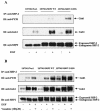
Reduced NF-κB activity in glioblastoma cells expressing SH2 domain mutants of SHP-2. (A and B) U87MG and LN229 cells were cotransfected with the 5× NF-κB-Luciferase promoter, the pSV-β-galactosidase vector, and 0.5 μg of vectors expressing either one of the two SH2 domain mutant of SHP-2: R32E (N-SH2) or R138E (C-SH2). Forty-eight after transfection, cells were placed in serum-free medium for 24 h followed by cell lysis. Cell lysates were assayed for luciferase and β-galactosidase activities according to the manufacturer's instruction (Promega). Luciferase values were normalized by β-galactosidase activities from three different experiments. The data are represented as means ± standard deviation, considering relative luciferase activity of cells transfected with empty vector as 1. P values were <0.05. (C) U87MG cells were stably transfected with either empty vector or vectors independently expressing wild-type [SHP-2(WT)], SHP-2(R32E), or SHP-2(R138E) cDNAs, serum starved for 24 h, and incubated with or without EGF (50 ng/ml) for 5 min. Equal amounts of whole lysates from each sample were subjected to immunoprecipitation by anti-SHP-2 antibody followed by SDS-PAGE and immunoblotting with antiphosphotyrosine and anti-Gab1 antibodies. The membrane was stripped and reprobed with anti-SHP-2 antibody to confirm consistent immunoprecipitation of SHP-2.
FIG. 9.
FIG. 9.
SHP-2 regulates Gab1 phosphorylation. U87MG cells were transfected with either empty vector, the vector containing wild-type SHP-2, or a phosphatase-inactive SHP-2(C459S) cDNA. The transfected cells were selected and pooled as described in Materials and Methods. (A) Cells were serum starved for 24 h and incubated with or without EGF (50 ng/ml) for 5 min. (B) Empty vector control, SHP-2(WT), and SHP-2(C459S) cells were preincubated with 100 μM sodium orthovanadate for 2 h, followed by EGF (50 ng/ml) treatment for 5 min. Equal amounts of whole lysates from each sample were subjected to immunoprecipitation by anti-SHP-2 antibody followed by SDS-PAGE and immunoblotting with antiphosphotyrosine and anti-Gab1 antibodies. The membrane was stripped and reprobed with anti-SHP-2 antibody to confirm consistent immunoprecipitation of SHP-2.
FIG. 10.
FIG. 10.
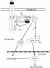
Model for regulation of EGFR-mediated NF-κB activation by SHP-2 through Gab1. EGFR activation by ligand or oncogenic mutation results in recruitment and tyrosine phosphorylation of the docking protein Gab1. Phosphorylation of Gab1 leads to recruitment of several signal relay molecules, including PI3-kinase and SHP-2. Current data suggest that association of Gab1 with SHP-2 and PI3-kinase is required for activation of Akt, which induces NF-κB activation and inhibition of proapoptotic factors, such as GSK-3. SHP-2 then acts to dephosphorylate Gab1 and downregulate the Gab1/PI3-kinase/Akt activation loop, thereby controlling the extent of EGFR-mediated NF-κB activation.

Similar articles

Cited by

References

    1. Allanson, J. E. 1987. Noonan syndrome. J. Med. Genet. 24:9-13. - PMC - PubMed
    1. Arrandale, J. M., A. Gore-Willse, S. Rocks, J. M. Ren, J. Zhu, A. Davis, J. N. Livingston, and D. U. Rabin. 1996. Insulin signaling in mice expressing reduced levels of Syp. J. Biol. Chem. 271:21353-21358. - PubMed
    1. Baeuerle, P. A., and T. Henkel. 1994. Function and activation of NF-kappa B in the immune system. Annu. Rev. Immunol. 12:141-179. - PubMed
    1. Baldwin, A. S., Jr. 1996. The NF-kappa B and I kappa B proteins: new discoveries and insights. Annu. Rev. Immunol. 14:649-683. - PubMed
    1. Basile, J. R., A. Eichten, V. Zacny, and K. Munger. 2003. NF-kappaB-mediated induction of p21(Cip1/Waf1) by tumor necrosis factor alpha induces growth arrest and cytoprotection in normal human keratinocytes. Mol. Cancer Res. 1:262-270. - PubMed

Publication types

MeSH terms

Substances

LinkOut - more resources