. 2004 Jan;78(2):1042-9.
doi: 10.1128/jvi.78.2.1042-1049.2004.

Incorporation of pol into human immunodeficiency virus type 1 Gag virus-like particles occurs independently of the upstream Gag domain in Gag-pol

Affiliations

Incorporation of pol into human immunodeficiency virus type 1 Gag virus-like particles occurs independently of the upstream Gag domain in Gag-pol

Shan Cen et al. J Virol. 2004 Jan.

Abstract

By using particle-associated reverse transcriptase (RT) activity as an assay for Pol incorporation into human immunodeficiency virus type 1 (HIV-1) Gag virus-like particles (VLPs), it has been found that truncated, protease-negative, Gag-Pol missing cis Gag sequences is still incorporated into Gag VLPs, albeit at significantly reduced levels (10 to 20% of the level of wild-type Gag-Pol). In this work, we have directly measured the incorporation of truncated Gag-Pol species into Gag VLPs and have found that truncated Gag-Pol that is missing all sequences upstream of RT is still incorporated into Gag VLPs at levels approximating 70% of that achieved by wild-type Gag-Pol. Neither protease nor integrase regions in Pol are required for its incorporation, implying an interaction between Gag and RT sequences in the Pol protein. While the incorporation of Gag-Pol into Gag VLPs is reduced 12-fold by the replacement of the nucleocapsid within Gag with a leucine zipper motif, this mutation does not affect Pol incorporation. However, the deletion of p6 in Gag reduces Pol incorporation into Gag VLPs four- to fivefold. Pol shows the same ability as Gag-Pol to selectively package tRNA(Lys) into Gag VLPs, and primer tRNA(3)(Lys) is found annealed to the viral genomic RNA. These data suggest that after the initial separation of Gag from Pol during cleavage of Gag-Pol by viral protease, the Pol species still retains the capacity to bind to both Gag and tRNA(3)(Lys), which may be required for Pol and tRNA(3)(Lys) to be retained in the assembling virion until budding is completed.


Figures
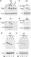
FIG.1.
FIG.1.
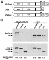
Incorporation of Pol and Gag-Pol into Gag VLPs. 293T cells were transfected or cotransfected with different plasmids coding for wild-type Gag and wild-type or mutant Gag-Pol proteins, using Lipofectamine. The plasmids used are listed along the top of each panel. BH10.P− is a simian virus 40-based vector that contains full-length wild-type HIV-1 proviral DNA containing an inactive viral PR (D25G) and was a gift from E. Cohen, University of Montreal. BH10.FS− contains mutations at the frameshift site, i.e., nucleic acid sequence 2082-TTTTTT-2087 replaces 2082-CTTCCT-2087, which prevents frameshifting during the translation of the Gag protein and generates particles that contain only Gag, not Gag-Pol (35). hGag-PolΔFSΔPR was constructed by deleting five thymidines in the frameshift site and codes for Gag-Pol. The humanized proteins have identical amino acid sequences to their viral counterparts, but the mRNAs coding for them have had their codons optimized for mammalian cell codon usage, which results in more efficient translation and protein production and also makes nuclear export of these mRNAs Rev-independent through modification of the multiple inhibitory sequences (26, 43). Both hPol and hGag-PolΔFSΔPR contain an inactive PR due to an R42G mutation in the active site. hPol-V5, hPol.ΔPR-V5, and hPol.ΔIN-V5 code for Pol, Pol missing PR, and Pol missing integrase, respectively; and all proteins contain a C-terminal V5 tag. Plasmid pSV.Myr1.3 h was a gift from R. Craven and J. Wills (University of Pennsylvania, Philadelphia) and was constructed as previously described (9). We have renamed it RSV.Gag.P−. It encodes a truncated Myr1 protein that carries RSV Gag sequences and only the first seven amino acids of RSV PR, followed by one foreign amino acid, and produces RSV extracellular Gag VLPs containing Gag but no Gag-Pol. HEK-293T cells were cultured as previously described (20), transfected with Lipofectamine 2000 (Invitrogen, Carlsbad, Calif.) according to the manufacturer's instructions. At 48 h posttransfection, Gag VLPs were purified from the media as previously described (14). Cells or Gag VLPs were lysed in radioimmunoprecipitation assay buffer (10 mM Tris [pH 7.4], 100 mM NaCl, 1% sodium deoxycholate, 0.1% sodium dodecyl sulfate [SDS], 1% NP-40, 2 mg of aprotinin/ml, 2 mg of leupeptin/ml, 1 mg of pepstatin A/ml, 100 mg of phenylmethylsulfonyl fluoride/ml). Proteins in the cell or viral lysates were resolved by SDS-PAGE (10% acrylamide), followed by blotting onto nitrocellulose membranes (Amersham Pharmacia). Detection of protein by Western blotting utilized monoclonal antibodies that are specifically reactive with HIV-1 CA (Zepto Metrocs Inc.), RT (National Institutes of Health, AIDS Research and Reference Reagent Program) and β-actin (Sigma). Rabbit antiserum to RSV was a kind gift from R. Craven (University of Pennsylvania). Detection of proteins was performed by enhanced chemiluminescence (NEN Life Sciences Products) by using as secondary antibodies anti-mouse (for CA, RT, and β-actin) and anti-rabbit (for RSV), both obtained from Amersham Life Sciences. (A and B) Lysates of cells transfected with various plasmids or VLPs produced by these cells were analyzed by Western blots probed with anti-RT (upper panels), anti-β-actin (A, lower panel), or anti-CA (B, lower panel). (C) Western blots of Gag VLPs, probed with anti-RT (upper panel) or anti-CA (lower panel). (D) Western blots of lysates of RSV Gag VLPs probed with either RSV anti-CA (lower panel) or HIV anti-RT (upper panel). (E and F) Lysates of cells transfected with various plasmids or Gag VLPs produced by these cells were analyzed by Western blots probed with anti-V5 and either anti β-actin (E) or anti-CA (F). Bands in Western blots were quantitated with the UN-SCAN-IT gel automated digitizing system.
FIG. 2.
FIG. 2.
Sequences in Gag binding Gag-Pol or Pol. 293T cells were cotransfected with a plasmid coding for wild-type or mutant Gag and with a plasmid coding for either hGag-Pol (hGag-PolΔFSΔPR) or hPol (hPol). ZWt and ZWt-p6 were a gift of Heinrich Göttlinger (Dana- Farber Cancer Institute) and were constructed as previously described (1). The plasmids used are listed along the top of each panel. At 48 h posttransfection, purified Gag VLPs were lysed in radioimmunoprecipitation assay buffer, and Gag and Gag-Pol proteins were detected with Western blots probed with antibodies to HIV-1 RT or CA. (A) Cartoon of wild-type and mutant Gag expressed. (B) Lysates of viruses analyzed by Western blots probed with anti-RT (upper panel) or anti-CA (lower panel). Bands were quantitated with the UN-SCAN-IT gel automated digitizing system.
FIG. 3.
FIG. 3.
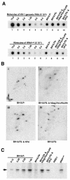
Incorporation of tRNA3Lys into Gag VLPs and its annealing to viral RNA. HEK-293T cells were transfected or cotransfected with different plasmids coding for wild-type Gag and wild-type or mutant Gag-Pol proteins with Lipofectamine. The plasmids used are listed along the top of each panel. At 48 h posttransfection, Gag VLPs were purified, and total viral RNA was extracted using guanidiumisothiocynate, as previously described (28). (A) Incorporation of tRNA3Lys into virions. Dot blots of viral RNA were hybridized with DNA probes complementary to either viral genomic RNA (upper panel) or tRNA3Lys (lower panel), as previously described (14). Hybridization signals were analyzed by phosphorimaging, and the ratio of tRNA3Lys/genomic RNA was determined for each sample (see Table 1). The standard curves shown in the left part of each blot contain known amounts of in vitro transcribed HIV-1 genomic RNA (5′ end fragment) or in vitro transcribed tRNA3Lys and were hybridized with the DNA probes complementary to either tRNA3Lys or genomic RNA to show the linearity of the signal. (B) 2D PAGE analysis of low-molecular-weight viral RNA. Total viral RNA was extracted from virions, 3′-end-labeled with [32P]pCp and electrophoresed in 11% polyacrylamide in the first dimension and 20% polyacrylamide in the second dimension, as previously described (28). Only low-molecular-weight RNA moves into the gel and is detected by autoradiography. Spot 3, tRNA3Lys; spots 1 and 2, tRNA1,2Lys. The text below each panel lists plasmids used to express each viral type analyzed. (C) tRNA3Lys annealing to viral RNA. Total viral RNA was used as the source of primer tRNA3Lys/genomic RNA template in an in vitro reverse transcription reaction, carried out in the presence of [α-32P]dGTP, dCTP, dTTP, and ddATP, as previously described (27). This process results in a six-base extension product since the first six bases incorporated are CTGCTA. Products were resolved by one-dimensional PAGE, with different samples containing equal amounts of genomic RNA. The standard curve shown in the left part of the blot in panel C contains a dilution series of total BH10 viral RNA, which is used as the source of primer or template to show the linearity of the signal.

Similar articles

Cited by

References

    1. Accola, M. A., B. Strack, and H. G. Göttlinger. 2000. Efficient particle production by minimal Gag constructs which retain the carboxy-terminal domain of human immunodeficiency virus type 1 capsid-p2 and a late assembly domain. J. Virol. 74:5395-5402. - PMC - PubMed
    1. Berkowitz, R., J. Fisher, and S. P. Goff. 1996. RNA packaging, p. 177-218. In H. G. Krausslich (ed.), Morphogenesis and maturation of retroviruses, vol. 214. Springer-Verlag, New York, N.Y.
    1. Berthet-Colominas, C., S. Monaco, A. Novelli, G. Sibai, F. Mallet, and S. Cusack. 1999. Head-to-tail dimers and interdomain flexibility revealed by the crystal structure of HIV-1 capsid protein (p24) complexed with a monoclonal antibody Fab. EMBO J. 18:1124-1136. - PMC - PubMed
    1. Bryant, M., and L. Ratner. 1990. Myristoylation-dependent replication and assembly of human immunodeficiency virus 1. Proc. Natl. Acad. Sci. USA 87:523-527. - PMC - PubMed
    1. Buchschacher, G. L., Jr., L. Yu, F. Murai, T. Friedmann, and A. Miyanohara. 1999. Association of murine leukemia virus pol with virions, independent of Gag-Pol expression. J. Virol. 73:9632-9637. - PMC - PubMed