A novel role for XIAP in copper homeostasis through regulation of MURR1
- PMID: 14685266
- PMCID: PMC1271669
- DOI: 10.1038/sj.emboj.7600031
A novel role for XIAP in copper homeostasis through regulation of MURR1
Abstract
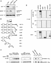
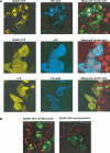
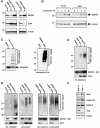
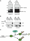
XIAP is a potent suppressor of apoptosis that directly inhibits specific members of the caspase family of cysteine proteases. Here we demonstrate a novel role for XIAP in the control of intracellular copper levels. XIAP was found to interact with MURR1, a factor recently implicated in copper homeostasis. XIAP binds to MURR1 in a manner that is distinct from that utilized by XIAP to bind caspases, and consistent with this, MURR1 did not affect the antiapoptotic properties of XIAP. However, cells and tissues derived from Xiap-deficient mice were found to contain reduced copper levels, while suppression of MURR1 resulted in increased intracellular copper in cultured cells. Consistent with these opposing effects, XIAP was observed to negatively regulate MURR1 protein levels by the formation of K48 polyubiquitin chains on MURR1 that promote its degradation. These findings represent the first described phenotypic alteration in Xiap-deficient mice and demonstrate that XIAP can function through MURR1 to regulate copper homeostasis.
Figures


Similar articles
-
Akt phosphorylation and stabilization of X-linked inhibitor of apoptosis protein (XIAP).J Biol Chem. 2004 Feb 13;279(7):5405-12. doi: 10.1074/jbc.M312044200. Epub 2003 Nov 25. J Biol Chem. 2004. Retraction in: J Biol Chem. 2016 Oct 21;291(43):22846. doi: 10.1074/jbc.A116.312044 PMID: 14645242 Retracted.
-
The copper toxicosis gene product Murr1 directly interacts with the Wilson disease protein.J Biol Chem. 2003 Oct 24;278(43):41593-6. doi: 10.1074/jbc.C300391200. Epub 2003 Sep 10. J Biol Chem. 2003. PMID: 12968035
-
Lead exposure disturbs ATP7B-mediated copper export from brain barrier cells by inhibiting XIAP-regulated COMMD1 protein degradation.Ecotoxicol Environ Saf. 2023 May;256:114861. doi: 10.1016/j.ecoenv.2023.114861. Epub 2023 Apr 5. Ecotoxicol Environ Saf. 2023. PMID: 37027943
-
XIAP: cell death regulation meets copper homeostasis.Arch Biochem Biophys. 2007 Jul 15;463(2):168-74. doi: 10.1016/j.abb.2007.01.033. Epub 2007 Feb 22. Arch Biochem Biophys. 2007. PMID: 17382285 Free PMC article. Review.
-
Molecular regulation of copper excretion in the liver.Proc Nutr Soc. 2004 Feb;63(1):31-9. doi: 10.1079/pns2003316. Proc Nutr Soc. 2004. PMID: 15099406 Review.
Cited by
-
A novel COMMD1 mutation Thr174Met associated with elevated urinary copper and signs of enhanced apoptotic cell death in a Wilson Disease patient.Behav Brain Funct. 2010 Jun 15;6:33. doi: 10.1186/1744-9081-6-33. Behav Brain Funct. 2010. PMID: 20550661 Free PMC article.
-
COMMD1 disrupts HIF-1alpha/beta dimerization and inhibits human tumor cell invasion.J Clin Invest. 2010 Jun;120(6):2119-30. doi: 10.1172/JCI40583. Epub 2010 May 10. J Clin Invest. 2010. PMID: 20458141 Free PMC article.
-
Third BIR domain of XIAP binds to both Cu(II) and Cu(I) in multiple sites and with diverse affinities characterized at atomic resolution.Sci Rep. 2019 May 15;9(1):7428. doi: 10.1038/s41598-019-42875-7. Sci Rep. 2019. PMID: 31092843 Free PMC article.
-
HSCARG regulates NF-kappaB activation by promoting the ubiquitination of RelA or COMMD1.J Biol Chem. 2009 Jul 3;284(27):17998-8006. doi: 10.1074/jbc.M809752200. Epub 2009 May 11. J Biol Chem. 2009. PMID: 19433587 Free PMC article.
-
COMMD1 forms oligomeric complexes targeted to the endocytic membranes via specific interactions with phosphatidylinositol 4,5-bisphosphate.J Biol Chem. 2009 Jan 2;284(1):696-707. doi: 10.1074/jbc.M804766200. Epub 2008 Oct 21. J Biol Chem. 2009. PMID: 18940794 Free PMC article.
References
-
- Aguilar RC, Wendland B (2003) Ubiquitin: not just for proteasomes anymore. Curr Opin Cell Biol 15: 184–190 - PubMed
-
- Birkey Reffey S, Wurthner JU, Parks WT, Roberts AB, Duckett CS (2001) X-linked inhibitor of apoptosis protein functions as a cofactor in transforming growth factor-β signaling. J Biol Chem 276: 26542–26549 - PubMed
-
- Bratton SB, Lewis J, Butterworth M, Duckett CS, Cohen GM (2002) XIAP inhibition of caspase-3 preserves its association with the Apaf-1 apoptosome and prevents CD95- and Bax-induced apoptosis. Cell Death Differ 9: 881–892 - PubMed
-
- Chai J, Shiozaki E, Srinivasula SM, Wu Q, Dataa P, Alnemri ES, Shi Y (2001) Structural basis of caspase-7 inhibition by XIAP. Cell 104: 769–780 - PubMed
Publication types
MeSH terms
Substances
Grants and funding
LinkOut - more resources
Full Text Sources
Other Literature Sources
Molecular Biology Databases

