. 2004 Jan;78(1):441-53.
doi: 10.1128/jvi.78.1.441-453.2004.

The Rep protein of adeno-associated virus type 2 interacts with single-stranded DNA-binding proteins that enhance viral replication

Affiliations

The Rep protein of adeno-associated virus type 2 interacts with single-stranded DNA-binding proteins that enhance viral replication

Travis H Stracker et al. J Virol. 2004 Jan.

Abstract

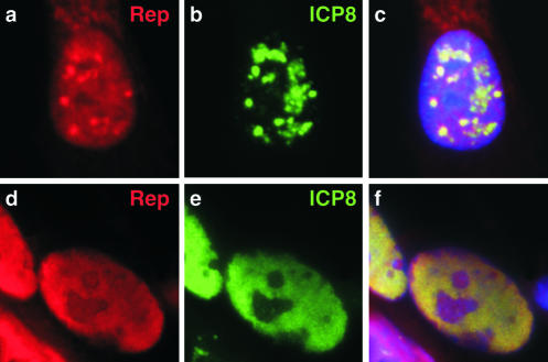
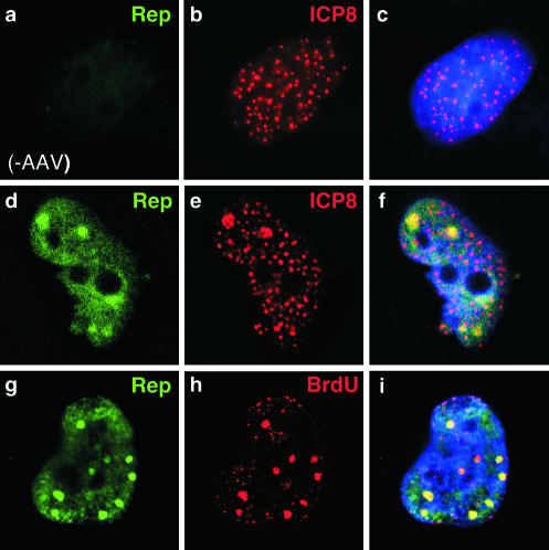
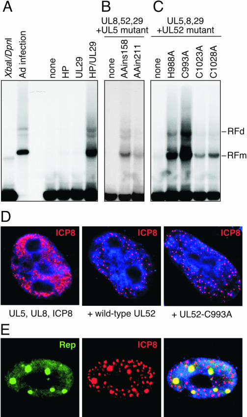
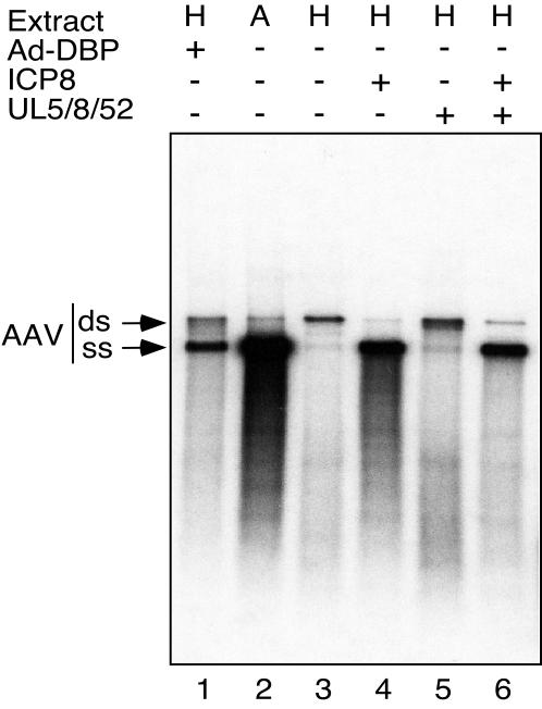
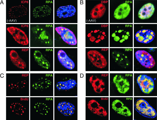
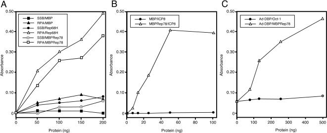
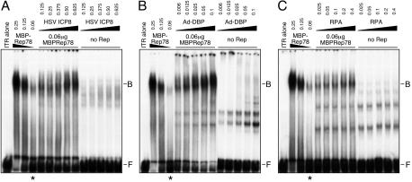
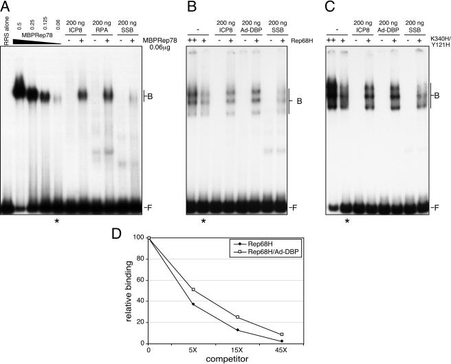
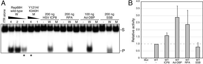
Adeno-associated virus (AAV) type 2 is a human parvovirus whose replication is dependent upon cellular proteins as well as functions supplied by helper viruses. The minimal herpes simplex virus type 1 (HSV-1) proteins that support AAV replication in cell culture are the helicase-primase complex of UL5, UL8, and UL52, together with the UL29 gene product ICP8. We show that AAV and HSV-1 replication proteins colocalize at discrete intranuclear sites. Transfections with mutant genes demonstrate that enzymatic functions of the helicase-primase are not essential. The ICP8 protein alone enhances AAV replication in an in vitro assay. We also show localization of the cellular replication protein A (RPA) at AAV centers under a variety of conditions that support replication. In vitro assays demonstrate that the AAV Rep68 and Rep78 proteins interact with the single-stranded DNA-binding proteins (ssDBPs) of Ad (Ad-DBP), HSV-1 (ICP8), and the cell (RPA) and that these proteins enhance binding and nicking of Rep proteins at the origin. These results highlight the importance of intranuclear localization and suggest that Rep interaction with multiple ssDBPs allows AAV to replicate under a diverse set of conditions.


Figures

FIG. 1.
FIG. 1.
AAV colocalizes with HSV-1 sites in coinfected cells. Vero cells were coinfected with AAV-2 (1,000 genomes/cell) and HSV-1 (multiplicity of infection of 0.1 PFU/cell). Subcellular localization of viral proteins was visualized by indirect immunofluorescence at 12 h postinfection (a to c) and 16 h postinfection (d to f). The AAV-2 Rep proteins were detected with a rabbit polyclonal antibody and ICP8 of HSV-1 was visualized with a mouse monoclonal antibody. Nuclei were located by costaining DNA with DAPI, as shown in the merged images in the right columns.
FIG. 2.
FIG. 2.
AAV colocalizes with ICP8 at discrete sites formed by transfection of HSV-1 helper genes. Transfection of HeLa cells with plasmids expressing UL5, UL8, UL52, and ICP8 (a to c) resulted in formation of discrete sites of ICP8 accumulation, as detected with a mouse monoclonal antibody. Cotransfection of the AAV genome with ICP8 and the helicase-primase (d to i) results in the formation of larger foci, to which Rep (d) and ICP8 (e) localize in the nucleus (f). Pulse-labeling with BrdU followed by immunofluorescence using a mouse monoclonal antibody demonstrated that sites of Rep accumulation (g) are active sites of DNA synthesis (h). Nuclei were located by costaining DNA with DAPI, as shown in the merged images in the right columns.
FIG. 3.
FIG. 3.
The helicase-primase activities of the UL5-UL8-UL52 complex are not required for AAV helper activity. Human HeLa cells were transfected with a clone of AAV-2 (pNTC244) alone or together with various combinations of vectors expressing wild-type or mutant versions of the four HSV-1 helper genes for UL5, UL8, UL52, and ICP8. Cells were harvested at 40 h posttransfection, and low-molecular-weight DNA was extracted and digested with XbaI and DpnI to remove the input plasmid. A Southern blot of the samples run on an agarose gel was hybridized with an AAV probe. The replicative intermediates in monomer or dimer form (RFm and RFd) are indicated on the right. (A) AAV replication requires both the UL5, UL8, and UL52 genes that encode the helicase-primase complex (HP) and the UL29 gene that encodes the ICP8 protein. Controls included the input AAV plasmid digested with XbaI and DpnI, the AAV plasmid transfected into Ad-infected cells as a positive control for AAV replication, and a transfection of the AAV plasmid without any HSV-1 helper genes (none). (B) AAV replication in transfections of HSV-1 helper genes without UL5 (none) or with mutants of UL5 (AAins158 and AAins211). (C) AAV replication in transfections of HSV-1 helper genes without UL52 (none) or with mutants of UL52 (H988A, C993A, C1023A, and C1028A). (D) The mutants of UL5 and UL52 are still able to assemble complexes at HSV prereplicative sites. Cells were transfected with the indicated viral genes and processed for immunofluorescence after 40 h. The UL29 gene product (ICP8) was detected by indirect immunofluorescence. In the absence of UL52, the ICP8 protein is detected in a diffuse pattern, but with either wild-type or mutant UL52, it is localized to discrete foci. All mutants tested showedformation of these discrete ICP8 centers. Nuclei were stained for DNA with DAPI, as shown in blue. (E) AAV Rep proteins colocalize with ICP8 at foci formed with mutant primase-helicase complexes. Cells were transfected with the AAV genome together with constructs expressing wild-type or mutant versions of the four HSV-1 genes. At 40 h, cells were processed for immunofluorescence with a rabbit polyclonal antibody against Rep and a monoclonal antibody against ICP8. The merged image is shown to the right, with costaining of nuclear DNA using DAPI. The example shown is for the UL52-C993A mutant, but all mutants tested showed similar discrete sites where Rep and ICP8 proteins colocalized.
FIG. 4.
FIG. 4.
The product of the HSV-1 UL29 gene (ICP8) and the Ad-DBP protein fulfill analogous functions and provide a direct helper effect to enhance AAV DNA replication in an in vitro assay. In vitro replication assays were performed as described in Materials and Methods. Extracts used were made from Ad-infected HeLa cells (A) or uninfected HeLa cells (H). Extracts were supplemented with recombinant Rep78 protein, and replication of a linear AAV DNA molecule was monitored by the incorporation of radiolabeled nucleotides. Replication products were separated by gel electrophoresis and two full-length products were visible: the upper band represents the full-length duplex genome (ds) and the lower band represents single-stranded DNA progeny genomes (ss), as previously described (55). The effect of proteins from the helper viruses was assessed by the addition of purified Ad-DBP or the HSV-1 proteins UL5, UL8, UL52, and ICP8, as indicated.
FIG. 5.
FIG. 5.
Accumulation of RPA at sites of AAV replication. Various combinations of Ad and HSV-1 proteins that allow replication of AAV were analyzed by indirect immunofluorescence after transfection of HeLa cells. (A) RPA colocalizes with sites formed by transfection of the helicase-primase and ICP8 of HSV-1. The ICP8 protein colocalized with RPA in cells transfected with the HSV-1 genes for UL5, UL8, UL52, and ICP8 (top row). The addition of an AAV plasmid resulted in some enlarged foci in which Rep and RPA colocalize. (B) RPA colocalizes at replication centers formed by Ad-DBP and AAV. Transfection of Ad-DBP alone results in diffuse nuclear expression and RPA is unaltered (top row). Cotransfection with an AAV genome results in the formation of discrete centers of replication in which RPA colocalizes with both DBP and Rep. (C and D) Transfection of expression vectors for the Ad helper proteins E1b55K and E4orf6 enables AAV replication in the absence of a viral ssDBP. Two major patterns of replication were observed in an equal number of cells, as shown. In both cases, Rep colocalized with RPA (top rows) and the replication centers were stained by Rep and BrdU (bottom rows). These images represent a single focal plane from a 0.5-μm cross section of the cell after deconvolution.
FIG. 6.
FIG. 6.
The AAV Rep protein interacts with RPA, ICP8, and Ad-DBP. ELISAs were performed in which purified protein was immobilized in 96-well plates and then challenged with increasing amounts of a second purified protein. In each case, the first protein listed is the immobilized one and the second is the challenging protein. (A) Rep proteins specifically bind to immobilized RPA. Recombinant human RPA (250 ng) or E. coli SSB (250 ng) was immobilized and challenged with increasing amounts of bacterially purified MBP, MBPRep78, or Rep68H. Interactions were detected with rabbit polyclonal antibodies against MBP or Rep and horseradish peroxidase anti-rabbit antibody. (B) Recombinant ICP8 binding to immobilized MBPRep78. Purified MBPRep78 (250 ng) or MBP (250 ng) was bound to ELISA wells and challenged with ICP8. Binding was detected with an anti-ICP8 monoclonal antibody. (C) Rep binding to immobilized Ad-DBP. Purified Ad-DBP (500 ng) was bound to ELISA wells and challenged with increasing amounts of MBPRep78 or the POU homeodomain of Oct-1 as a control protein. Binding was detected with an anti-MBP monoclonal antibody or an Oct-1 antibody (21a).
FIG. 7.
FIG. 7.
ssDBPs enhance Rep binding to the AAV hairpin ITR. EMSAs were performed with the 32P-labeled AAV terminal repeat hairpin DNA (1,000 cpm) incubated with the indicated amounts of MBPRep78 protein in the presence or absence of the ssDBPs. The numbers at the top represent the amounts of added protein, in micrograms. A constant amount of Rep (indicated by asterisks) was assessed for the effect of the ssDBPs. Rep binding was enhanced with HSV-1 ICP8 (A), Ad-DBP (B), and RPA (C). The lane marked “ITR alone” contains no added proteins. The positions of free DNA probe (F) and DNA probe bound in a Rep complex (B) are shown to the right.
FIG. 8.
FIG. 8.
Enhanced binding by Rep proteins is specific and does not require helicase activity. (A) Effect of ssDBPs on binding of MBPRep78 to a linear DNA substrate containing the RRS. The radiolabeled RRS fragment (1,000 cpm) was incubated with MBPRep78 (the numbers at the top indicate the amounts of protein, in micrograms). The ssDBPs were incubated with the probe in the absence (−) or presence (+) of MBPRep78. Binding was enhanced by the HSV-1 ICP8 and RPA proteins but not by the E. coli SSB protein. A constant amount of Rep (indicated by the asterisk below) was assessed for the effect of the ssDBPs. The lane marked “RRS alone” does not contain any added protein. The positions of free DNA probe (F) and DNA probe bound in a Rep complex (B) are shown to the right. (B) Enhanced binding of Rep68 to the RRS fragment. Either 1 ng (+) or 2 ng (++) of His-tagged recombinant Rep68 protein (Rep68H) was incubated with a radiolabeled RRS fragment together with the indicated ssDBP. (C) ssDBPs increase binding of a mutant Rep protein that has lost helicase activity. A mutant Rep protein (Y121H/K340H) was incubated in an EMSA with a 32P-labeled linear DNA fragment containing the RRS, in the presence or absence of the indicated ssDBPs. (D) Complex dissociation was assessed with Rep68H alone or in the presence of Ad-DBP. The Rep68H protein was bound to the RRS fragment and then dissociation was assessed by the addition of increasing amounts of unlabeled competitor DNA to assembled complexes. Binding assays were analyzed by gel electrophoresis and relative binding was quantitated by PhosphorImager analysis of gels.
FIG. 9.
FIG. 9.
Enhancement of Rep endonuclease activity by ssDBPs. The trs nicking assay included 32P-labeled AAV terminal repeat hairpin DNA (1,000 cpm) and either wild-type or mutant purified Rep68H in the presence or absence of the ssDBPs. (A) A representative experiment demonstrating increased endonuclease activity of Rep68H in the presence of ICP8, RPA, and Ad-DBP, but not E. coli SSB. Purified recombinant proteins of wild-type Rep68H (W) or the Y121H/K340H mutant (M) were incubated with the ITR in the hairpin configuration. A titration of wild-type Rep68H showed nicking of the hairpin substrate (S) and release of the cleavage product (P). The mutant failed to nick the double-stranded hairpin ITR. The amount of Rep protein (in nanograms) is indicated above the lanes, and the asterisks below indicate the amounts of wild-type and mutant Rep proteins incubated with the ssDBPs. The amount of each ssDBP included is indicated at the top, in nanograms. Samples of the hairpin (ITR alone) and the ssDBPs in the absence of Rep protein were included as negative controls. (B) Quantitation of endonuclease activities. The amount of nicked product in each reaction was quantitated by PhosphorImager analysis and plotted relative to a constant amount of Rep protein (1 ng). At least three independent reactions were quantitated for each experimental condition.

Similar articles

Cited by

References

    1. Arana, M. E., B. Haq, N. Tanguy Le Gac, and P. E. Boehmer. 2001. Modulation of the herpes simplex virus type-1 UL9 DNA helicase by its cognate single-strand DNA-binding protein, ICP8. J. Biol. Chem. 276:6840-6845. - PubMed
    1. Bashir, T., J. Rommelaere, and C. Cziepluch. 2001. In vivo accumulation of cyclin A and cellular replication factors in autonomous parvovirus minute virus of mice-associated replication bodies. J. Virol. 75:4394-4398. - PMC - PubMed
    1. Biswas, N., and S. K. Weller. 1999. A mutation in the C-terminal putative Zn2+ finger motif of UL52 severely affects the biochemical activities of the HSV-1 helicase-primase subcomplex. J. Biol. Chem. 274:8068-8076. - PubMed
    1. Biswas, N., and S. K. Weller. 2001. The UL5 and UL52 subunits of the herpes simplex virus type 1 helicase-primase subcomplex exhibit a complex interdependence for DNA binding. J. Biol. Chem. 276:17610-17619. - PubMed
    1. Boehmer, P. E. 1998. The herpes simplex virus type-1 single-strand DNA-binding protein, ICP8, increases the processivity of the UL9 protein DNA helicase. J. Biol. Chem. 273:2676-2683. - PubMed

Publication types

LinkOut - more resources