. 2004 Feb;15(2):827-37.
doi: 10.1091/mbc.e03-07-0524. Epub 2003 Dec 2.

Telomere attachment, meiotic chromosome condensation, pairing, and bouquet stage duration are modified in spermatocytes lacking axial elements

Affiliations

Telomere attachment, meiotic chromosome condensation, pairing, and bouquet stage duration are modified in spermatocytes lacking axial elements

Bodo Liebe et al. Mol Biol Cell. 2004 Feb.

Abstract

During the extended prophase to the meiosis I division, chromosomes assemble axial elements (AE) along replicated sister chromatids whose ends attach to the inner nuclear membrane (NM) via a specialized conical thickening. Here, we show at the EM level that in Sycp3(-/-) spermatocyte chromosomes lack the AE and the conical end thickening, but still they attach their telomeres to the inner NM with an electron-dense plate that contains T(2)AG(3) repeats. Immunofluorescence detected telomere proteins, SCP2, and the meiosis-specific cohesin STAG3 at the Sycp3(-/-) telomere. Bouquet stage spermatocytes were approximately threefold enriched, and the number of telomere but not centromere signals was reduced to the haploid in advanced Sycp3(-/-) spermatocytes, which indicates a special mode of homolog pairing at the mammalian telomere. Fluorescence in situ hybridization with mouse chromosome 8- and 12-specific subsatellite probes uncovered reduced levels of regional homolog pairing, whereas painting of chromosomes 13 revealed partial or complete juxtapositioning of homologs; however, condensation of Sycp3(-/-) bivalents was defective. Electron microscopic analysis of AE-deficient spermatocytes revealed that transverse filaments formed short structures reminiscent of the synaptonemal complex central region, which likely mediate stable homolog pairing. It appears that the AE is required for chromosome condensation, rapid exit from the bouquet stage, and fine-tuning of homolog pairing.


Figures

Figure 1.
Figure 1.
Immunofluorescence staining of cohesin cores (STAG3, green) and telomeres (TRF2, red) in structurally preserved (A) Sycp3-/- and (B) Sycp3+/+ spermatocytes I. (Ai) Sycp3-/- leptotene nucleus with a tight telomere cluster at a sector of the nuclear periphery (*) and STAG3 cohesin speckles (all focal planes in A at nuclear equator). (Aii) Sycp3-/- zygotene nucleus with STAG3 speckles and cores; telomeres are clustered at a limited sector of the NE (*). (Aiii) Advanced Sycp3-/- zygotene nucleus with STAG3 cores and telomeres dispersed about the nuclear periphery. (Bi) Wild-type leptotene nucleus with numerous STAG3 speckles and most telomeres accumulated at the lower left of the nuclear top. (Bii) Late wild-type zygotene nucleus with long STAG3 cores and telomeres clustered at a limited sector of the NE (zygotene bouquet; *, focal plane at nuclear equator). (Biii) Wild-type pachytene nucleus with synapsed STAG3 cohesin cores and telomeres dispersed about the nuclear periphery (focus at nuclear equator). Nuclear DNA was revealed with DAPI (blue). Note that telomere clustering is tighter in the Sycp3 mutant. Bar (B): 10 μm; it applies to all details.
Figure 2.
Figure 2.
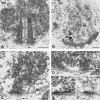
EM of telomere attachments in Sycp3+/+ (A) and Sycp3-/- spermatocytes (B-F). (A) Electron-dense LEs of the SC (CE, central element) terminate with a conical thickening (arrowheads) at the inner NM of a wild-type (WT) spermatocyte I nucleus. An electron-dense plate connects the wide end of the LE thickening with the inner NM (indented arrow). Note: the double-stranded nature of the LEs was only seen in this preparation but is known from hamster LEs (Dresser and Moses, 1980). (B) Overview of a Sycp3-/- bouquet spermatocyte at the leptotene/zygotene transition as indicated by the clustered, electron-dense heterochromatin in the lower region of the nucleus. Several telomere attachment plates are seen (arrowheads; No, nucleolus). (C and D) Details of Sycp3-/- attachment plates of proximal telomeres at the inner NM (demarcated by arrowheads). A stalk of dense chromatin extends from the adjacent bulk heterochromatin to the nuclear envelope and terminates in a flat electron-dense attachment plate (arrowheads). (D) Two telomere attachment plates that are ∼330 nm apart. (E and F) Distal telomere attachments (without adjacent heterochromatin) show solely an electron-dense plate at the nucleoplasmic face of the inner NM with the adjunct chromatin being unstructured. Fibrillar material spanning the nuclear envelope emanates into the cytoplasm opposite of all telomere attachments (small arrows). Bars (A, C-F) 0.2 μm; (B) 2 μm.
Figure 3.
Figure 3.
In situ DNA end labeling of Sycp3-/- nuclear sections (A) reveals DNA-specific gold grains over a detail of a spermatocyte I nucleus (N), which includes the attachment plates (demarcated by arrow heads). (B-D) Details of telomere attachments after EM-ISH with T2AG3 repeat probes. Specific telomere-repeat signals (black gold grains; arrows) are located exclusively at the attachment plates at the inner nuclear membrane (upper arrowheads), indicating that telomere repeats are contained in the attachment plate. This type of treatment also revealed an electron-dense “plate” at the cytoplasmic face of the NE, which was a particularity of EM-ISH and may represent a technical artifact due to collapse of the cytoplasmic fibrils opposite the telomere attachment at the inner NE. Bars, 0.2 μm.
Figure 4.
Figure 4.
(A) SCP2 (green, FITC) and telomeres (red, T2AG3 FISH) colocalize in a Sycp3-/- spermatocyte. The detail shows the gray channels of the enlarged region. Colors for SCP2 protein and telomeres as indicated. (B) Telomere (TRF1 red) and kinetochore staining (CREST, green) in a Sycp3-/- spermatocyte. Several separated centromere signals have a common telomere signal between them (arrows). Note the increased telomere signal size at CREST signal doublets, which indicates pairing of telomeres. Bar, 5 μm. (C and D) Integrity of cohesin cores as determined in three-dimensionally preserved nuclei. (C) Wild-type telomere with a continuous cohesin core in its vicinity. (D) Detail of a Sycp3-/- cohesin core (STAG3, green) that has several disruptions in the vicinity of the associated telomere signal (TRF2, red). Bar, 2 μm. (E and F) SCP1 staining (red, Cy3) and painting of mouse chromosomes MMU13 (green, FITC). (E) A single MMU13 FISH signal delineates a MMU13 bivalent associated with a central SCP1 fiber in a wild-type spermatocyte. (F) An advanced Sycp3-/- spermatocyte displays an extended paint signal representing the MMU13 bivalent that contains a long interrupted SCP1 fiber along its center. (G) Sycp3-/- MMU13 bivalents are more extended than wild-type bivalents. The graph is based on the length of MMU13 bivalents from 20 wild-type pachytene and Sycp3-/- nuclei (MMU13 homologs were requested to be completely paired in the mutant to allow comparison with the wild-type bivalents). The bivalent length is expressed as the fraction of the maximal nuclear diameter (set to 1), as determined from its DAPI outline (blue). It is apparent that condensation of MMU13 bivalents is defective in the Sycp3-/- knockout, the difference being highly significant (t test, p < 0.001).
Figure 5.
Figure 5.
Telomere (green, T2AG3) and centromere (red, major sat.) FISH patterns in structurally preserved nuclei from Sycp3-/- and wild-type (Sycp3+/+) testes suspensions. For staging according to FISH patterns, see Scherthan et al. (1996). (Ai and Bi) Premeiotic nuclei (blue, focal plane at nuclear equator) with numerous internal telomere and satellite DNA clusters. (Aii and Bii) Mid-preleptotene nuclei with peripheral satellite DNA and internal telomere signals (nuclear equator). (Aiii and Biii) Leptotene/zygotene nuclei (focal plane at nuclear top) with telomere clustering (bouquet topology). (Aiv) Wild-type pachytene and (Biv) advanced late zygotene Sycp3-/- nucleus with dispersed peripheral telomeres and satellite DNA clusters (focal plane at nuclear equator). Bar (B), 10 μm, it applies to all details. (C) Frequency of midpreleptotene spermatocytes is similar in the wild-type and Sycp3 knockout, whereas bouquet frequency is 2.8-fold elevated (2.2%, n = 2028 nuclei) compared with the wild type (0.8%; n = 2005 nuclei), which is statistically highly significant (χ2, p < 0.001). (D) Mouse telomeres (TRF1, red) cluster in the vicinity of the centrosome (arrow) as marked by γ-tubulin IF (green) in three-dimensionally preserved bouquet stage spermatocytes. Bar (D), 10 μm.
Figure 6.
Figure 6.
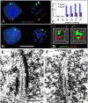
Homolog pairing in Sycp3-/- and wild-type (Sycp3+/+) spermatocytes detected by FISH with MMU8 (red, Cy3) and 12 (green, FITC) region-specific DNA probes in combination with IF for SCP1 (blue, Cy5, false colored). (A) Sycp3-/- nucleus (DNA blue, left detail) with paired MMU8 and MMU12 signals (focal plane near the nuclear equator). Paired MMU12 signals (arrow) colocalize with a SCP1 fiber fragment signal (blue, right detail) (arrow). (B) Wild-type spermatocyte with paired MMU8 and MMU12 signals (focal plane near the nuclear equator). SCP1 fibers (blue) and FISH signals of the same nucleus are shown to the right. Bar (A), 10 μm, it applies to A and B. (C) Frequencies of pairing of homologous MMU8 and 12 signals in spermatogonia (G), zygotene (Z), and pachytene nuclei (P*); the asterisk refers to Sycp3-/- spermatocyte nuclei with long SCP1 fibers. In round spermatogonia homologous signal pairing occur red in ≤10% of Sycp3-/- and WT nuclei (n = 56 and 48, respectively). Zygotene spermatocytes: pairing of MMU8 or 12 homologous regions is seen in 84% of wild-type nuclei, but only in 17 and 23% of Sycp3-/- nuclei (n = 31 and 74, respectively). Pairing is reduced but not abolished in advanced Sycp3-/- zygotene nuclei (P*; n = 86). P* equals pachytene in the wild-type. (D) 3D reconstruction of a deconvoluted nuclear region of a Sycp3-/- spermatocyte that shows a SCP1 core (red) surrounded by paired MMU12-specific chromatin (green; FISH). Two angles (0° and 90°) of the same region are shown after computational rotation. Wild-type SCP1/MMU12 chromatin associations were similar (our unpublished data). (E) EM of a detail of a sectioned wild-type spermatocyte showing tripartite SC with transverse filaments connecting electron-dense lateral elements (arrow). Note the central element. (F) EM of a detail of a Sycp3-/- spermatocyte I showing a succession of transverse filaments forming a central element. A row of ordered chromatin grains follows the course of this central region-like structure (arrow) giving raise to a delicate SC-like structure. Bar (E), 0.2 μm, and it also applies to F.

Similar articles

Cited by

References

    1. Alsheimer, M., and Benavente, R. (1996). Change of karyoskeleton during mammalian spermatogenesis: expression pattern of nuclear lamin C2 and its regulation. Exp. Cell Res. 228, 181-188. - PubMed
    1. Alsheimer, M., von Glasenapp, E., Hock, R., and Benavente, R. (1999). Architecture of the nuclear periphery of rat pachytene spermatocytes: distribution of nuclear envelope proteins in relation to synaptonemal complex attachment sites. Mol. Biol. Cell 10, 1235-1245. - PMC - PubMed
    1. Barlow, A.L., and Hultén, M.A. (1997). Combined immunocytogenetic and molecular cytogenetic analysis of meiosis I oocytes from normal human females. Zygote 6, 27-38. - PubMed
    1. Bass, H.W., Bordoli, S.J., and Foss, E.M. (2003). The desynaptic (dy) and desynaptic1 (dsy1) mutations in maize (Zea mays L.) cause distinct telomere-misplacement phenotypes during meiotic prophase. J. Exp. Bot. 54, 39-46. - PubMed
    1. Benavente, R., Alsheimer, M., and von Glasenapp, E. (2004). The nuclear envelope at the attachment sites of mammalian meiotic telomeres. Chrom. Today 14 (in press).

Publication types

LinkOut - more resources