. 2003 Dec 15;22(24):6642-52.
doi: 10.1093/emboj/cdg617.

IAP-antagonists exhibit non-redundant modes of action through differential DIAP1 binding

Affiliations

IAP-antagonists exhibit non-redundant modes of action through differential DIAP1 binding

Anna Zachariou et al. EMBO J. .

Abstract

The Drosophila inhibitor of apoptosis protein DIAP1 ensures cell viability by directly inhibiting caspases. In cells destined to die this IAP-mediated inhibition of caspases is overcome by IAP-antagonists. Genetic evidence indicates that IAP-antagonists are non-equivalent and function synergistically to promote apoptosis. Here we provide biochemical evidence for the non-equivalent mode of action of Reaper, Grim, Hid and Jafrac2. We find that these IAP-antagonists display differential and selective binding to specific DIAP1 BIR domains. Consistently, we show that each DIAP1 BIR region associates with distinct caspases. The differential DIAP1 BIR interaction seen both between initiator and effector caspases and within IAP-antagonist family members suggests that different IAP-antagonists inhibit distinct caspases from interacting with DIAP1. Surprisingly, we also find that the caspase-binding residues of XIAP predicted to be strictly conserved in caspase-binding IAPs, are absent in DIAP1. In contrast to XIAP, residues C-terminal to the DIAP1 BIR1 domain are indispensable for caspase association. Our studies on DIAP1 and caspases expose significant differences between DIAP1 and XIAP suggesting that DIAP1 and XIAP inhibit caspases in different ways.


Figures

None
Fig. 1. Co-purification of drICE-V5 or DCP-1-V5 with DIAP1–GST. (A) Schematic representation of the DIAP1 mutants used in this study. (B) The gain-of-function mutant DIAP16–3s binds more efficiently to caspases than wild-type (wt) DIAP1. Top right panel: DIAP1–caspase co-purification; affinity-purified DIAP1–GST was used to precipitate drICE-V5 (lanes 3–6) or DCP-1-V5 (lanes 7–10) from cellular extracts. Top left panel: total extracts of 293 cells expressing the indicated caspases. Caspase expression (top left panel) and caspase–DIAP1 binding (top right panel) were detected by immunoblot analysis using anti-V5 antibody. Bottom right panel: the purification of DIAP1–GST was confirmed by western blot analysis of the eluate using anti-DIAP1 RING antibody. (C and D) The BIR1 region of DIAP1 is necessary and sufficient for caspase binding. (E and F) DIAP111–3e fails to bind to drICE and DCP-1. (C–F) Experiments were conducted as in (B). Top panels: expression (lane 1) and co-purification (lanes 2–end) of drICE-V5 (C and E) and DCP-1-V5 (D and F) with the indicated DIAP1–GST fragments. Bottom panel: purification of the DIAP1–GST fragments was confirmed by immunoblot analysis of the eluate using anti-GST antibody. Note, the purified wild-type and 6–3s mutant DIAP1 BIR1 fragments (E and F; lanes 2 and 3) are cleaved at position 20 by drICE and DCP-1 while BIR111–3e that failed to bind to caspases was only partially processed.
None
Fig. 2. DIAP1 simultaneously binds to initiator and effector caspases. DRONC-FLAG was co-expressed with DIAP1 or controls in S2 cells. DRONC-FLAG was purified by immunoprecipitation using anti-FLAG antibody-coupled agarose beads. Resin-bound DRONC-FLAG was subsequently incubated with extracts of 293 cells expressing the indicated caspases. Following incubation of the DRONC-FLAG beads with the extracts, the bound proteins were eluted with FLAG peptides. Co-immunoprecipitation (lanes 1–4) and expression (lanes 5 and 6) of the indicated proteins were determined by immunoblot analysis using the indicated antibodies.
None
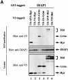
Fig. 3. IAP-antagonists bind to distinct regions of DIAP1. DIAP1–GST fragments were used to purify Rpr-V5 (A), Grim-TAP (B), Hid-V5 (C) or Jafrac2 (D) from S2/p35 cellular extracts. Top panel: DIAP1–GST deletion mutants were affinity purified from cellular extracts using glutathione–Sepharose beads and associated IAP-antagonists were detected by immunoblotting using anti-V5 (A and C), anti-protein A (B) or anti-Jafrac2 (D) antibodies, respectively. Middle panel: purification of the DIAP1–GST fragments was verified by western blot analysis using anti-GST antibody. Bottom panel: equal expression of the indicated IAP-antagonist was examined by immunoblotting the S2 extracts using the indicated antibodies. The asterisk denotes a cross-reactive band.
None
Fig. 4. Rpr, Grim, Hid and Jafrac2 differentially interact with DIAP1. (A–D) Co-purification of Rpr, Grim, Hid and Jafrac2 with wild-type or mutant BIR fragments. Expression and purification of the indicated constructs were determined as in Figure 3. The BIR1 region of DIAP16–3s shows impaired binding to Rpr (A) and Hid (C), while its association to Grim (B) remains unaffected. The 11–3e mutation does not affect the binding of Rpr, Grim or Hid to DIAP1 (A–C, top left panel). (A–D) The th4 and 23–4 BIR2 mutations impair DIAP1’s ability to bind to Rpr (A), Grim (B), Hid (C) and Jafrac2 (D).
None
Fig. 5. Rpr but not Hid directly competes with drICE for DIAP1 binding. (A and B) Co-purification of drICE with DIAP1–GST in the presence or absence of Rpr (A) or Hid (B). DIAP–GST was affinity purified from S2 cellular extracts. Resin-bound DIAP1 was subsequently incubated with a mixture of cellular extracts containing drICE-V5 and Rpr-V5 (A) or drICE-V5 and Hid-V5 (B). Bound proteins were eluted and analysed by immunoblotting using anti-V5 antibody (top right panel, lanes 4 and 5). Left panel: total extracts of cells expressing the indicated constructs; western blot analysis using anti-V5 antibody. Bottom right panel: effective DIAP1 purification was confirmed by immunoblotting the eluate with anti-DIAP1 antibody. (C) Rpr’s ability to compete with drICE for DIAP1 binding is contingent on its association with the BIR1 domain. Co-purification of drICE with the indicated DIAP1–GST proteins in the presence of wild-type or mutant Rpr or Jafrac2. Top right panel: DIAP1–GST co-purification. Top left panel: total extracts expressing the indicated constructs; immunoblot analysis using anti-V5 antibody. Bottom right panel: purification of DIAP1–GST was confirmed by western blot analysis of the eluate using anti-DIAP1 RING antibody. The asterisk denotes a cross-reactive band.
None
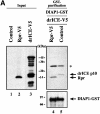
Fig. 6. Ala1 of the IBM of Rpr, Grim and Hid is indispensable for their binding to DIAP1. (A) Co-purification of Rpr-V5, Grim-V5 or Hid-V5 with DIAP1–GST from cellular extracts. Wild-type but not mutant Rpr, Grim or Hid lacking Ala1 efficiently interacted with DIAP1. Rpr, Grim and Hid were expressed using the ubiquitin fusion technique. Protein expression (bottom panel) and co-purification (top panel) was examined by immunoblot analysis with the indicated antibodies. Purification of DIAP1–GST was verified by western blot analysis of the eluate using anti-DIAP1 RING antibody (middle panel). (B) Rpr and Grim proteins that lack their entire IBM completely failed to bind to DIAP1. Note, while the difference in mobility between Grim and ΔIBM-Grim is readily detectable, Rpr and ΔIBM-Rpr appear to migrate at the same level due to the SDS–acrylamide gel used. (C) Detection of rpr and grim mRNAs by RT–PCR from total RNA of healthy S2 cells. (D) No residual binding of IA-Grim to DIAP1 is detectable in 293 cells suggesting that the residual binding shown in (A) may be due to the association of the Ala1-mutants with endogenous Rpr or Grim proteins.
None
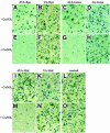
Fig. 7. IAP-antagonists induce apoptosis exclusively in an IAP-binding-dependent manner. Induced expression of AVA-Rpr, AIA-Grim and AVP-Hid promotes apoptosis, while expression of VA-Rpr, IA-Grim or VP-Hid fail to trigger cell death. The indicated Ub fusion constructs were co-transfected with lacZ reporter plasmids, and 24 h post-transfection cells from each well were divided into two dishes to avoid variations in transfection efficiencies. Expression of the constructs was induced by copper sulfate and cells were examined for β-galactosidase activity. (AD) and (IL), untreated cells; (EH) and (MO), cells treated with copper sulfate (induced state).
None
Fig. 8. Caspases, Rpr, Grim, Hid and Jafrac2 display differential and selective binding to specific DIAP1 BIR domains. The differential interaction seen both between DIAP1 and caspases and IAP-antagonists indicate that different IAP-antagonists compete with distinct caspases for DIAP1 binding. The relative affinity of individual IAP-antagonists to specific BIR domains is indicated by +.

Similar articles

Cited by

References

    1. Chai J., Du,C., Wu,J.W., Kyin,S., Wang,X. and Shi,Y. (2000) Structural and biochemical basis of apoptotic activation by Smac/DIABLO. Nature, 406, 855–862. - PubMed
    1. Chen P., Nordstrom,W., Gish,B. and Abrams,J.M. (1996) grim, a novel cell death gene in Drosophila. Genes Dev., 10, 1773–1782. - PubMed
    1. Christich A., Kauppila,S., Chen,P., Sogame,N., Ho,S.I. and Abrams,J.M. (2002) The damage-responsive Drosophila gene sickle encodes a novel IAP binding protein similar to but distinct from reaper, grim and hid. Curr. Biol., 12, 137–140. - PubMed
    1. Claveria C., Albar,J.P., Serrano,A., Buesa,J.M., Barbero,J.L., Martinez,A.C. and Torres,M. (1998) Drosophila grim induces apoptosis in mammalian cells. EMBO J., 17, 7199–7208. - PMC - PubMed
    1. Ditzel M., Wilson,R., Tenev,T., Zachariou,A., Paul,A., Deas,E. and Meier,P. (2003) Degradation of DIAP1 by the N-end rule pathway is essential for regulating apoptosis. Nat. Cell Biol., 5, 467–473. - PubMed

LinkOut - more resources