. 2003 Dec;77(24):13433-8.
doi: 10.1128/jvi.77.24.13433-13438.2003.

Folate receptor alpha and caveolae are not required for Ebola virus glycoprotein-mediated viral infection

Affiliations

Folate receptor alpha and caveolae are not required for Ebola virus glycoprotein-mediated viral infection

Graham Simmons et al. J Virol. 2003 Dec.

Abstract

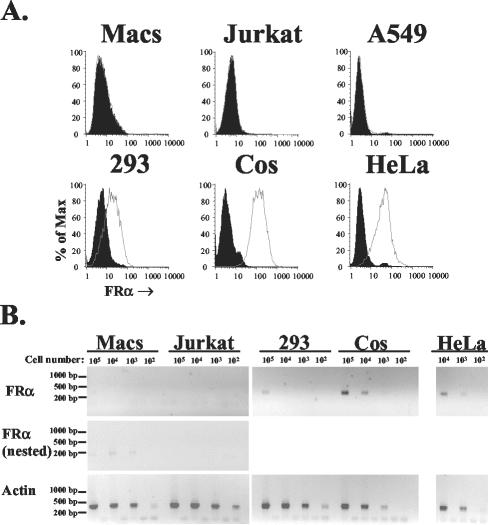
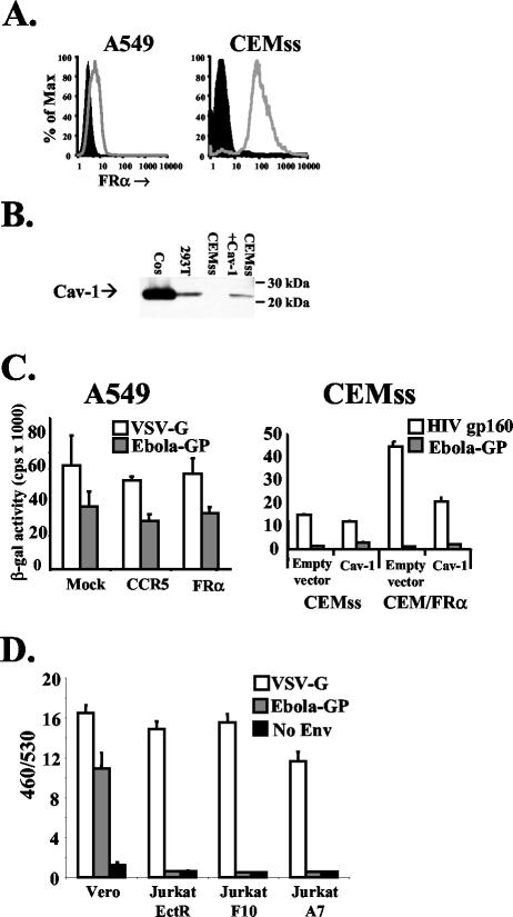
Folate receptor alpha (FRalpha) has been described as a factor involved in mediating Ebola virus entry into cells (6). Furthermore, it was suggested that interaction with FRalpha results in internalization and subsequent viral ingress into the cytoplasm via caveolae (9). Descriptions of cellular receptors for Ebola virus and its entry mechanisms are of fundamental importance, particularly with the advent of vectors bearing Ebola virus glycoprotein (GP) being utilized for gene transfer into cell types such as airway epithelial cells. Thus, the ability of FRalpha to mediate efficient entry of viral pseudotypes carrying GP was investigated. We identified cell lines and primary cell types such as macrophages that were readily infected by GP pseudotypes despite lacking detectable surface FRalpha, indicating that this receptor is not essential for Ebola virus infection. Furthermore, we find that T-cell lines stably expressing FRalpha are not infectible, suggesting that FRalpha is also not sufficient to mediate entry. T-cell lines lack caveolae, the predominant route of FRalpha-mediated folate metabolism. However, the coexpression of FRalpha with caveolin-1, the major structural protein of caveolae, was not able to rescue infectivity in a T-cell line. In addition, other cell types lacking caveolae are fully infectible by GP pseudotypes. Finally, a panel of ligands to and soluble analogues of FRalpha were unable to inhibit infection on a range of cell lines, questioning the role of FRalpha as an important factor for Ebola virus entry.


Figures

FIG. 1.
FIG. 1.
FRα expression on common cell lines and primary macrophages. (A) Surface levels of FRα on five cell lines and day 6 primary macrophages were determined by standard flow cytometry (33) with a murine monoclonal antibody directed against human FRα (LK26; Signet) (open histograms) and an isotype control (immunoglobulin G2a; Sigma, St. Louis, Mo.) (filled histograms). Macrophages were isolated from human leukocytes by plastic adherence, as previously described (32). (B) Levels of cellular mRNA for FRα and human β-actin were evaluated by RT-PCR, using an Express direct mRNA capture and RT system (Pierce, Rockford, Ill.). mRNA was extracted and cDNA synthesis was performed in the same well containing serial 10-fold dilutions of each cell type. PCR was performed with 16.5 μl of cDNA by using the conditions 95°C for 30 s followed by 30 cycles of 95°C for 30 s, 58°C for 30 s, and 72°C for 40 s for FRα (forward primer, TGGGTGGCTGTAGTAGGGGAG; reverse primer, CAGGGGCACGTTCAGTACC) and β-actin (forward primer, CTGGCACCACACCTTCTACAATG; reverse primer, AATGTCACGCACGATTTCCCGC), resulting in 359- and 381-bp products, respectively. Primers were designed so that products spanned intron and exon boundaries, such that the predicted products derived from genomic DNAs were 3,440 and 822 bp for FRα and β-actin, respectively. For cell types negative for FRα in the first round of PCR (macrophages [Macs] and the T-cell line, Jurkat), 5 μl was transferred to new tubes and nested PCR was performed with the same conditions as those described above and the following set of nested primers: forward primer GCCAAGCACCACAAGGAAAAG and reverse primer CCTGGATGAAATGCCGTTTG. This resulted in a 189-bp product with a 3,118-bp predicted genomic DNA product. Both first-round and nested primers recognized all seven isoforms of FRα (8). Gaps between each sample represent controls for which the whole mRNA extraction and RT-PCR procedure was carried out on empty wells (a total of seven controls for the nested PCR). Control experiments with the same reaction mixtures performed on mRNA from each cell line in the absence of reverse transcriptase were all negative (data not shown).
FIG. 2.
FIG. 2.
Expression of FRα and Cav-1 in cell lines does not enhance Ebola virus pseudotype infection. (A) Surface levels of FRα were assessed as described in the legend to Fig. 1 for A549 cells (left panel) transduced with lentiviral vectors (for protocols, see reference 31) expressing a control of CCR5 (filled histogram) or FRα (open histogram) and for CEMss cells (right panel, filled histogram) or CEM cells stably expressing FRα (open histogram). (B) Total protein levels of Cav-1 were assessed by Western blotting using a directly horseradish peroxidase-conjugated polyclonal rabbit antibody raised against the N terminus of Cav-1 (N-20; Santa Cruz Biotechnology, Santa Cruz, Calif.). Sodium dodecyl sulfate-polyacrylamide gel electrophoresis was performed on cell lysates from 106 cells. CEMss cells were transduced with MLV amphotropic envelope pseudotyped MLV particles carrying the MIGR1 packaging vector encoding human CD8 (27) or MLV MIGR1 encoding human Cav-1. Cos and 293 cells were also tested to determine endogenous levels of Cav-1. (C) Mock-transduced and CCR5- or FRα-transduced A549 cells (left panel) or CEMss and CEM/FRα cells transduced with control or Cav-1-encoding MLV pseudovirions (right panel) were challenged with HIV-based pseudovirions encoding LacZ (18) pseudotyped with Ebola virus GP, VSV-G, or HIV gp160 (HXB2 strain). Two days postchallenge, cells were lysed and soluble β-galactosidase (β-gal) levels were measured with a chemiluminescence kit (Galactostar; Tropix) as per the manufacturer's instructions. Similar results were gained by using pseudovirions encoding luciferase as a reporter gene (data not shown). (D) Vero cells, but not Jurkat cells stably expressing ecotropic MLV receptor or previously described derivates expressing FRα (6), were transducible with VLPs bearing Ebola virus GP. VLPs were made by cotransfecting293T cells with eqimolar amounts of a Bla-VP40 fusion protein and GP, VSV-G, or empty vector. Forty-eight hours posttransfection, filtered supernatant was collected and diluted. Cells (5 × 10⁵) were spin infected at 2,500 rpm for 2 h at 4°C and then incubated for 4 h at 37°C to allow entry. Cells were then labeled with CCF2-AM (Invitrogen, Carlsbad, Calif.) as per the manufacturer's instructions, washed, transferred to clear-bottomed, black-walled microtiter plates, and incubated overnight at room temperature to allow the enzymatic reaction to occur. The following morning, plates were read at 460 and 530 nm (Cytofluor 4000; Applied Biosystems, Foster City, Calif.). Background fluorescence from unlabeled cells was subtracted, and the ratio of fluorescence at 460 nm to that at 530 nm (460/530) was calculated for each well. Means and standard deviations shown are calculated from the results of four replicates, and the results presented are representative of three independent experiments.
FIG. 3.
FIG. 3.
Inhibition of Ebola virus pseudotype infection of GHOST cells by folic acid. (A) GHOST cells were incubated at 4°C for 30 min with M199 medium containing 5% charcoal-stripped fetal bovine serum alone (approximate basal folic acid concentration in M199 medium is 0.02 mM) or supplemented with various concentrations of folic acid dissolved in sodium hydroxide (1 M). The pH of all solutions was adjusted to 8.5. An equal volume of HIV-based pseudovirions encoding GFP pseudotyped with Ebola virus GP or VSV-G was then added to give final concentrations of folic acid of 0.02, 0.22, 0.73, and 2.2 mM. Cells were incubated for 6 h, the medium was replaced, and the cells were incubated for a further 48 h. GFP expression was then analyzed by flow cytometry, and results are displayed as percentages of the expression in the control (M199 medium alone [pH 8.5], i.e., 0.02 mM folic acid). The results of the experiment shown are representative of three independent experiments. Error bars represent standard deviations of results from three replicates. (B) Vero cells were incubated as described above with M199 medium supplemented with folic acid (broken lines) or 5-methyltetrafolic acid (solid lines). An equal volume of HIV-based pseudovirions encoding LacZ pseudotyped with Ebola virus GP (squares) or MLV amphotropic envelope (circles) was then added to give the plotted concentrations. Cells were incubated for 6 h, the medium was replaced, and the cells were incubated for a further 48 h. lacZ expression was then analyzed by quantification of X-galactosidase-stained cells as previously described (32), and results are displayed as percentages of the expression level in the control.

Similar articles

Cited by

References

    1. Baskerville, A., S. P. Fisher-Hoch, G. H. Neild, and A. B. Dowsett. 1985. Ultrastructural pathology of experimental Ebola haemorrhagic fever virus infection. J. Pathol. 147:199-209. - PubMed
    1. Bavari, S., C. M. Bosio, E. Wiegand, G. Ruthel, A. B. Will, T. W. Geisbert, M. Hevey, C. Schmaljohn, A. Schmaljohn, and M. J. Aman. 2002. Lipid raft microdomains: a gateway for compartmentalized trafficking of Ebola and Marburg viruses. J. Exp. Med. 195:593-602. - PMC - PubMed
    1. Brzezinska, A., P. Winska, and M. Balinska. 2000. Cellular aspects of folate and antifolate membrane transport. Acta Biochim. Pol. 47:735-749. - PubMed
    1. Cavrois, M., C. De Noronha, and W. C. Greene. 2002. A sensitive and specific enzyme-based assay detecting HIV-1 virion fusion in primary T lymphocytes. Nat. Biotechnol. 20:1151-1154. - PubMed
    1. Cecilia, D., V. N. KewalRamani, J. O'Leary, B. Volsky, P. Nyambi, S. Burda, S. Xu, D. R. Littman, and S. Zolla-Pazner. 1998. Neutralization profiles of primary human immunodeficiency virus type 1 isolates in the context of coreceptor usage. J. Virol. 72:6988-6996. - PMC - PubMed

Publication types