Glatiramer acetate-specific T cells in the brain express T helper 2/3 cytokines and brain-derived neurotrophic factor in situ
- PMID: 14614135
- PMCID: PMC283562
- DOI: 10.1073/pnas.2336171100
Glatiramer acetate-specific T cells in the brain express T helper 2/3 cytokines and brain-derived neurotrophic factor in situ
Erratum in
- Proc Natl Acad Sci U S A. 2005 Aug 23;102(34):12288
Abstract
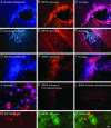
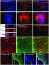
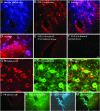
The ability of a remedy to modulate the pathological process in the target organ is crucial for its therapeutic activity. Glatiramer acetate (GA, Copaxone, Copolymer 1), a drug approved for the treatment of multiple sclerosis, induces regulatory T helper 2/3 cells that penetrate the CNS. Here we investigated whether these GA-specific T cells can function as suppressor cells with therapeutic potential in the target organ by in situ expression of T helper 2/3 cytokines and neurotrophic factors. GA-specific cells and their in situ expression were detected on the level of whole-brain tissue by using a two-stage double-labeling system: (i) labeling of the GA-specific T cells, followed by their adoptive transfer, and (ii) detection of the secreted factors in the brain by immunohistological methods. GA-specific T cells in the CNS demonstrated intense expression of the brain-derived neurotrophic factor and of two antiinflammatory cytokines, IL-10 and transforming growth factor beta. No expression of the inflammatory cytokine IFN-gamma was observed. This pattern of expression was manifested in brains of normal and experimental autoimmune encephalomyelitis-induced mice to which GA-specific cells were adoptively transferred, but not in control mice. Furthermore, infiltration of GA-induced cells to the brain resulted in bystander expression of IL-10 and transforming growth factor beta by resident astrocytes and microglia. The ability of infiltrating GA-specific cells to express antiinflammatory cytokines and neurotrophic factor in the organ in which the pathological processes occur correlates directly with the therapeutic activity of GA in experimental autoimmune encephalomyelitis/multiple sclerosis.
Figures


Similar articles
-
Do Th2 cells mediate the effects of glatiramer acetate in experimental autoimmune encephalomyelitis?Int Immunol. 2006 Apr;18(4):537-44. doi: 10.1093/intimm/dxh394. Epub 2006 Feb 15. Int Immunol. 2006. PMID: 16481342
-
Glatiramer acetate: mechanisms of action in multiple sclerosis.Int Rev Neurobiol. 2007;79:537-70. doi: 10.1016/S0074-7742(07)79024-4. Int Rev Neurobiol. 2007. PMID: 17531858 Review.
-
Secretion of brain-derived neurotrophic factor by glatiramer acetate-reactive T-helper cell lines: Implications for multiple sclerosis therapy.J Neurol Sci. 2005 Jun 15;233(1-2):109-12. doi: 10.1016/j.jns.2005.03.010. J Neurol Sci. 2005. PMID: 15869765
-
The therapeutic effect of glatiramer acetate in a murine model of inflammatory bowel disease is mediated by anti-inflammatory T-cells.Immunol Lett. 2007 Oct 15;112(2):110-9. doi: 10.1016/j.imlet.2007.07.009. Epub 2007 Aug 8. Immunol Lett. 2007. PMID: 17719654
-
Glatiramer acetate: mechanisms of action in multiple sclerosis.Autoimmun Rev. 2007 Aug;6(7):469-75. doi: 10.1016/j.autrev.2007.02.003. Epub 2007 Mar 6. Autoimmun Rev. 2007. PMID: 17643935 Review.
Cited by
-
Autoimmune concepts of multiple sclerosis as a basis for selective immunotherapy: from pipe dreams to (therapeutic) pipelines.Proc Natl Acad Sci U S A. 2004 Oct 5;101 Suppl 2(Suppl 2):14599-606. doi: 10.1073/pnas.0404874101. Epub 2004 Aug 11. Proc Natl Acad Sci U S A. 2004. PMID: 15306684 Free PMC article. Review.
-
Mechanism of action of glatiramer acetate in treatment of multiple sclerosis.Neurotherapeutics. 2007 Oct;4(4):647-53. doi: 10.1016/j.nurt.2007.08.002. Neurotherapeutics. 2007. PMID: 17920545 Free PMC article. Review.
-
Divergent Immunomodulation Capacity of Individual Myelin Peptides-Components of Liposomal Therapeutic against Multiple Sclerosis.Front Immunol. 2017 Oct 16;8:1335. doi: 10.3389/fimmu.2017.01335. eCollection 2017. Front Immunol. 2017. PMID: 29085375 Free PMC article.
-
Role of immunity in recovery from a peripheral nerve injury.J Neuroimmune Pharmacol. 2006 Mar;1(1):11-9. doi: 10.1007/s11481-005-9004-0. J Neuroimmune Pharmacol. 2006. PMID: 18040787 Review.
-
Inconsistent Effects of Glatiramer Acetate Treatment in the 5xFAD Mouse Model of Alzheimer's Disease.Pharmaceutics. 2023 Jun 24;15(7):1809. doi: 10.3390/pharmaceutics15071809. Pharmaceutics. 2023. PMID: 37513996 Free PMC article.
References
-
- Liblau, R. S., Singer, S. M. & McDevitt, H. O. (1995) Immunol. Today 16, 34–38. - PubMed
-
- Kennedy, M. K., Torrance, D. S., Picha, K. S. & Mohler, K. M. (1992) J. Immunol. 149, 2496–2505. - PubMed
-
- Ziemssen, T., Kumpfel, T., Klinkert, W. E., Neuhaus, O. & Hohlfeld, R. (2002) Brain 125, 2381–2391. - PubMed
-
- Teitelbaum, D., Aharoni, R., Fridkis-Hareli, M., Arnon, R. & Sela, M. (1998) in The Decade in Autoimmunity, ed. Shoenfeld, Y. (Elsevier, New York), pp. 183–188.
Publication types
MeSH terms
Substances
LinkOut - more resources
Full Text Sources
Other Literature Sources

