

Ovarian cystocytes can repopulate the embryonic germ line and produce functional gametes
- PMID: 14610282
- PMCID: PMC283542
- DOI: 10.1073/pnas.2235591100
Ovarian cystocytes can repopulate the embryonic germ line and produce functional gametes
Abstract
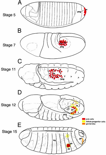
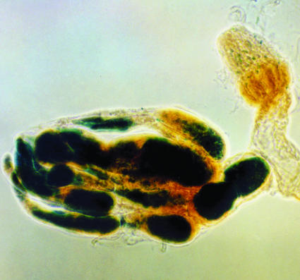
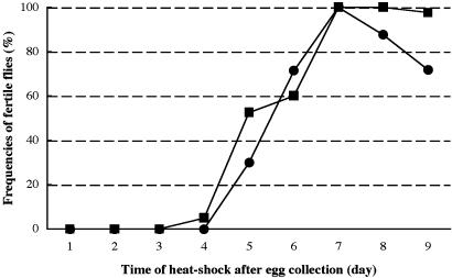
Ovarian tumors are formed either in the absence of Bam (bag-of-marbles) in germ-line cells or the overexpression of Dpp (decapentaplegic) in ovarian somatic cells. These tumor cells contain spectrosomes characteristic of ovarian germ-line stem cells and the immediate descendents called cystoblasts. We show that pole cells can successfully populate the gonad after transplantation to the dorsal mesoderm of host embryos following germ-band extension. By using this approach, we demonstrate that bam- cells can populate the gonad and become established as germ-line stem cells. Tumor cells containing the wild-type bam gene under heat shock transcriptional control are able to produce functional oocytes. Thus, stem cells/cystoblasts of the adult ovary are capable of forming stem cells in the embryonic ovary and recapitulating the development of the female germ line.
Figures
Similar articles
-
Ectopic expression of the Drosophila Bam protein eliminates oogenic germline stem cells.Development. 1997 Sep;124(18):3651-62. doi: 10.1242/dev.124.18.3651. Development. 1997. PMID: 9342057
-
Regulatory relationship among piwi, pumilio, and bag-of-marbles in Drosophila germline stem cell self-renewal and differentiation.Curr Biol. 2005 Jan 26;15(2):171-8. doi: 10.1016/j.cub.2005.01.005. Curr Biol. 2005. PMID: 15668175
-
The arrest gene is required for germline cyst formation during Drosophila oogenesis.Genesis. 2001 Apr;29(4):196-209. doi: 10.1002/gene.1024. Genesis. 2001. PMID: 11309853
-
Germ cell migration: as slow as molasses.Curr Biol. 2002 Sep 17;12(18):R612-4. doi: 10.1016/s0960-9822(02)01131-4. Curr Biol. 2002. PMID: 12372264 Review.
-
Organizing stem cell units in the Drosophila ovary.Curr Opin Genet Dev. 2015 Jun;32:31-6. doi: 10.1016/j.gde.2015.01.005. Epub 2015 Feb 19. Curr Opin Genet Dev. 2015. PMID: 25703842 Review.
Cited by
-
Molecular profiling of stem cell-like female germ line cells in Drosophila delineates networks important for stemness and differentiation.Biol Open. 2019 Nov 12;8(11):bio046789. doi: 10.1242/bio.046789. Biol Open. 2019. PMID: 31649115 Free PMC article.
-
Functional analysis of the Drosophila embryonic germ cell transcriptome by RNA interference.PLoS One. 2014 Jun 4;9(6):e98579. doi: 10.1371/journal.pone.0098579. eCollection 2014. PLoS One. 2014. PMID: 24896584 Free PMC article.
-
Establishment of stable cell lines of Drosophila germ-line stem cells.Proc Natl Acad Sci U S A. 2006 Oct 31;103(44):16325-30. doi: 10.1073/pnas.0607435103. Epub 2006 Oct 20. Proc Natl Acad Sci U S A. 2006. PMID: 17056713 Free PMC article.
-
The development of germline stem cells in Drosophila.Methods Mol Biol. 2008;450:3-26. doi: 10.1007/978-1-60327-214-8_1. Methods Mol Biol. 2008. PMID: 18370048 Free PMC article. Review.
-
Translational control in germline stem cell development.J Cell Biol. 2014 Oct 13;207(1):13-21. doi: 10.1083/jcb.201407102. J Cell Biol. 2014. PMID: 25313405 Free PMC article. Review.
References
-
- Lin, H. (2002) Nat. Rev. Genet. 3, 931–940. - PubMed
-
- Xie, T. & Spradling, A. (2001) in Stem Cell Biology, eds. Marshak, D. R., Gardner, R. L. & Gottlieb, D. (Cold Spring Harbor Lab. Press, Plainview, NY), pp. 129–148.
-
- Xie, T. & Spradling, A. C. (2000) Science 290, 328–330. - PubMed
-
- Spradling, A. C. (1993) in The Development of Drosophila, eds. Bate, M. & Maartinez-Arias, A. (Cold Spring Harbor Lab. Press, Plainview, NY), pp. 1–70.
-
- Lin, H., Yue, L. & Spradling, A. C. (1994) Development (Cambridge, U.K.) 120, 947–956. - PubMed
Publication types
MeSH terms
Substances
LinkOut - more resources
Full Text Sources
Molecular Biology Databases
Miscellaneous
