. 2003 Dec;77(23):12579-91.
doi: 10.1128/jvi.77.23.12579-12591.2003.

A ferret model of canine distemper virus virulence and immunosuppression

Affiliations

A ferret model of canine distemper virus virulence and immunosuppression

Veronika von Messling et al. J Virol. 2003 Dec.

Abstract

Canine distemper virus (CDV) infects many carnivores, including ferrets and dogs, and is the member of the Morbillivirus genus most easily amenable to experimentation in a homologous small-animal system. To gain insights into the determinants of CDV pathogenesis, we isolated a strain highly virulent for ferrets by repeated passaging in these animals. Sequence comparison of the genome of this strain with that of its highly attenuated precursor revealed 19 mutations distributed almost evenly in the six genes. We then recovered a virus from a cDNA copy of the virulent CDV strain's consensus sequence by using a modified reverse genetics system based on B cells. We infected ferrets with this virus and showed that it fully retained virulence as measured by the timing of rash appearance, disease onset, and death. Body temperature, leukocyte number, lymphocyte proliferation activity, and cell-associated viremia also had similar kinetics. We then addressed the question of the relative importance of the envelope and other viral constituents for virulence. Viruses in which the envelope genes (matrix, fusion, and hemagglutinin) of the virulent strain were combined with the other genes of the attenuated strain caused severe rash and fever even if the disease onset was delayed. Viruses in which the nucleocapsid, polymerase, and phosphoprotein genes (coding also for the V and C proteins) of the virulent strain were combined with the envelope genes of the attenuated strain caused milder signs of disease. Thus, virulence-inducing mutations have accumulated throughout the genome.


Figures

FIG. 1.
FIG. 1.
Generation of a CDV strain virulent for ferrets and sequence comparison with its precursor. (A) Selection of a virulent CDV strain. Onset of rash is indicated by dots, and time of death is indicated by triangles. Each pair of symbols represents one animal. (B) Nucleotide (NT) and amino acid (AA) differences between CDV5804 (5804) and CDV5804P (5804P). The amino acid exchanges are indicated with the one-letter code. The numbers refer to the residue position in the corresponding protein. The 50% label indicates that half of the clones had the A202S mutation while the other half carried the K208R mutation.
FIG. 2.
FIG. 2.
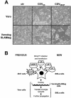
New recovery system for wild-type CDV. (A) Comparison of cytopathic effects observed after infection of Vero (top row) and VerodogSLAMtag cells (bottom row). Cell lines were infected with the attenuated CDV vaccine strain Onderstepoort as control (CDVOS, center column) or the virulent wild-type strain CDV5804P (CDV5804P, right column) at a multiplicity of infection of 0.01, and phase-contrast pictures were taken 48 h after infection. ctr, control. (B) New system allowing recovery of wild-type viruses. The expression plasmids for the N, P, and L proteins and the full-length CDV plasmid are shown with the MVA-T7 replication-deficient vaccinia virus providing the T7 polymerase. The conventional recovery process for the generation of vaccine strains is based on the transfection of 293T cells followed by the identification of infected foci and the subsequent virus propagation in Vero cells (indicated on the left). Wild-type CDV is recovered and propagated in B95a cells; foci are identified after overlaying the transfected B95a cells onto VerodogSLAMtag cells (indicated on the right).
FIG. 3.
FIG. 3.
Photograph of an animal infected with the recombinant attenuated (A and C) or virulent (B and D) strain. Chin and mouth (B) and abdominal rash (D) of a ferret at 12 d.p.i.
FIG. 4.
FIG. 4.
Course of disease in animals infected with parental or recombinant, attenuated or virulent viruses. (A) Comparison of disease progression in animals infected with the original (5804P) or recombinant (r5804P) virulent strains. Onset of rash is indicated by dots, and time of death is indicated by triangles. Each pair of symbols represents one animal. (B) Rectal temperatures of animals infected with the different attenuated or virulent strains (5804, n = 2; r5804, n = 4; 5804P, n = 4; r5804P, n = 4). Temperatures were recorded daily. The temperature (38.5°C) above which animals were considered to be pyrexic is indicated by a dotted line. Gray lines are used for attenuated viruses, and black lines are used for the pathogenic strains. Interrupted lines are used for the parental viruses, and continuous lines are used for strains recovered from infectious cDNA. Mean values are indicated. Leukocyte count (C) and neutralizing antibody titer (D) of animals infected with the different viruses (same animals as described in panel B) are shown. Blood samples were taken at the indicated time points. Lines are drawn with the same conventions as those used for panel B. Error bars are included for each data point. The leukocyte concentration (6,000 cells per mm3) below which animals are considered leukopenic and the protective neutralizing antibody titer (≥100) are indicated by a dotted line in panels C and D, respectively.
FIG. 5.
FIG. 5.
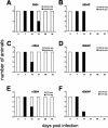
Cell-associated viremia and DTH responses of animals infected with attenuated and virulent strains. (A to D) Cell-associated viremia in the groups infected with the original viruses 5804 (A) and 5804P (B) and the recombinant viruses r5804 (C) and r5804P (D). PBMCs were isolated weekly from 0.5 ml of blood and were cocultivated with VerodogSLAMcells. Cell-associated viremia was detected by syncytium formation occurring between 1 and 3 days after the PBMC isolation. The white portions of the columns represent virus-negative animals, the black portions represent virus-positive animals. (E and F) DTH responses of animals infected with the recombinant attenuated (r5804) (E) and virulent (r5804P) (F) viruses. Animals were immunized against tetanus prior to the experiment, and a positive DTH response (diameter of induration, ≥10 mm) was detected at the time of infection. The white portions of the columns represent animals with a positive DTH response; the black portions represent DTH-negative animals.
FIG. 6.
FIG. 6.
In vitro lymphocyte proliferation activity of ferrets infected with the recombinant attenuated or virulent viruses. PBMC of each animal were isolated by Ficoll gradient centrifugation and split in four equal aliquots, stimulated with PHA or left untreated and incubated with BrdU. BrdU incorporation was detected by using a peroxidase-linked anti-BrdU antibody and was revealed with a chemiluminescent substrate. The proliferation activity was expressed as the ratio between stimulated and nonstimulated cells. The mean values of animals infected with r5804 (n = 4) are represented by squares connected by a continuous line, those of animals infected with r5804P (n = 4) are represented by dots connected by a continuous line, and those of three noninfected control animals are indicated by solid triangles connected by an interrupted line. Error bars are shown.
FIG. 7.
FIG. 7.
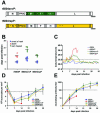
Scheme of the genome of viruses with reassorted genes and course of disease they elicited. (A) Scheme of the r5804envP genome consisting of the virulent envelope genes (M, F, and H) in combination with the attenuated replication complex genes (N, P, and L) and of the r5804repP genome with the inverse gene combination. The genes originating from strain r5804P are indicated in green for r5804envP or yellow for r5804repP. Locations of mutations are indicated by stars. (B) Disease progression in animals infected with the virulent strain r5804P and the chimeric viruses r5804envP and r5804repP. Onset of rash is indicated by dots, time of death by triangles, and healing of rash by rhombuses. Each pair of symbols represents one animal. (C) Rectal temperatures were recorded daily (r5804envP, n = 6; r5804repP, n = 4). The temperature (38.5°C) above which animals were considered to be pyrexic is indicated by a dotted line. The mean values of all animals infected with 5804 are represented by a blue line, and those of all animals infected with r5804P are represented by a red line. Values of animals infected with r5804envP are represented by a green line, and those of animals infected with r5804repP are represented by a yellow line. Leukocyte count (D) and neutralizing antibody titer (E) of animals infected with the different viruses are shown. Blood samples were taken at the indicated time points. The color coding is as described for panel C. Error bars are included for each data point. Total leukocyte counts and neutralizing antibody titers were determined as described in Materials and Methods. The leukocyte concentration (6,000 cells per mm3) below which animals are considered leukopenic and a protective neutralizing antibody titer (≥100) are indicated by a dotted line in panels D and E, respectively.
FIG. 8.
FIG. 8.
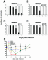
Cell-associated viremia, DTH responses, and in vitro lymphocyte proliferation activity of viruses with reassorted genes. (A and B) Cell-associated viremia detected after infection with r5804envP (A) and r5804repP (B). PBMCs were isolated weekly from 0.5 ml of blood and were cocultivated with VerodogSLAMcells. Cell-associated viremia was monitored by detection of syncytium formation between 1 and 3 days after the PBMC isolation. The white portions of the bars represent virus-negative animals, and the black portions represent virus-positive animals. (C and D) DTH responses of animals infected r5804envP (C) and r5804envP (D). Animals were immunized against tetanus prior to the experiment, and a positive DTH response was detected at the time of infection. The white portion of the columns represent animals with a positive DTH response, the black portions represent DTH-negative animals. (E) Comparison of in vitro proliferation activity of PBMC from animals infected the unaltered viruses r5804 (n = 4) and r5804P (n = 4) or the chimeric viruses r5804envP (n = 6) and r5804repP (n = 4). Methods were as described in the legend to Fig. 6. The color coding is as described for Fig. 7D. The proliferation activity of three noninfected control animals at each time point is indicated by black squares connected by an interrupted black line. Error bars are shown.

Similar articles

Cited by

References

    1. Anonymous. 2002. From the Centers for Disease Control. Measles—United States, 2000. JAMA 287:1105-1106. - PubMed
    1. Appel, M. J., W. R. Shek, and B. A. Summers. 1982. Lymphocyte-mediated immune cytotoxicity in dogs infected with virulent canine distemper virus. Infect. Immun. 37:592-600. - PMC - PubMed
    1. Aylward, R. B., J. Clements, and J. M. Olive. 1997. The impact of immunization control activities on measles outbreaks in middle and low income countries. Int. J. Epidemiol. 26:662-669. - PubMed
    1. Baranowski, E., C. M. Ruiz-Jarabo, and E. Domingo. 2001. Evolution of cell recognition by viruses. Science 292:1102-1105. - PubMed
    1. Baron, M. D., and T. Barrett. 1997. Rescue of rinderpest virus from cloned cDNA. J. Virol. 71:1265-1271. - PMC - PubMed

Publication types

Substances

LinkOut - more resources