. 2004 Jan 16;279(3):1810-8.
doi: 10.1074/jbc.M305796200. Epub 2003 Nov 3.

Prolonged kappa opioid receptor phosphorylation mediated by G-protein receptor kinase underlies sustained analgesic tolerance

Affiliations

Prolonged kappa opioid receptor phosphorylation mediated by G-protein receptor kinase underlies sustained analgesic tolerance

Jay P McLaughlin et al. J Biol Chem. .

Abstract

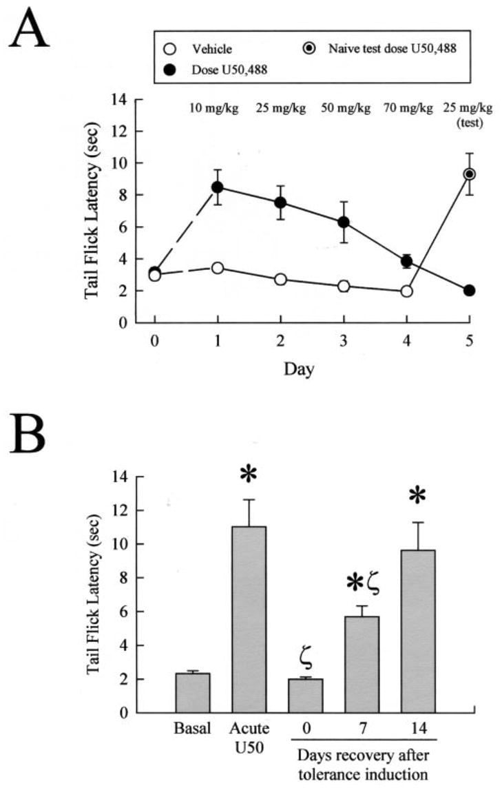
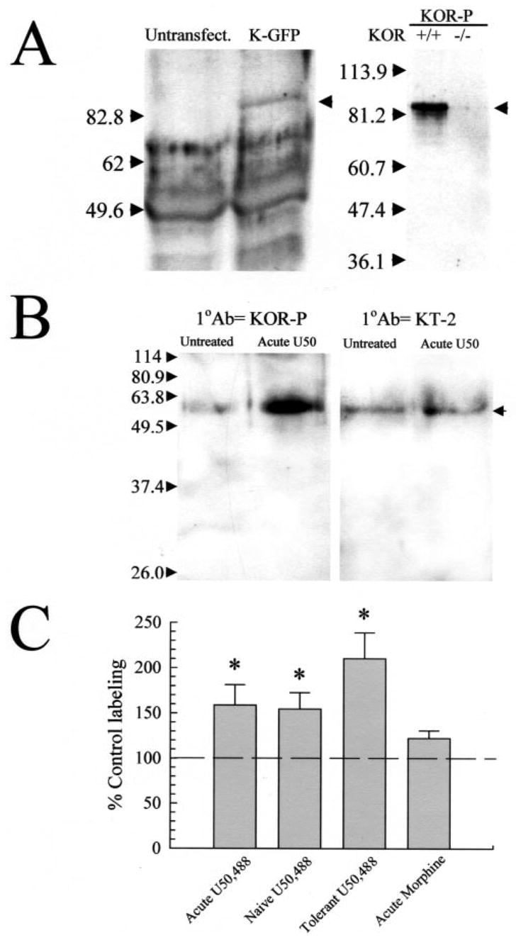
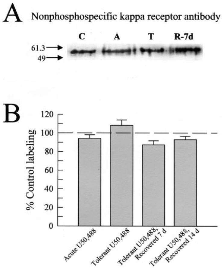
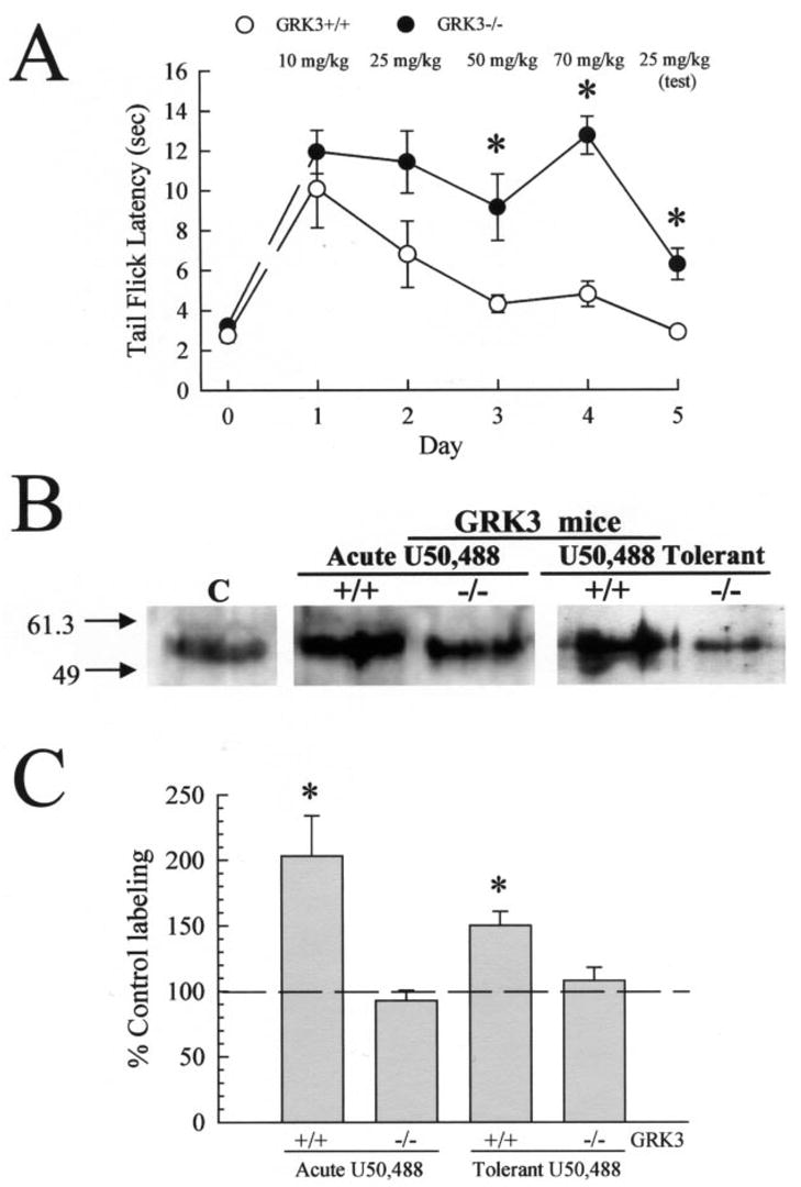
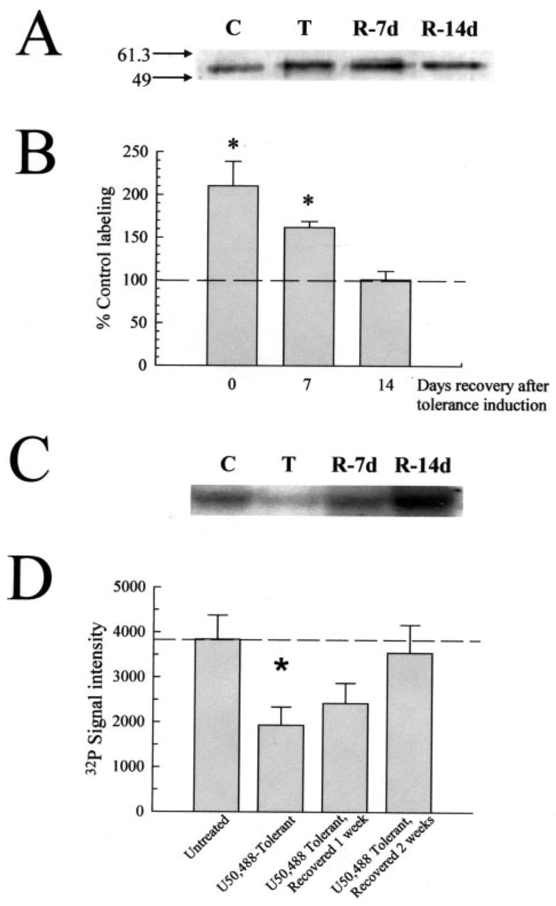
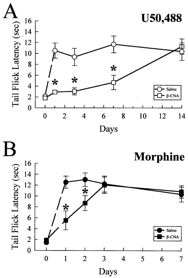
Kappa opioid receptor (KOR) desensitization was previously shown to follow agonist-dependent phosphorylation of serine 369 by G-protein receptor kinase (GRK) and beta-arrestin binding in transfected cells. To study the in vivo effects induced by phosphorylation of KOR(S369), C57Bl/6 mice were administered single or repeated doses of the KOR agonist, U50,488, and isolated brain glycoprotein was probed with an antibody, KOR-P, that specifically recognized phosphoserine 369 KOR. Western blot analysis using KOR-P antibody showed that labeling intensity increased after either single or repeated treatment of mice with U50,488 by 59 +/- 22% and 101 +/- 29%, respectively. In contrast, there was no change in labeling intensity by nonphosphoselective KOR antibodies following acute or chronic in vivo treatment with kappa agonist. Moreover, mice lacking GRK3 showed no increase in KOR-P labeling and developed significantly less analgesic tolerance following treatment with kappa agonist. The result suggests that tolerance to kappa agonists includes phosphorylation of serine 369 within KOR by GRK3. Recovery of analgesic potency and reduction of elevated KOR-P labeling in wild-type mice both required 2 weeks to return to base line. Consistent with these results, in vitro phosphorylation by GRK3 of KOR isolated from tolerant mice resulted in 46 +/- 7% less (32)P incorporation than in KOR isolated from untreated mice. In addition, in vitro (32)P incorporation returned to base line levels only in KOR isolated from tolerant mice allowed to recover for 2 weeks. The coincident reversal of analgesic tolerance and slow return to a basal phosphorylation state matched the regeneration rate of functional kappa receptors following irreversible antagonism and suggested that receptor replacement rather than dephosphorylation was required to restore sensitivity.


Figures

Fig. 1
Fig. 1. Reversal of analgesic tolerance following chronic kappa opioid agonist treatment is slow
A, mice developed tolerance to the antinociceptive effects of U50,488 following repeated administration. Latency of tail withdrawal from the 55 °C warm water was measured in untreated animals, which were then injected over 4 days with increasing doses of U50,488 (●) or vehicle (○) as detailed under “Experimental Procedures.” Antinociceptive response to morning injection of U50,488 was measured each day, 30 min after administration, to monitor the generation of tolerance. Animals pretreated for 5 days with U50,488 developed pronounced antinociceptive tolerance (day 5, ●), whereas naïve animals receiving only vehicle prior to the test dose showed a normal antinociceptive effect from 25 mg/kg U50,488 (⊙). Data points represent the means ± S.E. of 10 mice from two separate experiments. In some cases, symbol size exceeds error bar width. B, mice made tolerant to U50,488 recover sensitivity to U50,488 after 2 weeks being drug-free. Mice made tolerant to U50,488 as above were then allowed to recover 1 or 2 weeks (12 or 19 days after initial drug administration) without drug. After the recovery period, animals were tested by measuring the tail withdrawal latency response 30 min after intraperitoneal 25 mg/kg U50,488. Data points represent the means ± S.E. of 7–8 mice from two separate experiments. *, p < 0.05 compared with basal group;ζ, p < 0.05 compared with acute U50,488.
Fig. 2
Fig. 2. Opioid agonist-induced tolerance corresponds to an increase in kappa opioid receptor phosphorylation detected by the KOR-phosphoselective antibody, KOR-P
A, kappa opioid receptor selective binding by the KOR-P antibody. Left panel: representative gel shows immunoprecipitated KOR-GFP labeled by KOR-P. Lysates from untransfected HEK293 cells or cells transiently transfected with KOR-GFP cDNA were immunoprecipitated with 20 μg of KE-4 and then analyzed by Western blot with 5 μg of KOR-P. Cells were pre-treated with 10 μM U50,488 for 30 min at 37 °C. The arrow on the right side highlights an ~85-kDa band corresponding to the predicted molecular mass of the KOR-GFP fusion protein which was labeled in the KOR-GFP-transfected cell lysate but not in the untransfected cell lysates. Right panel: representative Western blot of isolated mouse brain membrane protein (40 μg/lane) taken from either KOR wild-type (WT) or knockout (KO) mice, and incubated with KOR-P. For these experiments, brain protein was resolved on nonreducing PAGE gels. The KOR-P antibody labeled protein in the WT, but not KO, brain protein samples. Identical specificity results were obtained with the KOR-selective antibody, KT-2, labeling of KOR(+/+) and KOR(−/−) brain membrane proteins (data not shown). B, representative Western blots of isolated mouse brain membrane protein (90 μg/lane), incubated with KOR-P (left) or KT-2 (right). For these and subsequent experiments, brain protein was resolved on reducing PAGE gels containing 0.01% SDS. Both antibodies recognized a protein of ~55 kDa corresponding to the KOR in samples from untreated control animals (Untreated). Pre-treatment with 30 mg/kg intraperitoneal U50,488 (Acute) increased blotting intensity with KOR-P compared with untreated controls. This increase in blotting intensity indicates an increase in KOR phosphorylation, as the nonphosphospecific KOR antibodies KT-2 (shown) or KE-4 (not shown) detected no significant change. C, phosphorylation state of the KOR is increased in animals following agonist treatment. A single administration of the kappa-selective agonist (Acute U50,488 group) increased KOR phosphorylation ~60%, whereas chronic administration more than doubles basal phosphorylation of the KOR (Tolerant). Mice treated with vehicle for 5 days then tested with a single injection of 25 mg/kg U50,488 on day 5 (Naïve U50,488 group) demonstrated a 63 ± 16% increase in KOR-P labeling, comparable to that produced by a single U50,488 injection. In contrast, while acute morphine (30 mg/kg, intraperitoneally) produced a tail withdrawal latency at the 15 s cutoff in the 55 °C warm water tail withdrawal assay 30 min after administration, it did not significantly increase the phosphorylation of KOR measured in subsequent Western blot analysis using KOR-P antibody. The lack of effect of morphine further demonstrates that specific kappa receptor activation was required for increased KOR-P labeling. The graph summarizes the analysis results of protein blots using NIH densitometry software of the ECL image, standardized against matching, untreated control protein from the same experiment. Data points represent the means ± S.E. of 26–33 independent experiments. Statistical significance was determined with Student’s paired t test. *, p < 0.05 against matching KE-4 antibody-labeled band.
Fig. 3
Fig. 3. Kappa receptor number is unchanged by acute or chronic U50,488 treatment
A, representative Western blot analysis with KE-4 antibody and 90 μg/lane mouse brain membrane protein isolated from untreated control mice (“C”), mice pretreated acutely with U50,488 (25 mg/kg intraperitoneal, “A”), mice made tolerant to U50,488 and sacrificed immediately (“T”), or after a recovery period of 7 days (“R-7d”). KE-4 antibody labeling intensity was consistent across all protein samples, suggesting the total number of KOR remained unchanged despite the different pretreatments with U50,488 and recovery time. B, summary graph of densitometry analysis of Western blots shows KE-4 antibody labeling of KOR from each set of pretreated mice. No significant differences were found in KE-4 antibody labeling across the tissue sets tested. Data points represent the means ± S.E. of 18 –24 independent Western blot experiments.
Fig. 4
Fig. 4. Kappa agonist administration induces KOR phosphorylation in mice mediated by GRK3
A, onset of U50,488 antinociceptive tolerance was impaired in GRK3(−/−) mice in the mouse 55 °C warm water, tail withdrawal assay. Latency to tail flick from a warm water stimulus was measured in untreated animals, and then increasing doses of U50,488 were administered over 4 days to GRK3(+/+) (○) or (−/−) (●) mice as detailed under “Experimental Procedures.” Antinociceptive response to morning injection of U50,488 was measured each day, 30 min after administration, to monitor the progress of antinociceptive tolerance. Development of antinociceptive tolerance was tested on day 5 with a test dose of 25 mg/kg U50,488. GRK3(+/+) animals pretreated for the week with U50,488 developed pronounced antinociceptive tolerance (day 5, ○), whereas GRK3(−/−) animals showed a reduced U50,488 antinociceptive tolerance with delayed onset of effect (●). Data points represent the means ± S.E. of 8 mice from two separate determinations. Both GRK3(+/+) and (−/−) mice were also injected acutely with 30 mg/kg intraperitoneal U50,488 and sacrificed 30 min later. B, representative Western blot analysis of 90 μg/lane brain membrane protein isolated from untreated C57Bl/6 mice (Control, C) or GRK3(+/+) or GRK3(−/−) mice treated acutely (center) or chronically (right) with U50,488 and blotted with 5 μg/ml KOR-P. KOR protein isolated from U50,488-pre-treated GRK(+/+), but not GRK(−/−), mice showed increased labeling by KOR-P. KE-4 showed no change in blotting intensity between the protein sets tested. A summary graph is shown in C, with data representing the means ± S.E. of 19–20 independent experiments analyzed with NIH Image software of the ECL image and standardized against matching, untreated control C57Bl/6 mouse brain protein from the same experiment. Brain membrane protein was isolated by WGA column purification in all cases as detailed under “Experimental Procedures.” Statistical significance was determined with Student’s paired t test. *, p < 0.05 against matching KE-4 antibody-labeled band.
Fig. 5
Fig. 5. Return to basal KOR phosphorylation state in U50,488-tolerant mice follows recovery from antinociceptive tolerance after 14 days
A, representative Western blot analysis with KOR-P and 90 μg/lane mouse brain membrane protein isolated from untreated control mice (“C”), or mice made tolerant to U50,488 and sacrificed immediately (“T”), or after a recovery period of 7 or 14 days (“R-7d” and “R-14d,” respectively). KOR-P labeling intensity was elevated in protein samples from tolerant and 1-week recovered animals, suggestive of prolonged KOR phosphorylation matching the demonstration of antinociceptive tolerance. B, summary graph of densitometry analysis of Western blots shows that the phosphorylation intensity of KOR from tolerant mice requires 2 weeks to return to basal values. Data points represent the means ± S.E. of 6 –24 independent experiments. *, p < 0.01 against matching KE-4-labeled band. C, representative PAGE gel with 90 mg/lane mouse brain protein isolated from untreated control mice (“C”), or mice made tolerant to U50,488 and sacrificed immediately (“T”), or after a recovery period of 7 or 14 days (“R-7d” and “R-14d,” respectively) following 32P incorporation with isolated GRK3 enzyme. Following GRK3/32P incubation, all samples were immunoprecipitated with the KOR-selective antibody, KT-2 as described under “Experimental Procedures.” Adjusting for background, 32P labeling intensity was decreased in protein samples from tolerant and 1-week recovered animals, suggestive of prolonged KOR phosphorylation. These results match the changes KOR-P labeling and antinociceptive tolerance, indicating that GRK3 phosphorylation sites were occupied by nonradioactive phosphate. D, summary graph of GRK3/32P incorporation experiments shows 32P signal intensity level of KOR from tolerant mice requires 2 weeks to return to basal values. Data points represent the means ± S.E. of five independent experiments. *, p < 0.05 against matching untreated control brain protein value.
Fig. 6
Fig. 6. Mice pretreated with β-CNA require prolonged recovery for kappa, but not mu, opioid-induced antinociception
C57Bl/6 mice were treated with saline (0.9% intraperitoneally, circles) or β-CNA (2.4 nmol intracerebroventricularly, squares) and returned to their home cages for 1–14 days prior to use in analgesic testing with opioid agonists. A, antinociception induced by the kappa-selective agonist, U50,488, is blocked by pretreatment with opioid antagonists up to 2 weeks. After recovering from antagonist pretreatment for 1–14 days, mice were administered U50,488 (25 mg/kg, intraperitoneally), and the antinociceptive effect was measured 30 min later. Pretreatment with the irreversible opioid antagonist β-CNA (open squares) produced significant antagonism of U50,488-induced antinociception up to 7 days after antagonist administration, with recovery observed after 14 days. In contrast, similar β-CNA pre-treatment (closed squares, B) significantly reduced the antinociception induced by subsequent administration of morphine (30 mg/kg, intraperitoneally, closed circles; B) only up to 2 days, with recovery observed after 3 days. Each animal received only a single dose of agonist (U50,488 or morphine). Data points represent the means ± S.E. of 9 –14 mice. *, p < 0.05 against matching saline pre-treated time point.

Similar articles

Cited by

References

    1. Gutstein HB, Akil H. Goodman and Gilman’s The Pharmacological Basis of Therapeutics. Vol. 10. McGraw-Hill Co.; New York, NY: 2001. pp. 569–619.
    1. Nestler EJ. Curr Opin Neurobiol. 1997;7:713–719. - PubMed
    1. Law PY, Wong YH, Loh HH. Annu Rev Pharmacol Toxicol. 2000;40:389 –430. - PubMed
    1. Lefkowitz RJ. J Biol Chem. 1998;273:18677–18680. - PubMed
    1. Pei G, Kieffer BL, Lefkowitz RJ, Freedman NJ. Mol Pharmacol. 1995;48:173–177. - PubMed

Publication types

MeSH terms

Substances

LinkOut - more resources