. 2004 Feb;15(2):870-82.
doi: 10.1091/mbc.e03-07-0517. Epub 2003 Oct 31.

GLUT4 is retained by an intracellular cycle of vesicle formation and fusion with endosomes

Affiliations

GLUT4 is retained by an intracellular cycle of vesicle formation and fusion with endosomes

Ola Karylowski et al. Mol Biol Cell. 2004 Feb.

Abstract

The intracellularly stored GLUT4 glucose transporter is rapidly translocated to the cell surface upon insulin stimulation. Regulation of GLUT4 distribution is key for the maintenance of whole body glucose homeostasis. We find that GLUT4 is excluded from the plasma membrane of adipocytes by a dynamic retention/retrieval mechanism. Our kinetic studies indicate that GLUT4-containing vesicles continually bud and fuse with endosomes in the absence of insulin and that these GLUT4 vesicles are 5 times as likely to fuse with an endosome as with the plasma membrane. We hypothesize that this intracellular cycle of vesicle budding and fusion is an element of the active mechanism by which GLUT4 is retained. The GLUT4 trafficking pathway does not extensively overlap with that of furin, indicating that the trans-Golgi network, a compartment in which furin accumulates, is not a significant storage reservoir of GLUT4. An intact microtubule cytoskeleton is required for insulin-stimulated recruitment to the cell surface, although it is not required for the basal budding/fusion cycle. Nocodazole disruption of the microtubule cytoskeleton reduces the insulin-stimulated exocytosis of GLUT4, accounting for the reduced insulin-stimulated translocation of GLUT4 to the cell surface.


Figures
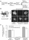
Figure 1.
Figure 1.
Anti-HA antibody binding does not alter trafficking of HA-GLUT4-GFP. (A) Outline of the experiment protocol. The incubation with anti-HA antibody was performed in the presence of insulin because the recycling time of GLUT4 in the absence of insulin is very slow (Figure 2). (B) A cartoon of the HA-GLUT4-GFP construct. The distributions of HA-GLUT4-GFP prebound by anti-HA antibody in the presence of insulin, after removal of insulin, and after restimulation with insulin are shown. Surface HA-GLUT4-GFP was detected by indirect IF with a Cy3-labeled anti-mouse IgG antibody. The images are from representative cells for each condition. (C) Quantification of surface-to-total distribution of HA-GLUT4-GFP unbound and prebound with anti-HA-antibody. Prebound HA-GLUT4-GFP on the surface was measured in IF with a Cy3-goat anti-mouse IgG, and unbound HA-GLUT4-GFP (control cells) was measured by first binding anti-HA antibody to fixed cells followed by Cy3-Goat anti-mouse IgG. The results shown are the averages of 20 cells per condition ± SEM from a representative experiment. The data have been normalized to the surface expression level in the presence of insulin (condition 1). Insulin (170 nM) was used for stimulation.
Figure 2.
Figure 2.
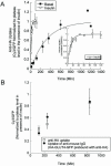
Insulin increases the cycling rate of HA-GLUT4-GFP between the interior and cell surface. (A) The uptake of anti-HA antibody from the medium at 37°C in the presence and absence of insulin. Total cell associated anti-HA was measured by IF of permeabilized cells. The data are the average of three experiments ± SEM in which the Cy3/GFP ratio per cell for each experiment were normalized to the plateau level for the samples treated with insulin in that experiment. The curves are (Cy3/GFP)t = 1 - exp(-ke*t), where the (Cy3/GFP)t ratio is the fraction of HA-GLUT4-GFP bound by anti-HA antibody at time t, and ke is the exocytic rate constant. The inset panel is the data in the presence of insulin plotted on an expanded time scale. Insulin (170 nM) was used for stimulation. (B) HA-GLUT4-GFP prebound by anti-HA antibody (filled symbol) cycles to the cell surface in the basal state with similar kinetics to HA-GLUT4-GFP (open symbol), establishing that the basal retention is a dynamic process involving slow exocytosis and rapid retrieval. The data are from a single experiment (average values for 20 cells per time point ± SEM) and have been normalized to the plateau Cy3/GFP level when cells are incubated with anti-HA for 60 min in the presence of insulin (A).
Figure 3.
Figure 3.
The IRAP-TR rapidly moves from the cell surface to the specialized compartment in the basal state. (A) Flow diagram of experiment shown in B. (B) IRAP-TR is rapidly transported from the cell surface to both the TR-containing endosomes and the specialized GLUT4/IRAP compartment. Cells coexpressing HA-GLUT4-GFP and the TR or the IRAP-TR chimera were incubated with unlabeled anti-HA antibody to label the complete cycling pool. The cells were pulse labeled with HRP-Tf, and at the indicated times, cells were incubated with DAB/H2O2 at 4°C. The amount of anti-HA epitope ablation was determined by staining saponin-permeabilized cells with Cy3-labeled anti-mouse IgG. The data are the averages of five experiments ± SEM. In each experiment the average Cy3/GFP ratio of 20 cells per time point were normalized to the Cy3/GFP ratio of cells that were not incubated with HRP-Tf (time 0). The curves are (Cy3/GFP)t = A*exp(-ke*t) + C, where A is the fraction of anti-HA antibody that can be ablated, ke is the rate constant for the epitope ablation, and C is the plateau level, which reflects the fraction of HA epitopes that are not ablated. The (Cy3/GFP)t value is the fraction of epitopes unablated at time t.
Figure 4.
Figure 4.
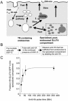
There is rapid bidirectional transport between endosomes and the specialized GLUT4/IRAP compartment. (A) Cartoon of the two models. GLUT4 equilibrates with the cell surface with a half-time of 230 min. If there is unidirectional traffic from endosomes to the specialized compartment(s), then the specialized compartment must be a stable compartment to which GLUT4 vesicles fuse and from which vesicles bud (pathway 1). In this model, exit of GLUT4 from endosomes is necessarily slow to achieve the near equal distribution between endosomes and the specialized compartment. The bidirectional model (pathway 2) is compatible with the specialized compartment(s) being fusion-fission competent (as in the unidirectional model) or a reservoir of GLUT4 vesicles. In the latter case, the mixing would occur as GLUT4 vesicles refuse with endosomes. In this model transport from endosomes can be rapid. (B) Flow diagram of experiment shown in C. (C) GLUT4 equilibrates between TR-containing endosomes and the specialized compartment with a half-time of ∼20 min. Adipocytes coexpressing HA-GLUT4-GFP and the TR or the IRAP-TR chimera were incubated with HRP-Tf for 4 h to load the endosomes, or endosomes and the specialized compartment, respectively. The cells were pulsed in the continued presence of HRP-Tf with anti-HA and at the times specified the codistribution of the anti-HA antibody and Tf-HRP were determined as in Figure 3. The fraction of HA-GLUT4-GFP in the specialized compartment at each time point is shown. The data are the averages ± SEM from four independent experiments. The curve is (Cy3/GFP)t = A0 - (A0*exp(-k*t)), where A0 is the fraction of HA-GLUT4-GFP in the specialized compartment at steady state and k is the rate constant for equilibration between endosomes and the specialized pool. The (Cy3/GFP)t value is the fraction of HA-GLUT4-GFP in the specialized compartment at time t.
Figure 5.
Figure 5.
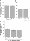
Overlap of the insulin-regulated pathway with the furin pathway is restricted to TR-containing endosomes. (A) IRAP-TR, like TR, has access to only 20% of the Alexa488-anti-Tac fluorescence internalized by Tac-furin. Cells coexpressing Tacfurin and the TR or the IRAP-TR chimera were incubated with an Alexa488-labeled anti-Tac-antibody and HRP-Tf for 5 h. The cells were chilled to 4°C and treated with DAB/H2O2. The Alexa488 fluorescence in cells incubated with HRP-Tf is plotted as a percentage of the Alexa488 fluorescence in cells that were not incubated with HRP-Tf. The data are from a representative experiment and are the averages ± SEM of 25 cells per condition. (B) Fifty percent of the TR, the IRAP-TR chimera, and HA-GLUT4-GFP are in intracellular compartments accessible to Tac-furin. Cells expressing Tac-furin and the TR, IRAP-TR chimera, or HA-GLUT4-GFP were incubated with an HRP-anti-Tac antibody and Cy3-Tf (in cells expressing TR or IRAP-TR) or Cy3-anti-HA-antibody (in cells expressing HA-GLUT4-GFP) for 6 h. The Cy3 fluorescence is plotted as a percentage of the Cy3 fluorescence in cells that were not incubated with HRP-anti-Tac antibody. The data are the averages ± SEM of four independent experiments. (C) Tacfurin and HA-GLUT4-GFP overlap in TR endosomes. Cells expressing Tac-furin, TR and HA-GLUT4-GFP were incubated for 6.5 h in medium containing saturating concentrations of Cy3-anti-HA and HRP-anti-Tac antibodies with or without HRP-Tf. The cells were chilled to 4°C and treated with DAB/H2O2. The data are plotted as the percentage of Cy3 fluorescence in cells that were not incubated with either HRP-anti-Tac or HRP-Tf. The Cy3 fluorescence was divided by GFP per cell to correct for expression of HA-GLUT4-GFP. The data shown are the averages ± SEM (at least 20 cells per condition) from a representative experiment.
Figure 6.
Figure 6.
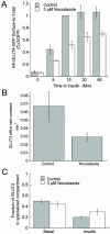
Nocodazole disruption of microtubules partially inhibits translocation of GLUT4 without affecting basal retention. (A) Insulin-stimulated recruitment of GLUT4 to the cell surface was inhibited by nocodazole. Cells were incubated with 3 μM nocodazole for 30 min at 37°C and then treated with 170 nM insulin and nocodazole for the times indicated. Control cells were similarly treated except nocodazole was not included in the incubations. Surface HA-GLUT4-GFP was measured by IF. To allow for direct comparison among experiments, the data from each experiment were normalized to the control 15-min insulin-stimulated surface-to-total value determined in that experiment. The data shown are the averages of at least four independent measurements ± SEM, except for the 60-min time point, which is the average of two experiments ± SD. Nocodazole (3 μM) depolymerized the microtubule cytoskeleton as determined by IF in fixed cells. (B) Insulin-stimulated exocytosis of GLUT4 was inhibited by nocodazole. Cells were incubated with 3 μM nocodazole for 30 min, followed by a 15-min incubation with 170 nM insulin and 3 μM nocodazole. After this incubation antibody was added to the cells in the continued presence of nocodazole and insulin, and the accumulation of anti-HA antibody was measured as described in Figure 2. The efflux rate constants for exocytosis, measured as described in Figure 2, are presented. Control cells were similarly treated except nocodazole was not included in the incubations. The data are the average efflux rate constants ± SEM calculated in three separate experiments. (C) Nocodazole does not alter distribution of GLUT4 between endosomes and the specialized compartment. Cells expressing HA-GLUT4-GFP and TR or IRAP-TR were preincubated with anti-HA antibody and with or without HRP-Tf for 390 min at 37°C in medium without insulin. Half of the samples received 3 μM nocodazole for the final 30 min of incubation. Cells were then incubated for an additional 30 min with or without HRP-Tf ± insulin ± 3 μM nocodazole. Colocalization of intracellular HA-GLUT4-GFP with TR and IRAP-TR was determined as described in MATERIALS AND METHODS. The data are the averages ± SEM of five independent measurements.
Figure 7.
Figure 7.
Model for the dynamic exclusion of GLUT4 from the plasma membrane by a retention-retrieval mechanism. In the basal state, GLUT4 vesicles preferentially fuse with endosomes relative to the plasma membrane, thereby depleting the cell surface of GLUT4. Any GLUT4 that is introduced into the plasma membrane is rapidly retrieved by clathrin-mediated endocytosis back to endosomes. Insulin increases the exocytic rate of GLUT4 and thereby induces an increase of GLUT4 in the plasma membrane. Compartments of the insulin-regulated pathway are in gray. The half-time for appearance of GLUT4 at the cell surface in the presence and absence of insulin were directly measured, as was the equilibration time between endosomes and the specialized compartment in the basal state. For kinetic modeling experiments, the following parameters were used: a half-time of GLUT4 internalization of 10 min (Tengholm et al., 2003), and a steady-state distribution of GLUT4 in the basal state of 4% in the plasma membrane and 48% each in endosomes and the specialized pool. A value of 4% on the surface was calculated from % GLUT4Surface = 1/(1 + (kInternalziation/kExocytosis)), with kInternalziation = 0.07 min-1 (Tengholm et al., 2003) and kExocytosis = 0.003 min-1. The values for k2 in insulin and k4 in basal and insulin conditions were determined using SAAM II compartmentalization modeling software (Barrett et al., 1998; Cobelli and Foster, 1998). See text for discussion of the model.

Similar articles

Cited by

References

    1. Barrett, P., Bell, B., Cobelli, C., Golde, H., Schumitzky, A., Vicini, P., and Foster, D. (1998). SAAM II: simulation, analysis, and modeling software for tracer and pharmacokinetic studies. Metabolism 47, 484-492. - PubMed
    1. Bosshart, H., Humphrey, J., Deignan, E., Davidson, J., Drazba, J., Yuan, L., Oorschot, V., Peters, P.J., and Bonifacino, J.S. (1994). The cytoplasmic domain mediates localization of furin to the trans-Golgi network en route to the endosomal/lysosomal system. J. Cell Biol. 126, 1157-1172. - PMC - PubMed
    1. Brown, D. (2003). The ins and outs of aquaporin-2 trafficking. Am. J. Physiol. Renal. Physiol. 284, F893-F901. - PubMed
    1. Bryant, N., Govers, R., and James, D. (2002). Regulated transport of the glucose transporter GLUT4. Nat. Rev. Mol. Cell. Biol. 3, p267-277. - PubMed
    1. Cobelli, C., and Foster, D. (1998). Compartmental models: theory and practice using the SAAM II software system. Adv. Exp. Med. Biol. 445, 79-101. - PubMed

Publication types

MeSH terms

LinkOut - more resources